
Introduction
Seborrheic keratoses (SKs) are among the most common benign skin tumours in adults, with prevalence increasing with age [1]. Studies estimate that a majority of elderly individuals develop at least a few SK lesions [2]. While SKs are harmless, patients frequently seek removal due to cosmetic concerns or symptoms (e.g. pruritus or recurrent mechanical trauma, including lesions on the collar line or scalp). Importantly, SKs in highly visible or sun-exposed areas pose a therapeutic challenge: clinicians must balance effective clearance with the risk of scarring or dyspigmentation on cosmetically sensitive skin [3, 4].
A variety of treatment options exist for SK, each with its own advantages and disadvantages. Traditional destructive methods include topical therapies, cryotherapy, curettage (with or without electrodessication), and ablative or non-ablative lasers [5–13]. Cryotherapy is widely used due to its simplicity, but may incompletely remove thicker lesions and often causes post-inflammatory hypopigmentation, especially in darker skin types [7–9]. Curettage (often combined with light electrocautery) can provide immediate removal, yet may leave scars or textural change, which is particularly undesirable on the head and neck [7–9]. Laser-based treatments can achieve high clearance, but outcomes depend on lesion thickness and technique, and access is limited by cost and the need for specialised expertise [9–13]. Lasers can offer good cosmetic outcomes in selected lesions (e.g. flatter SKs or dermatosis papulosa nigra), but are not universally available [5, 9–13]. Topical agents (including hydrogen peroxide 40% solution, retinoids, or chemical peels) have also been explored. However, efficacy is variable and typically inferior to procedural treatments [6]. Overall, conventional modalities often require a trade-off between efficacy and cosmetic risk, supporting the need for approaches that maximize clearance while minimizing adverse effects.
High-frequency focused ultrasound has emerged as a novel, non-invasive modality for dermatologic treatments [14–16]. High-intensity focused ultrasound (HIFU) delivers acoustic energy into a precisely defined focal volume, inducing thermal ablation of target tissue without an incision [15, 16]. In dermatology, a 20 MHz HIFU device has been introduced for superficial lesions [16]. Early studies reported use in benign lesions such as SKs, sebaceous hyperplasia, and neurofibromas, and in inflammatory dermatoses including vulvar lichen sclerosus and granuloma annulare [14, 17–22]. More recently, pilot studies extended HIFU to premalignant and malignant lesions [23–26], including significant clearance of facial actinic keratoses and selective ablation of epidermal lesions such as superficial basal cell carcinoma in open-label studies [23–25].
However, data on HIFU for SK remain limited to small case series, and questions persist regarding its optimal use, including whether lesion location or morphology affects outcomes [14]. In particular, the head and neck region has received little focused analysis, despite being a common site for cosmetically driven treatments.
Dermoscopic examination is routinely used to diagnose SK and distinguish it from malignancies such as melanoma [27]. Classic dermoscopic features include a well-demarcated border, milia-like cysts, comedo-like openings, and fissures and ridges producing a cerebriform appearance, along with the absence of features typical for melanocytic lesions [27–30]. Many SKs are uniformly pigmented, although some display multiple colours, especially in darker skin types or irritated lesions [29, 30]. While dermoscopy is primarily diagnostic, its prognostic value is increasingly explored [31–34]. Baseline dermoscopic features have been linked to treatment response in other dermatologic conditions, including warts, inflammatory dermatoses, and vascular lesions [31–34]. We hypothesized that, for SK, dermoscopic characteristics might similarly predict response to HIFU: features reflecting keratinous architecture and lesion confinement (e.g. sharp demarcation, pronounced fissures) could indicate lesions amenable to focused ultrasound ablation, whereas marked colour heterogeneity could be associated with lower clearance and/or a higher risk of post-treatment pigmentary change [27, 28, 35].
Aim
To assess the effectiveness and safety of 20 MHz HIFU for head-and-neck seborrheic keratoses, compare outcomes by anatomical site (scalp/face/neck), and identify baseline dermoscopic predictors of clearance and cosmetic outcome.
Material and methods
Study design and setting
We conducted a single-centre retrospective cohort study at a dermatology outpatient clinic, analysing archival medical records and dermoscopic photographs of patients treated with high-frequency HIFU for SK between 2021 and 2025. All procedures were performed as part of routine clinical care. The study was conducted in accordance with the Declaration of Helsinki and was reviewed and approved by the Bioethics Committee at the Lower Silesian Medical Chamber in Wroclaw, Poland (approval no. 23/BNBO/2025). Data were de-identified prior to analysis. Written informed consent for treatment (and for the use of de-identified clinical photographs, in line with the clinic policy) had been obtained from all patients.
Inclusion and exclusion criteria
We included adult patients (≥ 18 years) who underwent HIFU treatment for one or more clinically and dermoscopically diagnosed SK lesions located on the face, scalp, or neck. Lesions were eligible if they had: (1) baseline dermoscopic images and clinical photographs, (2) recorded HIFU treatment parameters, and (3) at least one follow-up evaluation with outcome assessment. To minimize misclassification related to early wound healing, the primary analysis focused on lesions with an outcome assessment documented at ≥ 2 months post-treatment. Lesions with missing follow-up or unclear outcome documentation were excluded. Lesions with any dermoscopic/clinical doubt were not treated destructively and were managed according to standard diagnostic pathways (biopsy/surgical excision).
Treatment procedure (HIFU)
All treatments were performed using a 20-MHz high-intensity focused ultrasound system (System ONE-M, TOOsonix A/S, Hørsholm, Denmark). Each lesion was treated in one session, except for one lesion that underwent a repeat HIFU session due to incomplete initial clearance. Before each procedure, the transducer chamber was filled with deionized (DI) water and sealed with a polyethylene membrane. Acoustic coupling between the transducer and the skin surface was achieved using Parker Aquasonic 100® ultrasound gel (Parker Laboratories Inc., Fairfield, NJ, USA). No local anaesthesia was used in any case. For head and neck lesions, a focal depth of 0.8 mm (targeting the epidermis and superficial dermis) was used, delivering 150 ms pulses at 1.1 J per pulse (range: 0.9–1.3 J; majority at 1.1 J). Exposures were placed shoulder-to-shoulder at approximately 1-mm spacing, providing contiguous coverage of the lesion while limiting overtreatment of the surrounding skin. These parameters were selected based on prior studies demonstrating effective ablation of superficial lesions at this energy [14, 16, 24]. The procedural endpoint was immediate visible greying and/or slight coagulation of the lesion surface as documented in post-procedure records. No curettage or ablative adjunct was used. Lesions healed by secondary intention. Patients were instructed to keep the area clean, avoid sun exposure and use photoprotection, and apply a gentle antiseptic if needed. Prophylactic bleaching creams were not routinely prescribed.
Data collection
Using the clinic’s electronic medical records and a dermoscopic image archive acquired with a digital dermoscopy system (FotoFinder Medicam 1000; FotoFinder Systems GmbH, Bad Birnbach, Germany), enabling high-resolution image acquisition under both non-polarized and polarized light. We extracted the following lesion-level data:
Patient demographics: age, sex, Fitzpatrick skin phototype.
Lesion characteristics: anatomical location (scalp, face, or neck; facial sub-sites: cheek, forehead, temple, chin), and lesion thickness rated by the investigator on a 4-point ordinal scale (0–3; 0 = flat/absent elevation, 3 = markedly elevated).
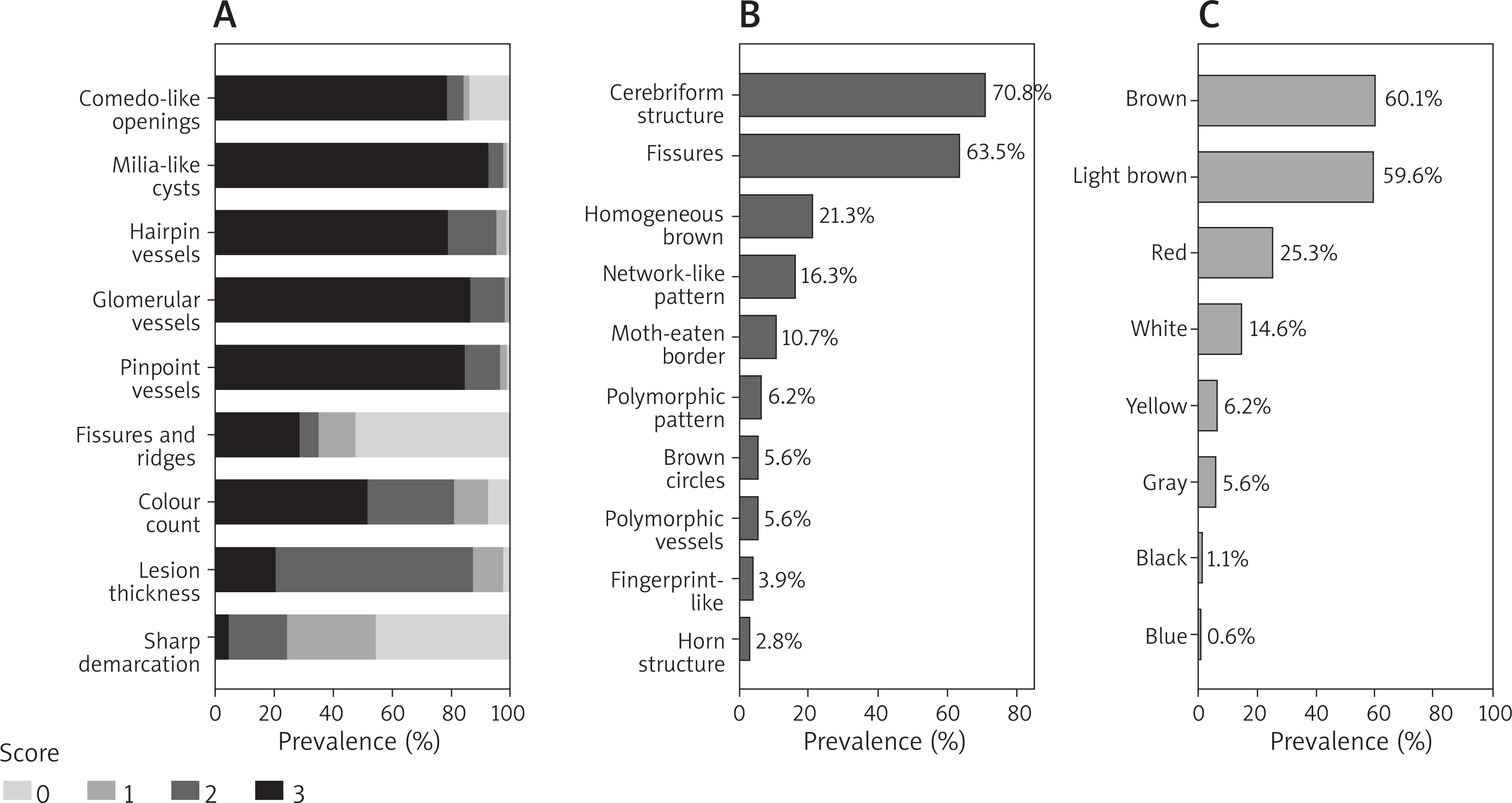
Dermoscopic features: baseline dermoscopic images were independently reviewed by three experienced dermatoscopists blinded to outcomes. Predefined features were scored either as binary (present/absent) or semi-quantitatively on an ordinal scale of 0–3 (0 = absent, 1 = mild, 2 = moderate, 3 = marked). Discrepancies were resolved by consensus. The full list and distribution of the assessed dermoscopic structures are presented in Figures 1 A, B. Colour assessment included documentation of colours present and a derived colour count variable representing the number of distinct colours observed on dermoscopy (scored 0–3, as assessed in Figure 1 C).
Figure 1
Frequency of baseline dermoscopic features in seborrheic keratoses (n = 178). A – Distribution of semi-quantitative dermoscopic variables scored on an ordinal scale of 0–3 (0 = absent, 1 = mild, 2 = moderate, 3 = marked), including comedo-like openings, milia-like cysts, hairpin vessels, glomerular vessels, pinpoint vessels, fissures and ridges, colour count, lesion thickness, and sharp demarcation. B – Prevalence of binary (present/absent) qualitative dermoscopic structures (e.g., cerebriform structure, fissures, homogeneous brown structures, network-like and ring-like patterns, monomorphic/polymorphic patterns, moth-eaten border, brown circles, polymorphic vessels, fingerprint-like structures, horn structure). C – Frequency of colours observed on baseline dermoscopy (brown, light brown, red, white, yellow, grey, black, blue)
The unit of analysis was the individual lesion (lesion-level data), with the patient treated as a clustering factor in all regression models. Analyses were performed using complete cases; no imputation was applied.
Outcomes
At follow-up, treatment response was assessed by clinical examination supported by dermoscopic comparison (performed routinely at each follow-up visit) and standardized photographic documentation. Clearance was graded on an ordinal scale of 0–3 based on visual assessment of the treated area relative to baseline: 0 = no clearance (0–25%), 1 = partial response (26–50%), 2 = near-complete clearance (51–99%), and 3 = complete clearance (100%). For regression analyses, the primary endpoint was complete clearance, defined as a clearance score of 3. For each lesion, the outcome was recorded from the last available follow-up visit. Cosmetic outcome and adverse effects were assessed using prespecified ordinal scales as documented in the medical records and photographic/dermoscopic review.
Statistical analysis
Continuous variables are presented as mean ± standard deviation or median (interquartile range, IQR), as appropriate; categorical variables are presented as counts and percentages. Two-sided p < 0.05 was considered statistically significant.
Because multiple lesions could originate from the same patient, within-patient clustering was evaluated using the intraclass correlation coefficient (ICC). The primary analysis of predictors of complete clearance used a multivariable mixed-effects logistic regression model (generalized linear mixed model, GLMM) with a random intercept for the patient. For ordinal outcomes (clearance score 0–3, cosmetic score 0–3), cumulative link mixed models (CLMM) were fitted. Prespecified fixed-effect covariates included anatomical location (scalp/face/neck), lesion thickness, sharp demarcation score, cerebriform pattern (binary), fissures (binary; present/absent), and colour count; follow-up duration (days) was included to adjust for observation time. To avoid redundancy, fissure-related predictors were not entered simultaneously with other closely overlapping fissure-based variables (e.g., semi-quantitative fissures/ridges scores) in the same multivariable model (Figure 1). Model assumptions were checked, including assessment of multicollinearity using variance inflation factors (VIF). Results are presented as odds ratios (OR) with 95% confidence intervals (CI). Missing data were < 3% for all variables; analyses used complete cases. Analyses were conducted using R (v4.2) with packages lme4 and ordinal.
Results
Patient and lesion characteristics
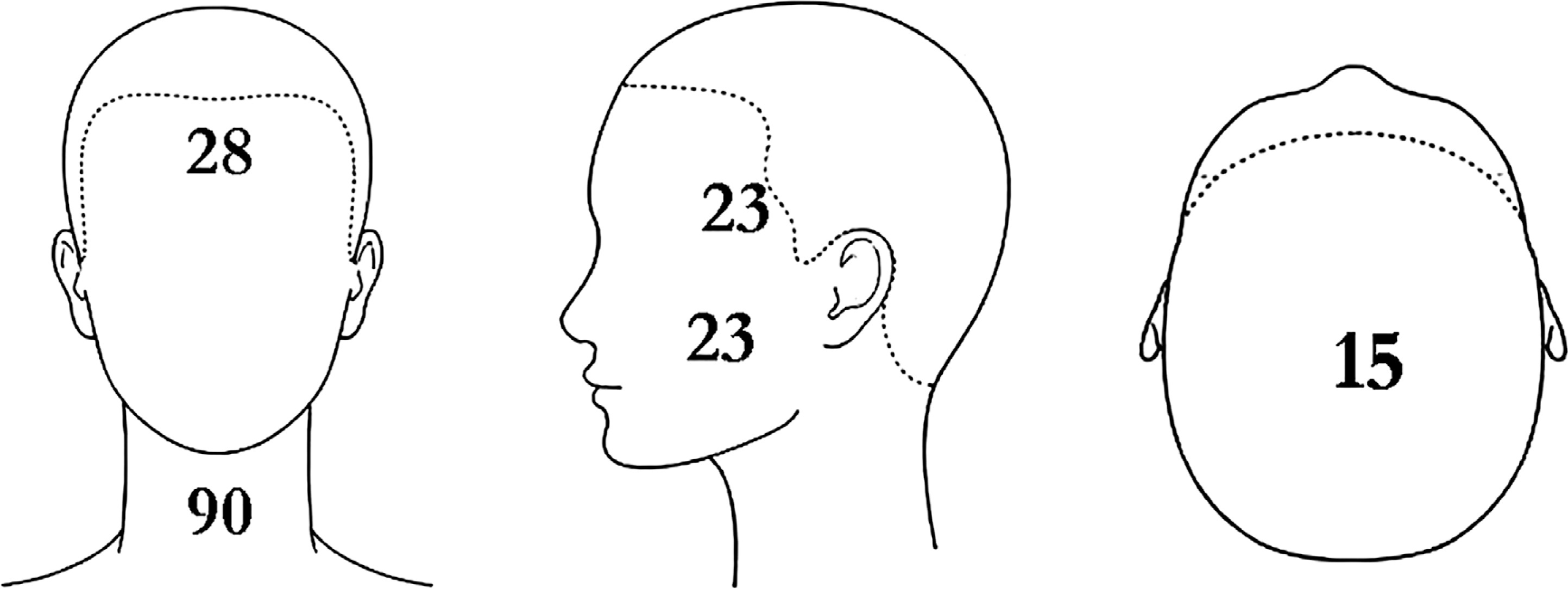
A total of 38 patients (mean age: 56 years, range: 32–81; 21 men and 17 women) with 178 seborrheic keratoses located on the face, scalp, or neck were included in the final analysis. Most patients presented with multiple lesions (median 4 lesions per patient, mean: 4.7 ±4.2). The follow-up period ranged from 67 to 1,432 days (median: 325 days, approximately 11 months). Fitzpatrick phototypes I–III were represented, with phototype II predominating, followed by phototype III. Lesions were located mainly on the neck (48.9%), with the remainder on the face (42.7%); and a smaller subgroup on the scalp (8.3%). The anatomical distribution of lesions is summarized in Figure 2.
Baseline dermoscopic phenotype
The full distribution of semi-quantitative (0–3), binary dermoscopic variables and colours are summarized in Figure 1.
On baseline dermoscopy, most lesions demonstrated a typical seborrheic keratosis phenotype characterized by a well-defined border and keratin-related surface architecture. Sharp demarcation was present in ~75% lesions, and fissures/ridges in ~65%, consistent with a cerebriform surface pattern. In contrast, features such as milia-like cysts, comedo-like openings, and vascular structures were observed less consistently. Chromatic heterogeneity was generally limited, with most lesions showing a low colour-count score and a predominance of brown tones.
Treatment outcomes
Effectiveness (overall)
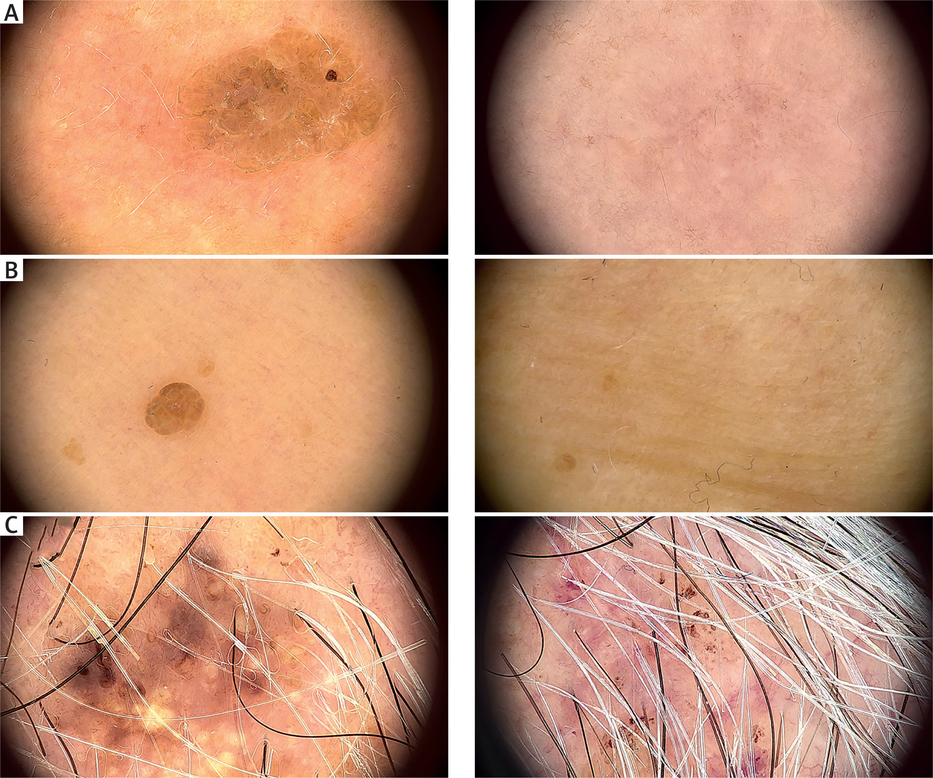
At the last documented follow-up, complete clearance (primary endpoint; clearance score 3, 100% resolution) was achieved in 74.7% of treated lesions (Table 1). An additional 14.0% showed near-complete response on the ordinal scale. Only a small fraction demonstrated minimal response or persistence. The mean clearance score was 2.6/3, indicating that, on average, lesions were nearly completely resolved. Representative “high responder” and “low responder” examples are presented in Figure 3.
Table 1
Treatment outcomes, cosmetic outcome, complete clearance by anatomical location, and adverse effects after HIFU for seborrheic keratoses. Panel A presents lesion clearance categories (0–3) for all treated lesions (n = 178). Panel B summarizes the cosmetic outcome score (0–3; n = 178). Panel C shows complete clearance rates stratified by anatomical location (n = 178), reported as n/N (%). Panel D details adverse effects by maximum severity grade observed per lesion (0–3; n = 178). Percentages are calculated per total lesions within each panel unless otherwise stated. Adverse effects are not mutually exclusive (more than one event could occur in the same lesion)
Figure 3
Representative dermoscopic images before (left) and at follow-up after 20-MHz HIFU (right), acquired with FotoFinder Medicam 1000. A – Good treatment response. Baseline seborrheic keratosis (SK) on the neck of a 45-yearold man (Fitzpatrick phototype III), showing a prominent brain-like (cerebriform) pattern with numerous fissures and ridges and sharp lesion demarcation. At 3-month follow-up, only minimal residual changes are visible. HIFU settings: 0.8-mm transducer, 1.10 J, 150 ms. B – Excellent treatment response with optimal cosmetic integration. Baseline SK on the neck of a 46-year-old woman (Fitzpatrick phototype I), slightly pedunculated and well demarcated, with fissures and ridges. At 2-year follow-up, complete clinical and dermoscopic clearance is observed with an excellent colour match to the surrounding skin. HIFU settings: 0.8-mm transducer, 1.02 J, 150 ms. C – Poor treatment response. Baseline SK on the scalp of a 70-year-old man (Fitzpatrick phototype III), heterogeneous in colour and structures, with milia-like cysts and comedo-like openings and poorly defined borders. At 3-month follow-up, partial response (~1/3 reduction) is seen, accompanied by persistent erythema, whitening/hypopigmentation and inflammatory changes. HIFU settings: 0.8-mm transducer, 1.10 J, 150 ms
Effectiveness by anatomical site
Scalp lesions showed lower complete clearance compared with lesions on the face and neck (OR = 0.15 vs. neck, 95% CI: 0.04–0.61, p = 0.008; Table 1). Facial sub-sites had broadly comparable outcomes; any apparent differences across the forehead, temple, and cheek should be interpreted cautiously due to smaller subgroup sizes.
Cosmetic outcomes
Cosmetic results were generally favourable (Table 1). Good/excellent cosmetic outcome (score 2–3) was achieved in 86.0% of lesions, with a mean cosmetic score of 2.4/3. Lower cosmetic scores were mainly attributable to persistent dyschromia and/or residual textural change at the treatment site. Importantly, lesions that did not fully clear often healed as flat residual macules; these were frequently rated cosmetically acceptable despite incomplete lesion removal, whereas poor cosmetic outcomes were concentrated among lesions with more visible pigmentary mismatch or textural alteration.
Adverse effects and safety
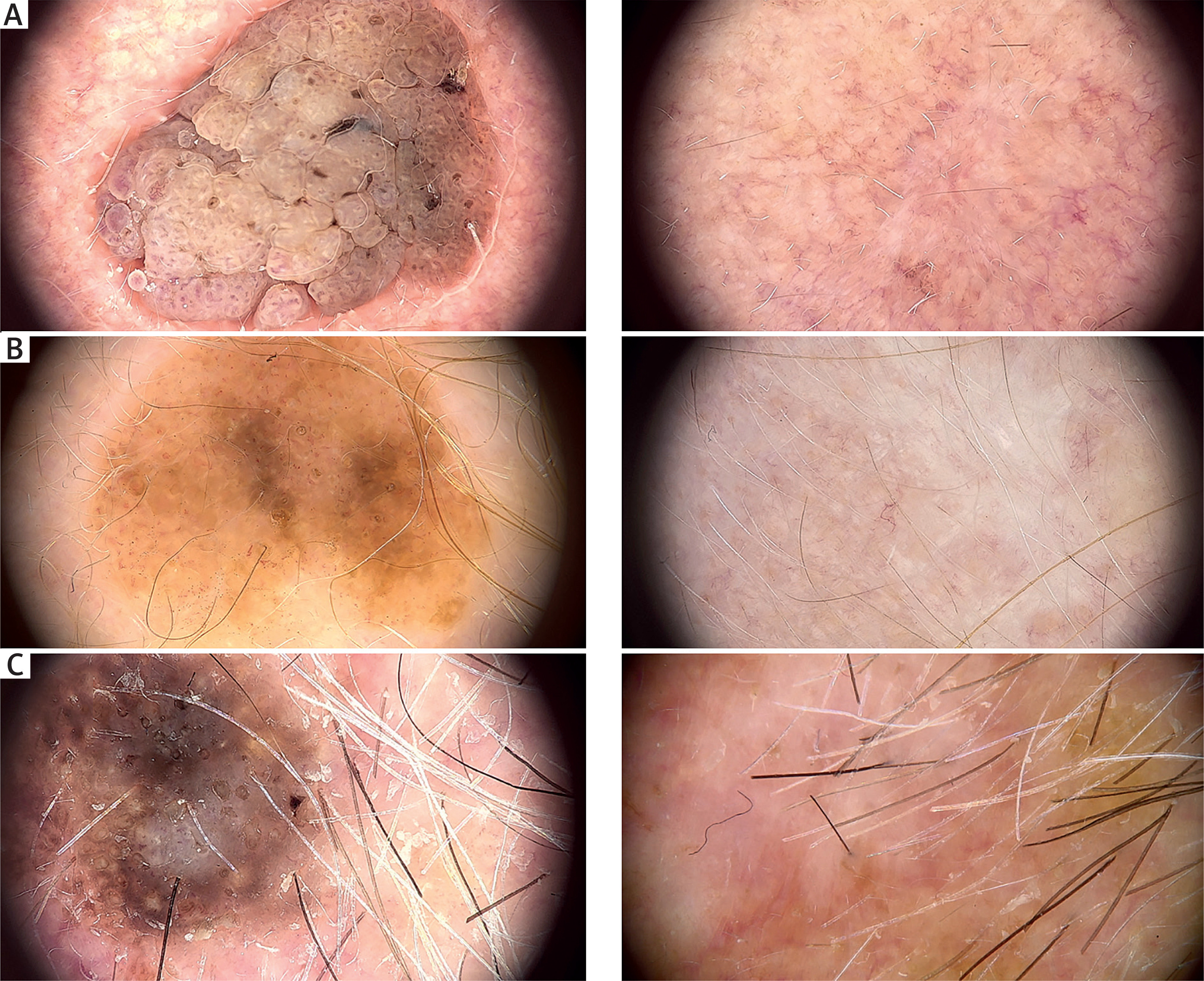
No serious adverse events were encountered. Early post-treatment erythema and crusting were common in the immediate period after HIFU and generally resolved over follow-up. Pigmentary change was the most frequent persistent sequela. Hypopigmentation was the most common adverse effect, observed in 42.7% of lesions, whereas post-inflammatory hyperpigmentation occurred less often (Table 1). Pigmentary alterations were assessed by videodermoscopy at 20× magnification, which may increase detection rates compared with unaided visual inspection; nevertheless, their severity was graded as mild (1/3) in > 80% of affected lesions, underscoring the need to treat the pigmentary change as a distinct endpoint from lesion clearance. Representative clinical examples of typical post-treatment hypopigmentation are shown in Figure 4. Clinically evident scarring was uncommon and, when observed, tended to be limited in extent. Importantly, most events recorded as “skin texture changes” in the statistical dataset corresponded to subtle textural alterations and minor surface irregularities, rather than clinically obvious scars. No infections, ulcerations, hypertrophic scars, or other clinically significant complications were documented.
Figure 4
Representative examples of post-treatment hypopigmentation following 20-MHz HIFU, graded according to severity (I–III). Dermoscopic images are shown before treatment (left) and at follow-up (right). A – Grade I hypopigmentation – mild, focal lightening with minimal contrast to the surrounding skin. B – Grade II hypopigmentation – moderate, clearly visible hypopigmented area with noticeable contrast to the adjacent skin. C – Grade III hypopigmentation – marked, extensive hypopigmentation with persistent erythema. The baseline lesion is a heterogeneous seborrheic keratosis (SK) on the temple, displaying features consistent with our predicted poor-response pattern. The follow-up image (12 months post-treatment) shows prominent hypopigmentation and erythema on photodamaged temporal skin in a 76-year-old man (Fitzpatrick phototype II)
Dermoscopic predictors of treatment success
Clustering and univariable patterns
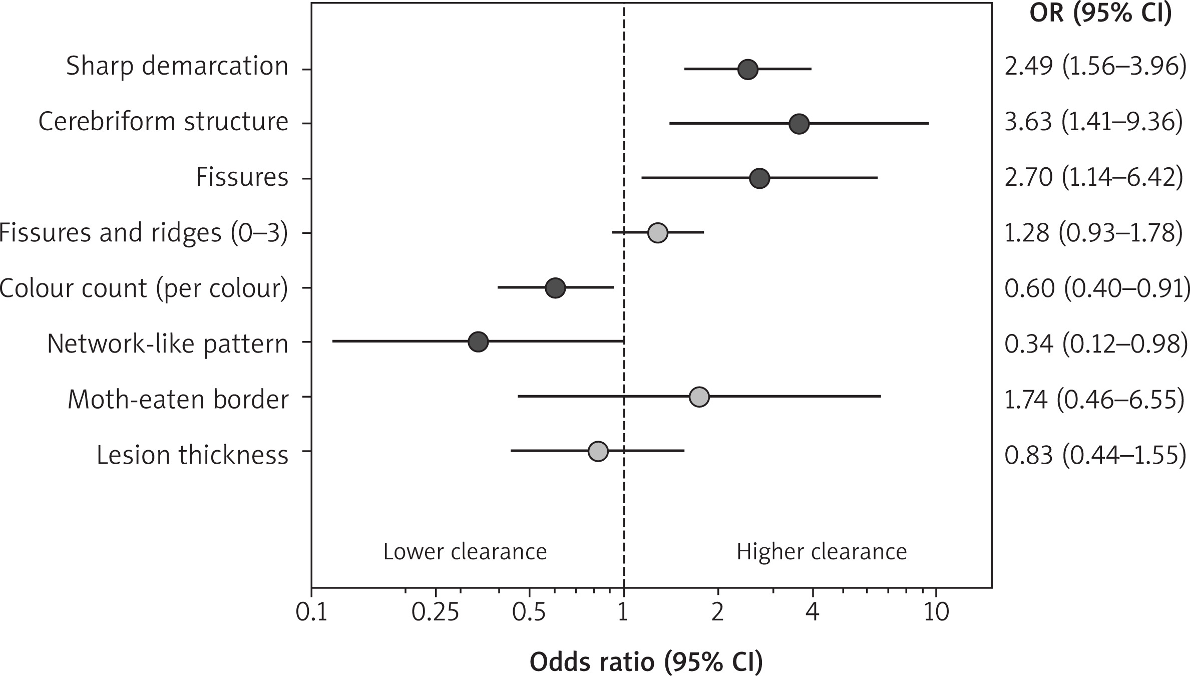
Because multiple lesions were treated per patient, clustering was assessed; the intraclass correlation coefficient (ICC) for lesion clearance was 0.09 (low clustering), supporting lesion-level inference with patient-level adjustment. In univariable assessments, lesions with a sharp border and cerebriform architecture tended to show higher clearance and more favourable cosmetic ratings, whereas lesions with a network-like pattern and greater baseline chromatic heterogeneity tended to show lower clearance.
Border sharpness stratification
Stratified analyses demonstrated a clinically meaningful gradient across border sharpness. Lesions lacking a sharp demarcation had low complete clearance rates (approximately one-quarter achieving complete clearance), whereas strongly demarcated lesions achieved complete clearance in the large majority (approximately 85–90%). A similar gradient was observed for cosmetic outcomes, with higher cosmetic scores among lesions with stronger border definition and lower adverse-effect burden overall.
Mixed-effects multivariable model
In the prespecified mixed-effects logistic regression model (patient random intercept), three baseline dermoscopic variables remained independently associated with complete clearance after adjustment for anatomical site, lesion thickness, and follow-up duration. A sharp border increased the odds of complete clearance (OR = 2.49, 95% CI: 1.56–3.96, p < 0.001), and a cerebriform pattern was also associated with higher odds of clearance (OR = 3.63, 95% CI: 1.41–9.36, p < 0.01). In contrast, greater dermoscopic colour count was inversely associated with complete clearance (OR = 0.61 per additional colour, 95% CI: 0.40–0.91, p = 0.017), indicating reduced likelihood of full response in more chromatically heterogeneous lesions. Together, these results suggest that sharply demarcated, cerebriform SKs with limited colour heterogeneity represent the most favourable candidates for HIFU, whereas multicoloured lesions have lower probability of complete clearance after a single standardised session (summarized in Figure 5).
Discussion
In this retrospective cohort of head-and-neck seborrheic keratoses, 20-MHz HIFU achieved clinically meaningful clearance in most treated lesions and was generally associated with favourable cosmetic outcomes. Pigmentary sequelae – particularly hypopigmentation – were common, underscoring that “lesion removal” and “cosmetic integration” are related but distinct endpoints in cosmetically sensitive areas [14]. These findings support HIFU as a pragmatic option for selected seborrheic keratoses of the head and neck, provided that patient selection and counselling address pigmentary risk and the possibility of incomplete clearance in specific phenotypes and anatomical sites.
A central contribution of this study is the identification of dermoscopic features independently associated with complete clearance after HIFU, reinforcing the concept that dermoscopy may inform not only diagnosis but also therapeutic stratification [31–34]. Sharp demarcation emerged as a robust positive predictor. A sharply delimited seborrheic keratosis likely represents a superficial, well-circumscribed keratotic proliferation with a discrete lesional–perilesional interface, which may facilitate uniform energy coverage and clearer treatment endpoints [27, 28]. Likewise, a cerebriform surface architecture predicted improved clearance. This pattern reflects prominent fissures and ridges with compact hyperkeratosis and may represent a more reproducible target for focal injury at superficial depths [27, 28]. In contrast, greater baseline colour heterogeneity was inversely associated with clearance. Dermoscopic variegation can reflect mixed keratin, melanin, vascular and irritation-related components, as well as heterogeneous epidermal architecture [27, 28, 30], potentially increasing the likelihood of residual lesional islands after a single standardized session. Clinically, multi-coloured lesions may therefore warrant alternative modalities, staged HIFU strategies, or planned reassessment with a low threshold for retreatment [5–14].
Taken together, these predictors suggest a pragmatic dermoscopic enrichment approach: sharply demarcated, cerebriform SKs with limited colour heterogeneity appear to be the most favourable candidates for HIFU, whereas lesions with pronounced multicolouration and network-like pattern may represent a lower-yield phenotype. Any lesion with diagnostic uncertainty or atypical dermoscopic features should undergo standard diagnostic pathways rather than energy-based destruction, particularly on the head and neck where malignant mimickers are common [30].
We also observed clinically relevant differences by anatomical site, with lower clearance rates on the scalp compared with the face and neck. Scalp SKs may be more hyperkeratotic, and consistent acoustic coupling can be technically more challenging in hair-bearing areas and on curved surfaces. In addition, operators may use more conservative dosing near follicles or in areas where overtreatment could be cosmetically undesirable. These site-specific constraints may reduce the effective delivered energy and contribute to incomplete ablation. Accordingly, scalp SKs may require closer follow-up, consideration of a second session, or alternative modalities when rapid and complete clearance is prioritized [5, 7, 9].
The safety profile was characterized mainly by local, predominantly mild adverse effects. Pigmentary change – especially hypopigmentation – was the most frequent sequela, whereas clinically significant scarring was uncommon. Counselling should therefore separate the probability of clearance from the probability of perfect colour match, and emphasize photoprotection and standardized aftercare; pigmentary changes may improve slowly and are not always fully reversible [14, 35].
From a broader patient-centred perspective, endpoints such as satisfaction, procedure-related discomfort, downtime, and cosmetic outcome are key determinants of treatment choice for seborrheic keratoses, particularly in facial and other cosmetically sensitive areas. Comparative studies consistently show higher patient satisfaction with laser-based treatments than with cryotherapy, largely driven by superior cosmetic outcomes and a lower risk of hypopigmentation, especially for facial lesions and in patients with darker skin types [9, 10]. Although procedure-related pain may be slightly higher with certain laser modalities – particularly 532-nm devices – pain intensity is generally mild, transient, and well tolerated, and overall downtime remains minimal for both approaches [11, 36]. These observations provide a relevant clinical benchmark for interpreting the present findings. While HIFU differs mechanistically from both cryotherapy and laser ablation, it shares several patient-valued attributes, including a non-incisional approach, limited downtime, and controlled, depth-selective tissue injury. In the present cohort, treatments were performed without anaesthesia in the head and neck region, highlighting the potential role of HIFU as a “procedure-light” option for selected lesions, even though pigmentary alteration – particularly hypopigmentation – remains an important limitation.
Evidence for dermatologic high-frequency HIFU in SK remains limited, but available case series suggest meaningful clearance rates with acceptable tolerability [14]. Differences across studies likely reflect variation in lesion spectrum, anatomical sites, energy settings, and outcome ascertainment [14]. Our data add value by focusing on head-and-neck lesions and by linking baseline dermoscopic phenotype to outcomes using models that account for within-patient clustering. Across indications, early HIFU reports highlight depth control and the ability to target superficial lesions while limiting collateral injury [17, 22–25]. However, evidence remains heterogeneous and largely non-randomized, precluding conclusions regarding superiority over established destructive modalities [5, 7, 10].
This study has limitations inherent to its retrospective, single-centre design, including non-standardized follow-up timing (we used the last available follow-up visit), reliance on ordinal visually assessed outcomes, and potential selection bias. Several variables were observer-dependent, and formal inter-rater reliability testing was not performed [37]. Strengths include lesion-level analysis with appropriate clustering adjustment and systematic dermoscopic feature assessment by blinded reviewers.
Future work should prospectively validate these dermoscopic predictors using standardized imaging and fixed follow-up windows, and assess whether dermoscopy-guided parameter tailoring or staged strategies can improve outcomes in unfavourable phenotypes while reducing dyspigmentation. Automated quantification of border sharpness, texture, and chromatic heterogeneity may further improve reproducibility and support clinically deployable decision tools [38].
Conclusions
High-frequency 20-MHz HIFU provided effective treatment of seborrheic keratoses in cosmetically sensitive head-and-neck areas, achieving high rates of lesion clearance with generally favourable cosmetic appearance. Outcomes varied by anatomical site, with lower clearance observed on the scalp than on the face or neck, suggesting that location-specific factors may influence treatment success. Adverse effects were frequent but usually mild; pigmentary changes – especially hypopigmentation – were the most common and should be discussed during pre-treatment counselling as cosmetic “colour match” may not fully parallel lesion removal. Baseline dermoscopy may support practical patient and lesion selection, helping identify SKs most likely to respond and guiding individualized management. Prospective studies are warranted to validate these observations and optimize treatment strategies.










