
Introduction
Blue nevi (BN), usually presenting as solitary, bluish, asymptomatic macules or nodules, are formed by collections of dermal melanocytes that failed to complete their migration from the neural crest to the dermo-epidermal junction. The term “agminated blue nevi” refers to multiple lesions grouped, linear, or arranged in a blashkoid distribution [1]. It is a relatively rare phenomenon with less than 35 cases reported in the literature, but only 14 cases with dermoscopic features. We report 4 additional patients who presented to the Department of Dermatology, Venereology and Allergology, Medical University of Gdansk (Poland) between May 2016 and May 2021 and summarize current literature on dermoscopic features of this entity.
Case series
Patient 1 was a 52-year-old man (phototype II) who presented with multiple, agminated blue, grey and whitish nodules within the right frontal and parietal region of the scalp (Figure 1 A). According to the patient, the lesions appeared around 20 years before, without a known causative factor. Due to their location within the hairy scalp, the stable history of most of the lesions could not be confirmed. The patient reported associated arterial hypertension and hypercholesterolemia. Additionally, on clinical examination there were multiple soft, subcutaneous tumours, described as lipomas based on subsequent ultrasound evaluation. Besides melanoma diagnosis in the patient’s aunt, his familial history was unremarkable. Dermoscopy of the scalp lesions showed a diverse spectrum of dermoscopic patterns including: blue structureless, blue-brown structureless, white structureless with serpentine vessels, blue-brown structureless with white lines, and blue-brown-white structureless with serpentine vessels (Figures 1 B–F). The largest tumours within the hairy scalp were excised and histopathological assessment confirmed the diagnosis of agminated blue nevi with no atypia. Total body dermoscopic assessment excluded cutaneous and mucosal neoplasms. The remaining blue lesions on the scalp revealed no clinical and dermoscopic evolution during 2-year follow-up.
Figure 1
Patient 1. Clinical presentation – multiple, agminated blue, grey and whitish nodules within the right frontal and parietal region of the scalp (A). Dermoscopy shows: combination of blue-brown structureless areas, dotted and short linear irregular vessels (B), combination of white-blue structureless areas with brown globules/clods, brown peripheral structureless areas and polymorphic vessels (C); combination of a blue-brown structureless area with white circles and a peripheral pink structureless area (D); polymorphic vessels over white-brownish background and a peripheral pink structureless area (E); blue-brown structureless areas in combination with linear irregular and branched vessels with a few ramifications (F) (FotoFinder, non-polarized videodermoscopy with immersion fluid, 20× magnification). Patient 2. Clinical presentation – multiple bluish, flesh-coloured and brownish lesions agminated in a plaque located within the left shoulder (G). Dermoscopy shows a combination of bluish-brown structureless areas (H, I) (FotoFinder, non-polarized videodermoscopy with immersion fluid, 20× magnification)
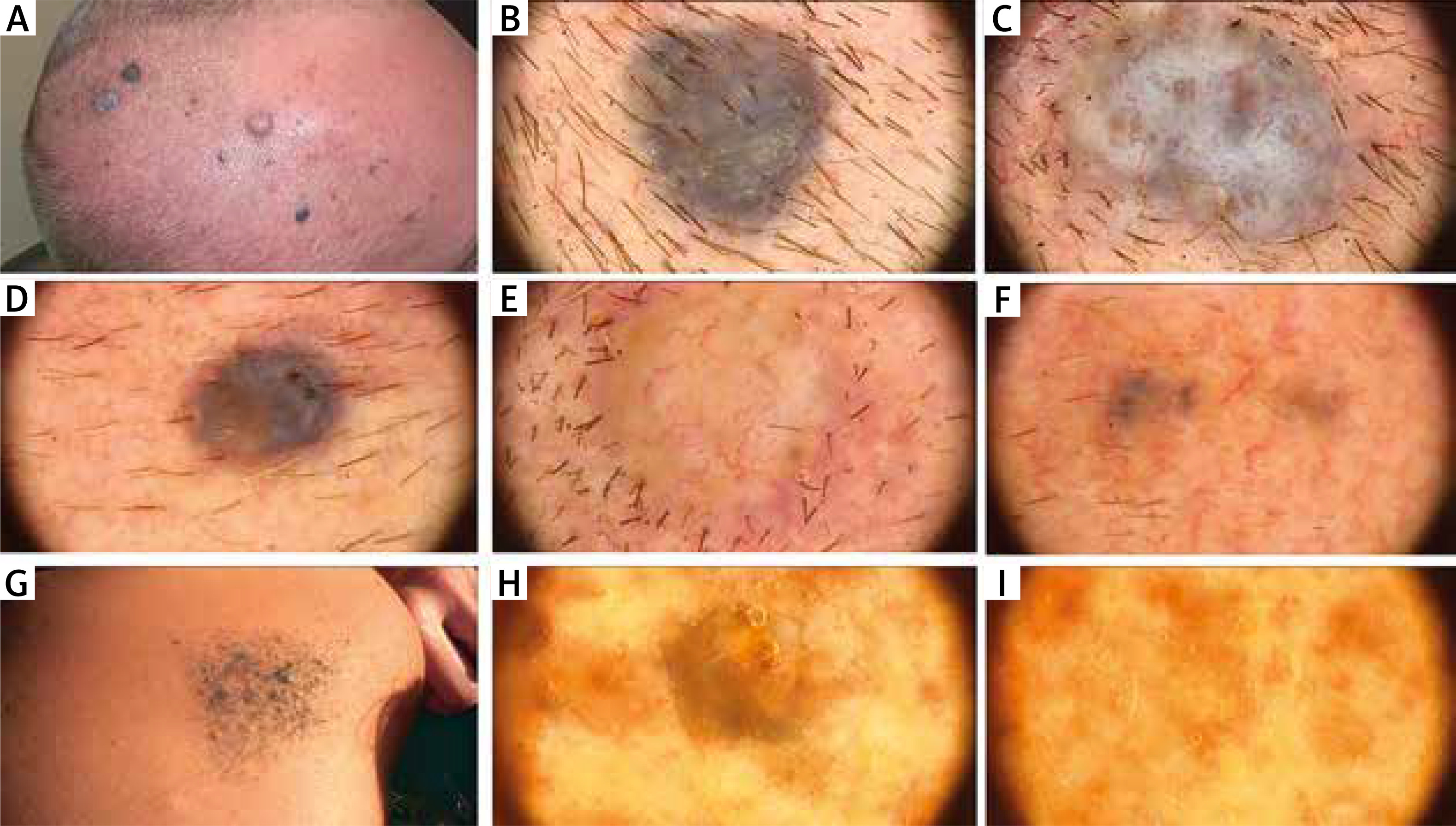
Figure 2
Patient 3. Clinical presentation – five bluish macules on both sides of the dorsum of the nose and the left ala nasi (A, B). Dermoscopy shows blue structureless areas (C–E) (FotoFinder, non-polarized videodermoscopy with immersion fluid, 20× magnification). Patient 4. Clinical presentation – two bluish nodules located on the dorsum of the right hand (F). Dermoscopy shows a combination of bluish-brown structureless areas (G) (FotoFinder, non-polarized videodermoscopy with immersion fluid, 20× magnification)

Patient 2 was a 37-year-old woman (phototype III) who presented with multiple bluish, flesh-coloured and brownish lesions agminated in a plaque located within the left shoulder (Figure 1 G). Dermoscopy showed a combination of blue, brown and flesh-coloured structureless areas (Figures 1 H–I). Skin lesions appeared at the age of 13, after an episode of sunburn. There were no associated comorbidities, and no dermatological disorders in personal and familial history. Three nodules were biopsied, what confirmed the diagnosis of BN. The remaining lesions were stable during subsequent clinical and dermoscopic follow-up within almost 3 years.
Patient 3 was a 40-year-old man (phototype III) who presented with five bluish macules on the nose, on dermoscopy revealing a structureless blue pattern (Figures 2 A–E). The lesions appeared more than 10 years before, without any known triggering factor. There were no associated comorbidities, and no dermatological disorders in personal and familial history. One of the lesions located on the left ala nasi was excised, what confirmed the diagnosis of BN. The remaining lesions were stable during subsequent 2-year clinical and dermoscopic follow-up.
Patient 4 was a 69-year-old woman (phototype II) who presented with two bluish nodules located on the dorsum of the right hand (Figure 2 F). Dermoscopy showed dark-blue structureless areas (Figure 2 G). According to the patient the lesions appeared 6 months before, without any known provoking factor. There were no associated comorbidities, and no dermatological disorders in personal and familial history. Diagnostic excision confirmed histopathologically the diagnosis of BN.
Discussion
Blue nevi usually present as solitary lesions. Agminated blue nevi (ABN), initially reported by Upshaw et al. [2] in 1947, currently are defined as multiple lesions arising within the area of a diameter ≤ 100 mm [3]. The term “agminate” derives from the Latin word “agminis” and indicates the army or troop [3]. ABN can be congenital or acquired, but they tend to occur earlier in life compared to blue nevi, which present most frequently in the second decade of life [1]. The occurrence of reported ABN is nearly identical in females and males. They tend to occur with a similar frequency on the trunk, extremities and head/neck area [1].
Pathogenesis of ABN is not fully elucidated. Hendricks [4] reported ABN which occurred after a sunburn, however in most cases no triggering factor could be identified. In 2018, Eichenfield et al. [1] identified GNAQ mutation in a patient with ABN, previously reported also in sporadic BN, in contrast Rodríguez-Jiménez et al. reported an association with CYSLTR2 mutation.
ABN usually present as an isolated feature, however coincidence with nevus spilus, exophthalmus, pre-tibial myxoedema, osteoarthropathia (EMO syndrome), dermatomyositis as well as Darier’s disease has been described [3].
The most important differential diagnosis of ABN includes melanoma metastases. It is known that they may present with various dermoscopic patterns and mimic other benign and malignant skin tumours [5]. According to the results of a case-control study by Avilés-Izquierdo et al. [5], blue melanoma metastases were more homogenous than blue nevi and usually did not present other dermoscopic focal features, however it has been underlined that in many cases differential diagnosis was very difficult. Thus, a careful clinical-dermoscopic correlation seems to be crucial in such cases. Additionally, cases of melanoma arising in plaque-type agminated blue nevus have been reported [6, 7]. Details concerning their dermoscopic presentation have not been given. Indicative histopathological features for malignant melanoma were the presence of cellular atypia, mitoses and focal necrosis [6, 7].
Moreover, a rare variant of blue nevus with satellitosis, in which multiple blue globules located on the skin close to the main tumour are found, should be considered in the differential diagnosis of AGM [8].
A dermoscopic review of cases described to date (Table 1) demonstrates the blue structureless pattern as a prevailing dermoscopic feature in most of them (17/18). The most common associated features were brown or grey structureless areas (both present in 7 cases) and white/flesh coloured areas (4 cases). The remaining structures were present in single cases [1–3, 9–15]. Dermoscopic presentation of our 4 cases generally are within the spectrum of previously described patterns of ABN, with structureless blue areas as a predominant feature. Completely amelanotic BN with serpentine vessels (Figure 1 E) was not previously noted in the spectrum of ABN.
Table 1
Agminated blue nevi – a review of dermoscopic features of previously described cases
| First author, year of publication | Age [years]/gender | Location | Size [cm] | Dermoscopy | Structureless blue areas | Structureless brown areas | Structure-less grey areas | White/flesh-coloured structureless areas | Black structureless areas | Radial blue lines | Black dots/globules | Brown parallel lines | White lines | Brown globules | Brown-blue pseudopods | Scales/crusts | Serpentine vessels |
|---|---|---|---|---|---|---|---|---|---|---|---|---|---|---|---|---|---|
| Pizzichetta et al., 2007 | 59/F | Left leg | 4 × 2 | Multiple, grouped, homogeneous, confluent, steel blue to brown-blue pigmented areas fading into the surrounding skin. On the surface of some steel blue areas, linear pigmented structures appear as darker sulci (diamond). In addition, blue areas, some globules and dots (black arrow), a brown veil (black arrowhead), and brown-blue and out-of-focus pseudopods (white arrows). A small rim of tan pigmentation (asterisk) | + | + | – | – | – | + | + | – | – | - | + | - | - |
| Skowron et al., 2009 | 25/F | Right chest wall involving the right breast | 14 × 10 | Homogenous, unstructured blue pigmentation, brownish-blue in the centre and steel blue at the periphery | + | + | – | – | – | – | – | – | – | - | - | - | - |
| Simonetti et al., 2013 | 30/M | Left thigh | 6 × 4 | Homogeneous blue steel colour | + | – | + | – | – | – | – | – | – | - | - | - | - |
| Rongioletti et al., 2015 | 60/F | Right flank | 7 | Plaque-like lesion was typified by areas of homogeneous blue pigmentation, brownish-blue in the centre and steel blue at the periphery | + | + | + | – | – | – | – | – | – | - | - | + | - |
| Ayal.a et al., 2016 | 79/F | Right lower leg | 10 × 6 | Brownish papules showed a globular melanocytic pattern while the bluish papules presented a homogenous pattern | + | – | – | – | – | – | – | – | – | + | - | - | - |
| Koba et al., 2016 | 11/M | Right sole | 1.2 × 0.5 | Homogenous, blue-grey pattern | + | – | + | – | – | – | – | – | – | - | - | - | - |
| Paolino et al., 2016 | 60/F | Forehead | 2 × 3 | Homogenous, blue or brown-blue colour in each of the pigmented islands | + | + | – | – | – | – | – | – | – | - | - | - | - |
| Paolino et al., 2016 | 58/M | Vertex | 2.5 central 1 peripheral | Homogenous blue-whitish pattern | + | – | – | + | – | – | – | – | – | - | - | - | - |
| Lisboa et al., 2016 | 64/M | Left upper back | 7 × 5 | Homogeneous bluish or greyish nevus with no structures | + | – | + | – | – | – | – | – | – | - | - | - | - |
| Collgros et al., 2016 | 65/M | Penis | NR | Multiple lesions of a homogeneous dark-brown colour, with some being a lighter colour at the periphery. On the glans, dermoscopy showed coalescence of brown homogeneous lesions in the centre of the glans and isolated lesions on the periphery, with a homogeneous pattern | – | + | – | – | – | – | – | – | – | - | - | - | - |
| Collgros et al., 2016 | 15/M | Penis | NR | Some areas with homogeneous blue pigmentation, and other areas with brown aligned globules and parallel lines | + | – | – | – | – | – | – | + | – | + | - | - | - |
| Hunjan et al., 2017 | 16/M | Left forehead | 7 × 13 | Dark blue macules appeared as a homogeneous steel-blue pigment pattern | + | – | + | – | – | – | – | – | – | - | - | - | - |
| Woo et al., 2019 | 49/M | Right shoulder | 5 × 4 | Blue homogenous areas with no network pattern or structures | + | – | – | – | – | – | – | – | – | - | - | - | - |
| Eichenfield, 2019 | 71/K | Left forearm | 7 × 4.5 | Multiple, homogenous, brown and blue-black nevi | + | + | – | +* | + | – | – | – | – | - | - | - | - |
| S³awiñska, 2021 | 52/M | Scalp (right frontal and parietal region) | Blue structureless, blue-brown structureless, white structureless nevi with serpentine vessels, blue-brown structureless nevi with white lines, blue-brown-white structureless nevi with serpentine vessels | + | + | + | + | – | – | – | – | + | - | - | - | + | |
| Sławińska, 2021 | 40/M | Nose | Structureless blue pattern | + | – | – | – | – | – | – | – | – | - | - | - | ||
| Sławińska, 2021 | 37/F | Left shoulder | Bluish, flesh-coloured and brownish lesions | + | – | + | + | – | – | – | – | – | - | - | - | - | |
| Sławińska, 2021 | 69/F | Dorsum of the right hand | Bluish nodules | + | – | – | – | – | – | – | – | – | - | - | - | - | |
| Number of cases with particular dermoscopic features | 17 | 7 | 7 | 4 | 1 | 1 | 1 | 1 | 1 | 2 | 1 | 1 | 1 |
Conclusions
ABN is a rare entity that needs to be considered in differential diagnosis in a patient who presents with multiple pigmented and sometimes also non-pigmented lesions in one anatomical region. Dermoscopy may provide an additional tool in the patient assessment, however if the history is inconclusive, biopsy will be needed to exclude melanoma metastases. Digital dermoscopy monitoring seems to be the optimal way of long-term management of patients with ABN.


