Terra firma-forme dermatosis (TFFD), also known as Duncan’s dirty dermatosis, is an acquired keratinization disorder characterised by brownish-gray, dirt-like plaques that persist despite adequate hygiene practices and can be readily removed with isopropyl alcohol. Although asymptomatic, it may cause cosmetic concern and is frequently misdiagnosed as other hyperkeratotic dermatoses [1].
TFFD is thought to result from delayed keratinocyte maturation and retention hyperkeratosis; however, its exact pathogenesis remains unclear. In contrast to dermatosis neglecta (DN), patients typically maintain normal hygiene. The condition predominantly affects children and young adults and most commonly involves the neck, trunk, and flexural areas, whereas facial involvement is relatively uncommon [2].
We report a 63-year-old woman who was referred to our dermatology clinic for evaluation of facial lesions during preoperative assessment for endometrial carcinoma. She described a 2-year history of gradually progressive, asymptomatic lesions over both cheeks which were resistant to routine washing with soap and water. Previous topical treatments had been ineffective.
Dermatological examination revealed bilaterally symmetrical, brownish, velvety, hyperkeratotic verrucous plaques over the malar regions (Figure 1). Initially, seborrheic keratosis and verruca vulgaris were considered in the differential diagnosis. Given the presence of malignancy and the atypical facial location, paraneoplastic acanthosis nigricans was also considered and a biopsy was initially planned. However, prior to biopsy, a diagnostic alcohol swab test was performed. Gentle rubbing with 70% isopropyl alcohol resulted in complete clearance of the lesions, revealing normal underlying skin and supporting the diagnosis of TFFD, thereby avoiding an unnecessary invasive procedure (Figures 2 A, B). No recurrence was observed during follow-up.
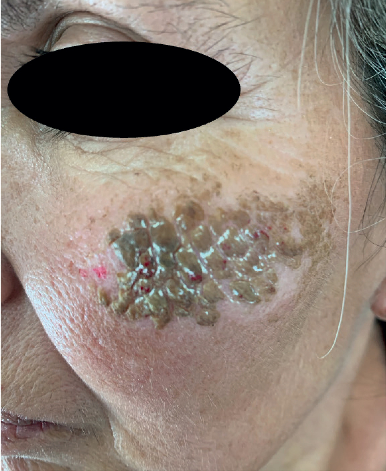
Figure 2
Partial and complete removal of the hyperkeratotic plaque on the left malar region following application of 70% isopropyl alcohol: A – partial clearance after initial swabbing, with minimal pinpoint bleeding likely related to mechanical friction during the procedure; B – complete resolution of the lesion, revealing normal underlying skin
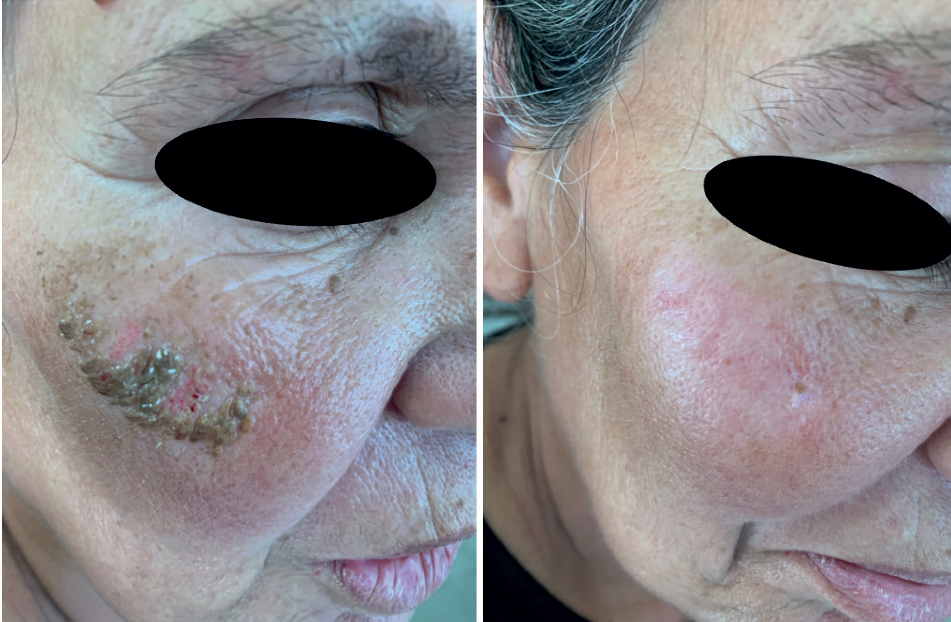
The diagnosis was supported by three key findings: (1) persistence of lesions despite adequate hygiene practices, (2) complete clearance with isopropyl alcohol, and (3) normal underlying skin after removal. The alcohol swab test is a simple, non-invasive, and highly characteristic diagnostic tool.
Facial involvement of TFFD is uncommon but has been reported, including bilateral malar presentations mimicking seborrheic keratosis or verruca. Most reported cases involve younger individuals without systemic confounding factors [3, 4]. However, TFFD can also occur in older adults, as demonstrated by case series including patients aged 54–86 years [5].
In our case, the presence of malignancy and atypical facial location led to a bias in the diagnosis toward paraneoplastic dermatoses, particularly acanthosis nigricans. This condition is a recognised indicator of internal malignancy, including endometrial carcinoma [6]. This clinical context increased suspicion and led to the initial plan for biopsy.
TFFD has been associated with dermatologic conditions such as xerosis and atopic dermatitis, and less frequently with systemic diseases including allergic disorders and hypothyroidism; however, its relationship with malignancy remains unclear and is most likely incidental [2]. Similarly, malignancies, including both non-melanoma and melanoma skin cancers, have been interpreted as age-related comorbidities rather than causal associations [5].
The distinction between TFFD and DN is clinically important and may be challenging in atypical presentations. DN is typically associated with inadequate hygiene, accumulation of dirt, sebum, and keratinocytes, and usually resolves with routine washing using soap and water. In contrast, TFFD occurs despite normal hygiene practices and is characteristically resistant to conventional cleansing but responds promptly to alcohol application. Previous studies have emphasised that TFFD lesions cannot be removed by routine washing, unlike DN [7]. In addition, DN often presents with more loosely adherent, patchy or crusted scales that can be cleared with simple cleansing, whereas TFFD demonstrates a more compact, adherent hyperkeratotic plaque. Thus, the alcohol swab test serves as a simple bedside diagnostic tool that effectively differentiates between these two entities [7]. In our patient, the presence of adequate hygiene, chronic persistence of lesions, and resistance to repeated washing strongly argued against DN and supported the diagnosis of TFFD.
Compared to previously reported cases, our patient represents a distinctive combination of advanced age, bilateral symmetrical facial involvement, and malignancy-associated diagnostic concern, all of which contributed to a higher likelihood of misinterpretation. This case highlights how the presence of malignancy may lead to diagnostic anchoring toward paraneoplastic dermatoses, potentially resulting in unnecessary invasive procedures. A simple alcohol swab test can prevent such interventions.
Management of TFFD is straightforward and typically requires no additional treatment beyond mechanical removal with alcohol. Patient education regarding the benign nature of the condition and appropriate skin cleansing practices is important to prevent recurrence.
A limitation of this report is the absence of histopathological confirmation; however, the complete resolution of lesions with alcohol application is considered pathognomonic and sufficient for diagnosis.
In conclusion, TFFD should be considered in atypical clinical settings, particularly in verrucous hyperkeratotic facial lesions, as early recognition enables prompt diagnosis and avoids unnecessary invasive procedures. Clinicians should maintain a high index of suspicion for TFFD even in elderly patients and in the presence of malignancy.