


Introduction
Hilar cholangiocarcinoma (HC) is a rare but aggressive and complex pathological condition that is characterized by its location and spread to local and distant organs [1–3]. With a prevalence of approximately 7000 cases per year in North America [2], less than 50% of these malignancies are resectable, and most of them succumb to the disease within a year of diagnosis [4]. The incidence of HC has remained relatively stable over the past decades in Western countries but has rapidly increased in China over the same period [5]. In China, it is estimated that more than 65 thousand patients with HC are diagnosed annually [6, 7].
Radical resection has been well recognized as the most effective treatment for HC and is also the only choice that confers long-term survival [8–10]. However, curative surgical resection with negative margins can pose great challenges due to the involvement of the biliary confluence, including the right and left hepatic ducts, which are in close proximity to the portal vein, hepatic artery, and liver parenchyma [11]. It is clearly evident from the literature that aggressive curative surgical resection can offer a better long-term survival rate. For example, major liver resection combined with an extended portion of the hepatic duct could significantly improve the R0 resection rate [12, 13]. Additionally, total removal of the caudate lobe has also been shown to decrease the recurrence rate and improve long-term survival [14, 15]. Thus, in recent years, the operative treatment of HC has continued to develop, with the surgical strategy shifting from local excision of the affected bile duct to extended resections involving combined major liver resection. This also makes the surgical procedure of radical resection for HC technically more challenging and highly demanding. Thus, even conventional open surgical approaches can only be performed in selected centers with special expertise in the management of HC.
The landscape of surgical treatments for hepatobiliary disease has significantly changed following the advent and evolution of laparoscopy [16]. In recent decades, we have witnessed the development of surgical devices, accumulated experience with laparoscopic surgery and spread of this minimally invasive surgery worldwide. Oncological outcomes for complicated laparoscopic maneuvers have been addressed in high-quality prospective studies for liver cancer [17, 18] and colon and rectal cancer [19, 20]. For example, laparoscopic liver resections are confirmed to be safe, with acceptable morbidity and mortality in experienced hands [21]. Additionally, the survival rates reported are comparable to those of open resection [21].
However, for the treatment of cancers occurring in the biliary tract, including HC, a primary laparoscopic approach is still rejected by the surgical community [22, 23]. In the past, the reasons cited have included oncological concerns, such as intraoperative peritoneal dissemination and possible port-site metastasis (PSM). Historically, there have indeed been an alarming number of reports of PSM at the initial utilization of laparoscopic surgery in biliary tract carcinoma. As shown in previous studies [24, 25], approximately 0.2–3.3% of patients were found to be gallbladder cancer positive postoperatively after laparoscopic cholecystectomy. Moreover, 50% of gallbladder cancers are diagnosed incidentally [26]. Although their number is not large, the risk of PSM in these patients is estimated to be up to 14–30% [27]. There is obvious statistical bias that has resulted in a strong concern about the ‘aerosol’ effect of pneumoperitoneum. Increasing evidence suggests that the incidence of PSM is approximately 1.1% in laparoscopic cancer surgery, which is comparable to open surgery [28]. Nevertheless, the current recommendations for radical resection of biliary tract carcinoma, especially HC, stipulate that an open approach should be based largely on expert opinion rather than on evidence of superior outcomes.
Today, real concerns are associated with the technical demands involved in performing total laparoscopic radical resection. Technically, for radical resection of HC, a surgeon must have experience in laparoscopic major liver resection with caudate lobectomy, hepatoduodenal lymphadenectomy and anastomosis between the hepatic duct and jejunum at the same time. Even for the experienced hepatobiliary surgeon, each of the abovementioned operative procedures is very challenging. Concerns about the safety, feasibility and outcomes have delayed the use of laparoscopic surgery in HC. In the past decade, with the accumulation of surgical experience in complicated hepatobiliary operations, the individual procedure that is essential for laparoscopic radical resection of HC has already been reported [21, 29]. Some studies recently described the application of minimally invasive surgery in patients with HC [30–36]. However, most of these studies have either enrolled a small number of patients or only included patients with HC in early stages. More importantly, few of the previous studies reported the long-term outcomes of HC patients following total laparoscopic radical resection.
Aim
The purpose of the present study is to determine the safety and feasibility of laparoscopic techniques in the treatment of HC patients and to investigate the oncological outcomes of patients who underwent total laparoscopic radical resection for HC at a single center specializing in hepatobiliary surgery at the current stage.
Material and methods
Patient cohort and data collection
Thirty-two patients who planned to undergo curative-intent laparoscopic resection for HC between December 2015 and November 2019 were enrolled. No patient had any signs of metastasis on clinical assessment at the time of enrollment that were deemed unresectable. The patients were nonconsecutive. The inclusion criteria for laparoscopic curative-intent resection were basically the same as those for open surgery described in a previous study [37, 38]. Briefly, the criteria for nonresectability were defined as overt distant metastasis, intrahepatic metastasis, peritoneal seeding or para-aortic lymph node metastasis. Patients demonstrating the involvement of vascular structures in the hilum were not considered to be unresectable. Informed acknowledgement concerning the advanced nature of this procedure was obtained from all patients. All cases were reviewed and approved by the Medical Ethics Committee of the Affiliated Hospital of North Sichuan Medical College (NSMC). All operations were performed by one surgeon and his team. The protocol of this study was designed and conducted by the authors of this manuscript and was approved by the Institutional Review Board of the Affiliated Hospital of NSMC (No. NSMC2015-07). All the states that the research work has been reported in are in line with the PROCESS criteria.
Preoperative management
Multiple imaging assessments are frequently required for appropriate preoperative imaging evaluations of a patient with suspected HC. For tumor staging, ultrasonography, contrast-enhanced multidetector-row CT (MDCT) and cholangiography were typically performed. If necessary, some patients were selected for magnetic resonance imaging (MRI) and positron emission tomography (PET) scans, and patients with imaging signs of distant metastasis (M1), including the liver, peritoneum and lymph nodes, were deemed unresectable.
The Bismuth-Corlette classification was applied to patients with imaging modalities that confirmed HC. A multidisciplinary team (MDT) meeting was held to evaluate the probability of R0 resection. In this meeting, the involvement of the vasculature around the hilar, the longitudinal and radial extension of the tumor, and the presence of radiologically proven metastases were comprehensively discussed by hepatobiliary surgeons, consultant oncologists, hepatologists, radiologists and specialist nurses. All tumors (HC) that were shown to be potentially resectable were later subjected to laparoscopic exploration and curative resection.
Laboratory examinations included measurements of CEA, Ca19-9 and liver function tests. Not all patients with overt jaundice required biliary drainage; the decision was balanced with the benefit and increased risk of perioperative complications and was made in a multidisciplinary setting. Any required presurgical biliary drainage was performed only into the reserved liver remnant.
Surgery was not continued in cases of the unexpected presence of metastatic disease. However, if the planned operation was not considered too risky and was expected to likely improve patient survival and quality of life, even if positive periaortic nodes were detected on frozen sections during the operation, resection was also undertaken according to the plan.
Technical aspects of the surgical approach
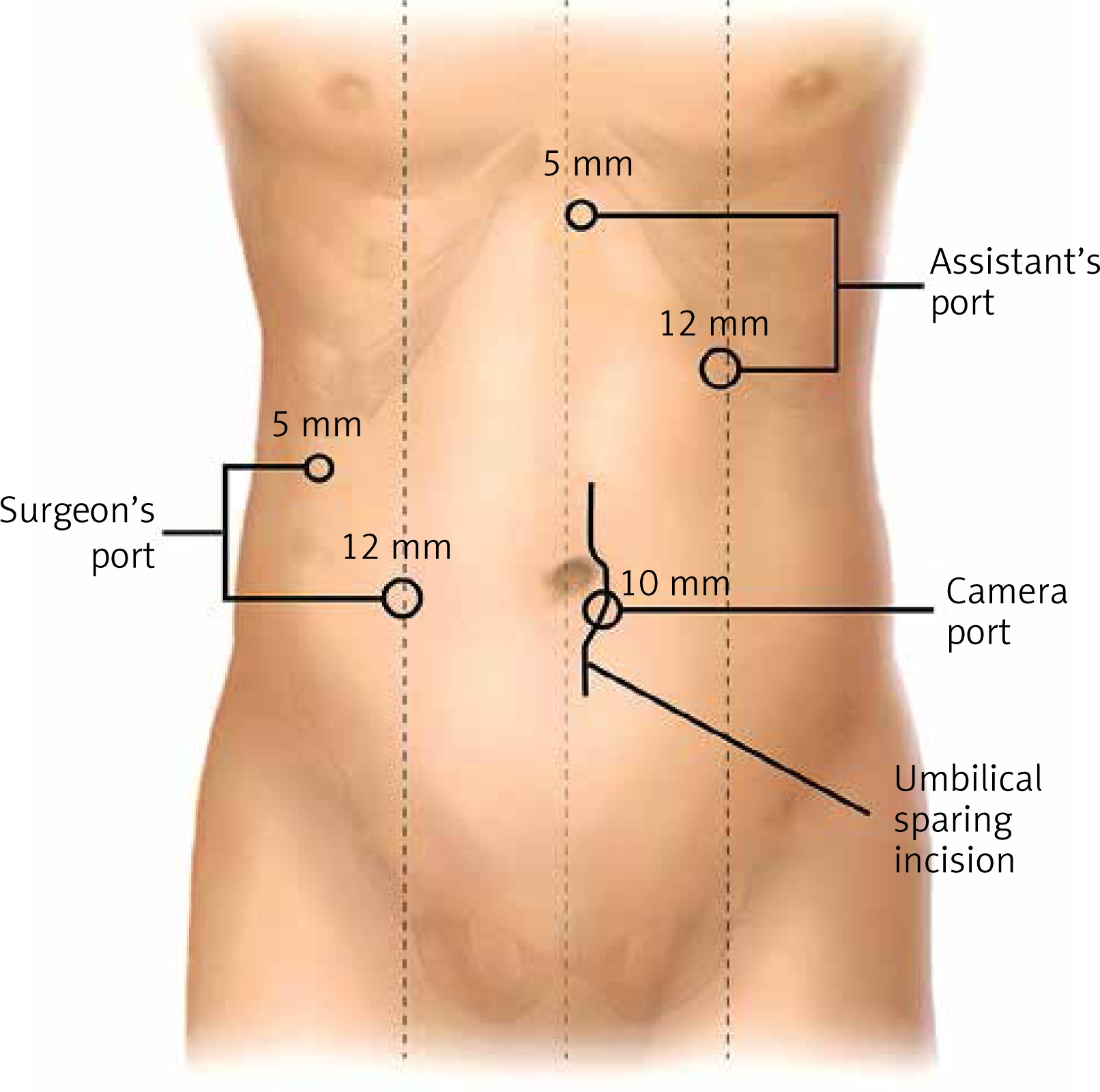
After general anesthesia, patients were placed in the supine reverse Trendelenburg position with the possibility to be tilted during the operation. The patient was firmly secured to the table. Both arms were abducted at a 90-degree angle to the body, and both legs were kept apart, slightly bent, and abducted to provide stability to the patient. All patient pressure areas were protected by soft devices. The surgeon stood either at the patient’s the right side or between the patient’s legs depending on the technical requirements during the operation. Pneumoperitoneum was established by a port positioned below the umbilicus and maintained at < 13 mm Hg. Staging laparoscopy was achieved as previously reported [32]. For patients with no signs of metastatic disease, the remaining ports were placed as shown in Figure 1.
Intraoperative ultrasonography was used to check and evaluate possible intrahepatic metastasis at the beginning of the laparoscopic HC resection surgery. Then, the Kocher maneuver was used to mobilize the duodenum and dissect the regional No. 16 a2/b1 lymph nodes. The lymph nodes scattered along the entire length of the hepatic artery from the celiac axis to the level of its bifurcation and the length of the right or left hepatic arteries as well as the pericholedochal, peri/retroportal and retropancreatic lymph nodes were routinely dissected. A circumferential excision of the hepatoduodenal ligament was performed, and the entire lymph nodal tissue was removed en bloc (Photo 1 A). The distal common bile duct at the upper border of the pancreatic head was subsequently transected for frozen biopsy (Photo 1 B). Subsequently, the inflow of the left or right liver was occluded and divided (Photos 1 C, D). The right/left lobes and caudate lobe were completely mobilized and excised en bloc (Photos 1 E, F). A specimen retrieval bag was used to remove the specimen through an additional 5- to 10-cm umbilical sparing median incision. This incision was closed immediately, and the pneumoperitoneum was resumed. Saline was used to irrigate the surgical field, and then bile leakage and bleeding were checked. After achieving hemostasis and biliostasis, the raw surface was covered with Surgicel SNoW (ETHICON, Cincinnati, Ohio, USA) (Photo 1 G). The biliary flow into the alimentary system was reconstructed by a Roux-en-Y bilioenteric anastomosis. A single-layer and interrupted bilioenteric anastomosis was performed to restore the biliary flow (Photo 1 H). At the end of the procedure, the surgical field was systemically explored for potential complications. Then, a drainage tube was placed around the hepaticojejunostomy (Photo 1 I).
Photo 1
Laparoscopic right hemihepatectomy with caudate lobectomy, hepatoduodenal lymphadenectomy and bilioenteric anastomosis to achieve radical resection for patients with Bismuth IV HC. A – Skeletonization of the vasculature and dissection of lymph nodes in the suprapancreatic area. B – The distal common bile duct was transected with scissors at the upper border of the pancreas, and the specimen was obtained for frozen biopsy to confirm R0 resection. C, D – The right hepatic artery (C) and portal vein (D) were occluded and divided E – Right hemiliver and total caudate lobe were completely mobilized and excised en bloc. F – A general overview of the surgical field is shown following removal of the specimen. G – The raw surface was covered with Surgicel SNoW (ETHICON, Cincinnati, Ohio, USA). H – Hepaticojejunostomy was performed with a single- layer and interrupted suture in the anterior and posterior walls. I – The drainage tube was placed around the hepaticojejunostomy
CBD – common bile duct, CHA – common hepatic artery, PV – portal vein, RHA – right hepatic artery, LHA – left hepatic artery, HA – hepatic artery, RPV – right portal vein, LPV – left portal vein, GDA – gastroduodenal artery.
For liver transection, a crush/clamp technique using a laparoscopic Harmonic Scalpel and bipolar forceps was adopted, along with associated saline pulses and aspiration. This process was performed with the alternate Pringle maneuver with decreased central venous pressure as previously described [39]. For the Pringle maneuver, cotton tape is used to pass around the hepatic pedicle. Then, a dedicated 5-mm port was created around the umbilicus that did not obstruct the field, and a tubular drain was applied to snugly attach the cotton tape with the help of a grasper. Another technique that allows for direct encircling of the hepatic pedicle was also utilized as necessary. The Pringle maneuver can be safely performed for 15 to 20 min and could be repeated many times during the whole procedure, with 5- to 10-minute perfusion between each maneuver.
Data collection and statistical analysis
Perioperative results included the laboratory examination, Bismuth-Corlette classification, blood loss, operative time, need for blood transfusion, biopsy of resection margins, 90-day mortality, postoperative morbidity, and the duration and cost of hospital stay. Operative time was determined from the beginning of pneumoperitoneum to the last port incision closure, including the instrumental preparation time. Postoperative morbidity was described according to the Clavien-Dindo classification. Patients were followed up for tumor condition and long-term prognosis through telephone interviews or individual clinic visits.
Additionally, a systematic search of all English literature was performed in PubMed and EMBASE based on the following search strategy: “hilar cholangiocarcinoma OR Klatskin tumor”. The retrieved articles were manually checked one by one to exclude studies without original data and that did not focus on curative surgical resection. Studies and series reports that included fewer than 40 resections were not included. Finally, the results of radical resection of patients with HC from 25 studies published from 1993 to 2014 were extracted and are detailed in Table I. Publicly available data about the long-term outcomes of HC patients who underwent curative-intent resection were obtained from 10 U.S. academic medical centers [40]. The Kaplan-Meier method was used for survival analysis. All statistical analyses were processed using R software (version 3.2.2).
Table I
Outcomes of radical resection for HC in series from 1993 to 2014
Results
Preoperative characteristics (Table II)
During the 45-month study period, 32 patients underwent radical laparoscopic resection surgery for HC. The detailed baseline characteristics of the patients are detailed in Table I. The female-to-male ratio was 11 : 21, and the median (range) age of the patients was 60.6 (38.8–76.7) years. The body mass index was 22.6 (18.4–29.1) kg/m2; 23 (71.8%) patients were in the normal range. The preoperative American Society of Anesthesiologists (ASA) score, which assesses the physical status before surgery, was 1 for most (71.8%) patients. Twenty-two (68.7%) patients had preoperative jaundice, and 14 (43.8%) patients with irreversible severe jaundice underwent preoperative biliary drainage. A percutaneous transhepatic biliary drain was inserted in 12 (37.6%) patients, and endoscopic nasogastric drainage was performed in 2 (6.2%) patients. Based on the preoperative imaging findings and MDT discussion, Bismuth types I, II, IIIa, IIIb, and IV cholangiocarcinoma were diagnosed in 0, 6 (18.7%), 4 (12.5%), 8 (25.0%), and 14 (43.8%) patients, respectively. The preoperative total bilirubin, direct bilirubin, alanine aminotransferase (ALT), aspartate aminotransferase (AST), ALB, CEA, and CA 199 levels are detailed in Table II.
Table II
Patients’ preoperative characteristics
Intraoperative parameters (Table III)
Among the 32 included patients, laparoscopic surgery with radical resection was ultimately performed in 24 (75.0%) patients. Three (9.3%) patients were found to be unresectable at the time of preliminary exploration, among whom 2 had peritoneal carcinomatosis and 1 had extensive duct spread with evidence of contralateral hepatic artery involvement. Five (15.7%) patients converted from laparoscopy to laparotomy due to accidental severe bleeding, anatomical variation or tumors invading the proximal trunk of the portal vein. Laparoscopic hemihepatectomy with caudate lobectomy was performed in 14 (65.6%) patients, with the right lobe resected in 6 patients and the left lobe resected in 8. The operation time and blood loss were 476.95 ±133.89 min and 568.75 ±324.01 ml, respectively. A Pringle maneuver was performed for 21 (77.8%) patients, with a median time (range) of 30 (15–60) min. A negative margin (R0) was achieved in 19 (79.1%) patients. Three (12.5%) patients were identified with a microscopic positive margin (R1). Because of macrovascular infiltration, 2 (8.4%) patients underwent macroscopic residual tumor resection (R2). After surgery, Bismuth types II, IIIa, IIIb, and IV cholangiocarcinoma were diagnosed in 2 (8.4%), 4 (16.6%), 5 (20.8%), and 13 (54.2%) patients, respectively. The tumor diameter was 3.076 ±0.38 cm, and the number of dissected lymph nodes was 8.93 ±5.26. All of the abovementioned parameters are summarized in Table III.
Table III
Intraoperative data
Postoperative and oncological outcomes (Table IV)
The length of postoperative stay was 23.3 ±11.7 days, including patients who suffered complications. There was 1 (4.1%) patient death within 30 days after surgery; the patient suffered serious intra-abdominal bleeding and consequent disseminated intravascular coagulation (DIC). The pathologic results showed that cholangiocarcinoma and cholangiocarcinoma combined with mucinous adenocarcinoma were confirmed in 21 (87.5%) and 3 (12.5%) patients, respectively. Postoperative complications were stratified according to the Clavien-Dindo classification. Grade I–II complications occurred in 15 (62.5%) patients. Severe morbidity was defined as grade III–V, which occurred in 4 (16.6%) patients. The hospital expenditure was USD 14348 ±4779. After a median (range) follow-up of 9 (1.5–45.1) months, 16 (66.7%) patients were alive without any sign of recurrence. Liver recurrence occurred in 3 (12.5%) patients, retropancreatic lymph node metastasis in 1 (4.1%) patient and multisite metastasis in 1 (4.1%) patient, including the liver and peritoneum (Table IV).
Table IV
Patients’ postoperative parameters and pathological results
Comparisons with conventional surgery results (real-world evidence)
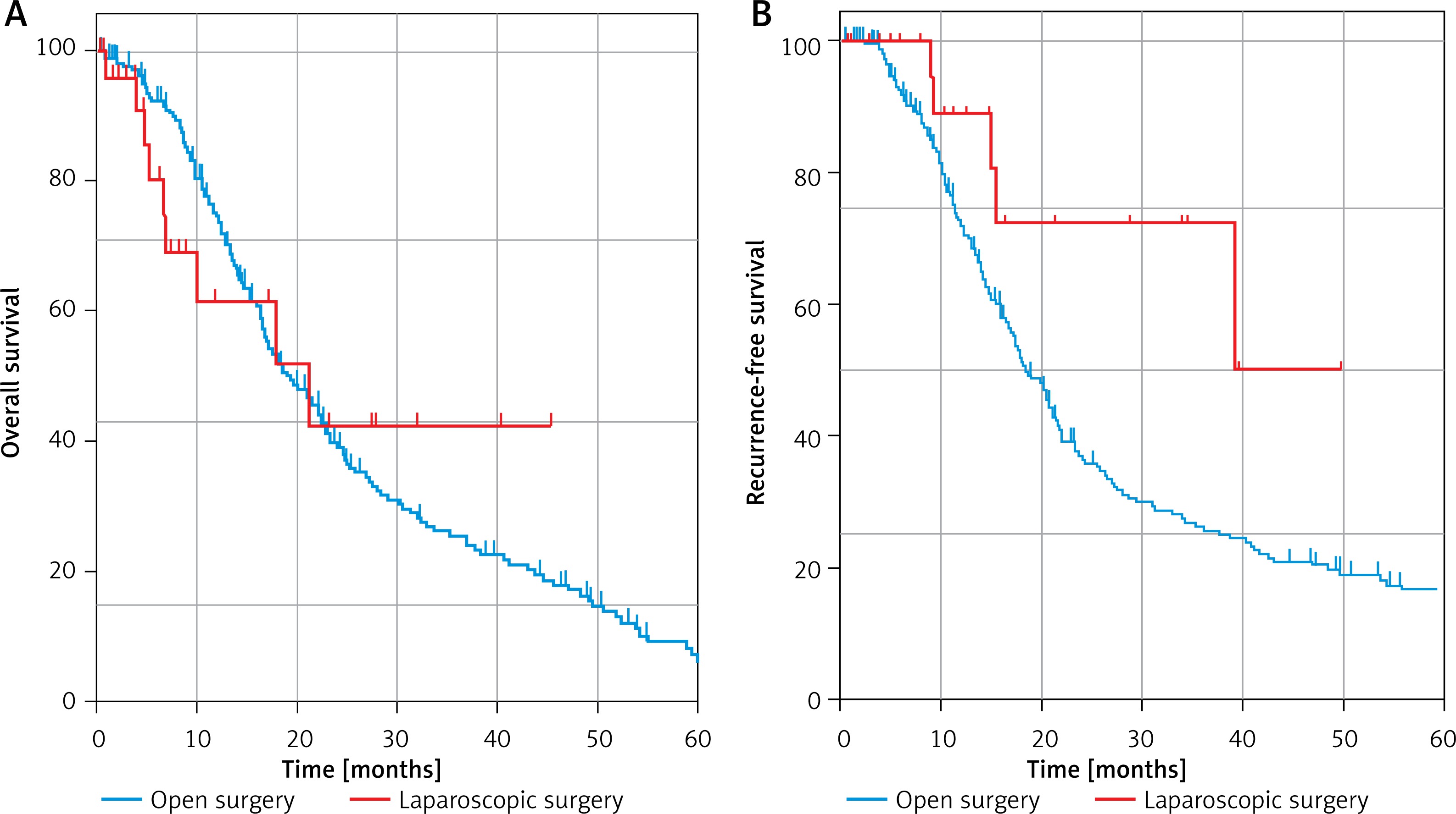
The extent of surgical resection for HC with the aim of curative treatment is already controversial. To date, numerous studies have published the oncological outcomes of the radical resection of HC though open surgery. These studies included a large number of patients with long-term oncological follow-up. The final results have tended towards stability and can reflect the actual effect of conventional open surgery. Thus, the outcomes of surgical resection from 25 studies published during the period 1993–2014 were extracted and compared with our results in the present study in Table I. Additionally, we further compared the long-term outcomes with publicly available data obtained from 10 U.S. academic medical centers [40]. As shown in Figure 2, HC patients who underwent curative-intent resection though open surgery had actuarial 3-year overall survival (OS) and recurrence-free survival (RFS) rates of 36.2% and 27.8%, respectively. However, the actuarial 3-year OS and RFS for laparoscopic surgery in the present study were 49.1% and 47.0%, respectively.

Discussion
Due to the overt benefits over open surgery, including decreased wound-related complications, early ambulation, quicker recovery time and much smaller scarring, laparoscopic surgery has been routinely used in the management of benign and malignant disease. Over the past decades, many kinds of laparoscopic surgery have evolved; at first unsuccessful, subsequent but still early procedures began showing some benefit, and now they are becoming the gold standard form of surgery. These promising results have encouraged surgeons to further explore the application of laparoscopic techniques in complex surgery. In fact, a similar process can also be observed in laparoscopic pancreaticoduodenectomy (LPD) [41] and laparoscopic hepatectomy [42], which was initially controversial but has been gradually accepted. For example, LPD was once regarded as the Mt. Everest of general surgery [43]. Even conventional laparotomy is considered hard to perform, let alone laparoscopic resection. However, after nearly 25 years of endeavor, recently published studies have finally confirmed that LPD is a safe and feasible procedure from a technical standpoint, with acceptable rates of mortality and morbidity [44, 45]. Wang et al. retrospectively analyzed 1029 consecutive patients who had undergone LPD in China and found that median operation time, major complications, and conversion rate improved significantly with surgeon’s experience [44]. More importantly, as one of the most complicated operations, LPD has a learning curve that is significantly longer than expected. As demonstrated in a previous study, only after completing at least 140 cases can competence for LPD be achieved [44]. Additionally, another study published at the same time reported their experience from 500 patients in a single center. This study revealed that LPD is a feasible choice for selected patients [45]. After overcoming the learning curve, perioperative outcomes and the prognosis of LPD can be significantly optimized [45]. Taken together, the abovementioned studies highlight that even the Mt. Everest of general surgery can be climbed with the accumulation of surgical experience and the development of laparoscopic techniques. At present, LPD, an incredibly difficult operation, has been transformed into routine surgery in high volume hospitals.
If LPD is the Mt. Everest of general surgery, laparoscopic radical resection of HC is the brightest jewel at the top of the mountain. As the greatest challenge for hepatobiliary surgeons, the exploration of minimally invasive surgery for HC was started by Giulianotti et al. in 2010 [46], 16 years after the invention of LPD. This in itself reflects the extremely high technical demands of laparoscopic radical resection for HC. HC is completely different from other cholangiocarcinomas in terms of the therapeutic strategy and prognosis [1, 47]. This is because HC arises from the biliary confluence or the left or right hepatic duct, which are close to vascular structures. There are variable, complex and intimate relationships between the biliary and vascular structures at this location. Meanwhile, the tumor properties allow HC to invade vascular structures, infiltrate adjacent liver tissues, spread along the bile ducts and metastasize to the lymph nodes and distant organs [48–50]. Undoubtedly, margin negative (R0) resection is the most effective treatment and probably the only approach that provides a chance of cure. However, radical resection with a negative margin is extremely challenging. The standard operations for resectable HC include extended liver resections in conjunction with extrahepatic bile duct resection, hepatoduodenal lymphadenectomy, caudate lobectomy and bilioenteric anastomosis [3, 10]. Increasing evidence has demonstrated that this ‘‘aggressive’’ surgical strategy, although technically challenging, has increased the rate of curative resection and long-term survival. Unfortunately, 29–40% of HC patients had locally advanced or metastatic disease at initial presentation, making them ineligible for surgical resection [13, 51]. Moreover, at the time of the curative-intent operation, only 50-60% of patients have been ultimately found to be resectable [51, 52]. In the current study, all enrolled patients were highly selected and considered suitable for curative-intent surgery. In contrast to what was observed in the above studies, only 9.3% (3/32) of the HC patients were found to be unresectable at the time of surgical exploration in the present study. This result may, in part, be due to the comprehensive use of various imaging techniques (thinly sliced CT, MRI, and USG) to evaluate tumor resectability and the selection of patients who could undergo laparoscopic surgery by the MDT approach.
With the accumulated experience of laparoscopic surgery and the development of laparoscopic devices, an increasing number of studies have reported the safety and feasibility of laparoscopic extended hemihepatectomy [53, 54], laparoscopic caudate lobectomy [55, 56], laparoscopic hepatoduodenal lymphadenectomy and bilioenteric anastomosis [44, 45]. Thus, from a technical perspective, it is obvious that the essential surgical techniques for laparoscopic resection of HC have become more reliable and diverse. To date, however, very few experienced surgeons have attempted to challenge the laparoscopic approach for the radical resection of HC. According to a systematic review published recently, only 21 studies have been published, reporting on a total of 142 minimally invasive procedures for HC, among which 1 hybrid, 59 robot-assisted and 82 laparoscopic procedures were included [57]. Margin-negative resection (R0) was achieved in almost 80% of patients, and only 4.9% of patients converted from laparoscopy to laparotomy. Furthermore, the total of 30 complications and 4 deaths among 126 patients suggests a postoperative morbidity rate of 24% and an overall 90-day mortality rate of 3%. These results indicated that laparoscopic radical resection of HC is technically feasible for highly selected patients in the hands of experienced surgeons. In the current study, an R0 resection with adequate lymphadenectomy was achieved in 79.1% of patients. Additionally, the postoperative morbidity rate and overall 90-day mortality rate were 16.6% and 4.1%, respectively. Obviously, for major perioperative indicators, such as R0 resection rate, 90-day mortality rate, and postoperative morbidity rate, the results from our current study and the systematic review [58] are very similar.
However, the rate of conversion to laparotomy in laparoscopic hepatectomy ranged from 9% to 42% [57], and even in laparoscopic cholecystectomy, the rate of conversion from laparoscopy to laparotomy remains between 5% and 10% [59]. Additionally, at the initial and exploratory stages, the conversion rate of LPD in high volume centers can reach 17.3%. Even when surgeons overcome the learning curve (performing more than 104 cases), the average conversion rate can only be reduced to 5.9%, which is higher than the result in the systematic review (4.9%). In the present study, 15.7% of patients converted from laparoscopy to laparotomy due to accidental severe bleeding, anatomical variation and tumors invading the proximal trunk of the portal vein, which is similar to the result for LPD (17.3%) in its infancy. Outcomes of open surgery for HC have been reported by several multicenter studies and single-institutional series, and these larger case series are detailed in Table I. The morbidity and mortality of open surgery in patients with HC are notoriously high and have been reported to reach 18% and 68%, respectively. Additionally, publicly available data on the long-term outcomes of HC patients who underwent curative-intent surgery, obtained from 10 U.S. medical centers, were comparatively analyzed in this study. As shown in Figure 2, HC patients who underwent curative-intent resection though open surgery had an actuarial 3-year OS and RFS of 36.2% and 27.8%, respectively. However, the actuarial 3-year OS and RFS for laparoscopic surgery in the present study were 49.1% and 47.0%, respectively.
These comparisons with the literature (Table I) demonstrate a benefit of laparoscopic radical resection for HC compared with open surgery. However, the abovementioned findings should be interpreted with extreme caution. These preliminary results may not be truly representative of current practice and are very likely to be influenced by strict patient selection, which is also a limitation of this current study. Therefore, the results cannot be widely reproduced and the use of laparoscopic radical resection of HC for this specific patient population should be limited to experienced centers only.
To the best of our knowledge, the current study is one of the largest series regarding the feasibility and safety of laparoscopic resection for HC. Combining the results from previously published articles and those of the present study, it is safe to say that the laparoscopic radical resection of HC is feasible from a technical point of view but is still in its initial stages. However, before the laparoscopic approach for patients with HC becomes routinely implemented, the questions that need to be solved involve not only the safety and technical feasibility but also oncological concerns. For example, is there any increase in the incidence of port-site metastases? Can the long-term oncological outcomes be compared to those of an open procedure?
In the present study, throughout a median (range) follow-up of 9 (1.5–45.1) months, no patients with HC who were treated by laparoscopic radical resection suffered recurrence at the port site. Similar to what we observed in this series, earlier studies also indicated that implanted metastasis or port site recurrence (PSR) did not occur in any patients during a median follow-up of 48 and 60 months, respectively [31, 35]. Previous studies have also demonstrated that the median time of PSR is 6 months [60]. Thus, it is reasonable to assume that laparoscopic radical resection for HC does not increase the incidence of PSR. Combining both the former experimental studies and our own observations, the effect of pneumoperitoneum alone on PSR and implanted metastasis is almost negligible. However, due to the possibility of cell exfoliation during tumor handling, which may result in the dissemination or direct contamination of the small wound in the process of specimen extraction, appropriate surgical techniques, such as averting bile leakage, using a specimen retrieval bag and minimal tumor treatment, are necessary.
In our study, laparoscopic hemihepatectomy with caudate lobectomy was performed in 14 (65.6%) patients, with the right lobe resected in 6 patients and the left lobe resected in 8 patients. The anatomical characteristics of the hepatic duct at least partly affect the difficulty of hepatectomy. The right hepatic duct is short and has a variable position, which increases the complexity of right hemihepatectomy, whereas the left hepatic duct is more predictable and longer in length. This makes resection of the left half of the liver more straightforward. Among the 24 patients who underwent complete laparoscopic radical resection, 3 patients with a Bismuth IIIa cholangiocarcinoma and 2 patients with a Bismuth II cholangiocarcinoma underwent only right hemihepatectomy. However, they all underwent R0 resection. In fact, the bile of the caudate lobe generally drains into the biliary confluence or left biliary tree. The hepatic duct of the caudate lobe has a close anatomical relationship with the left hepatic artery. Therefore, en bloc caudate lobectomy should be performed for all left-sided and centrally located tumors but only selectively for right-sided tumors if the duct of the caudate lobe joins the biliary system from the left side. Our experience partly supports this surgical strategy [10]. In the current study, we followed this strategy to develop individualized operation plans. In fact, following these criteria for caudate lobectomy may not only minimize the risk of biliary leak but also reduce the risk of local recurrence from a positive margin, but further clinical research is needed for validation.
Conclusions
Laparoscopic radical resection for HC is presently at an early stage but is rapidly developing. From the current study, it is obvious that favorable perioperative outcomes could be achieved by experienced hepatobiliary surgeons, and the benefits of laparoscopic surgery could be provided to highly selected patients. However, laparoscopic radical resection for HC remains a technical challenge, and it should be performed only in centers with sufficient experience in advanced laparoscopic hepatobiliary surgery. A limitation of this current study is that it is a noncomparative and retrospective case series, which may result in a potential bias. However, the current study has demonstrated a promising future. Randomized trials with long-term follow-up should be performed to address the question of whether laparoscopic procedures can achieve the same results as open surgery. The results of this retrospective case series study could form the foundation for a future randomized trial.