
We present the case of a 70-year-old female diagnosed with peripheral T-cell lymphoma, not otherwise specified (PTCL-NOS), admitted to the Department of Bone Marrow Transplantation and Onco-Haematology due to disease progression confirmed in a recent positron emission tomography (PET) scan. Previous treatments included 6 cycles of CHOP-21 (cyclophosphamide, doxorubicin, vincristine, and prednisone), autologous hematopoietic stem cell transplantation (AHSCT), and 4 cycles of BGD (bendamustine, gemcitabine, and dexamethasone).
On admission, she exhibited a confluent, bright red maculopapular rash covering over 50% of her body, sparing the hands, face, and mucosae (Figure 1 A). Peripheral lymph nodes were not palpable. The suspicion of drug reaction to recent trimethoprim-sulfamethoxazole or bendamustine was raised. Control peripheral blood flow cytometry demonstrated 42.9% T-lymphoid cells with an aberrant immunophenotype (reduced expression of CD3 and CD45), with CD30 coexpression on 5% of cells, which supported the progression of PTCL.
Figure 1
Clinical presentation of maculopapular rash on admission (A), with gradual progression to confluent erythematous-infiltrative changes (B), with the final formation of nodules (C, D)
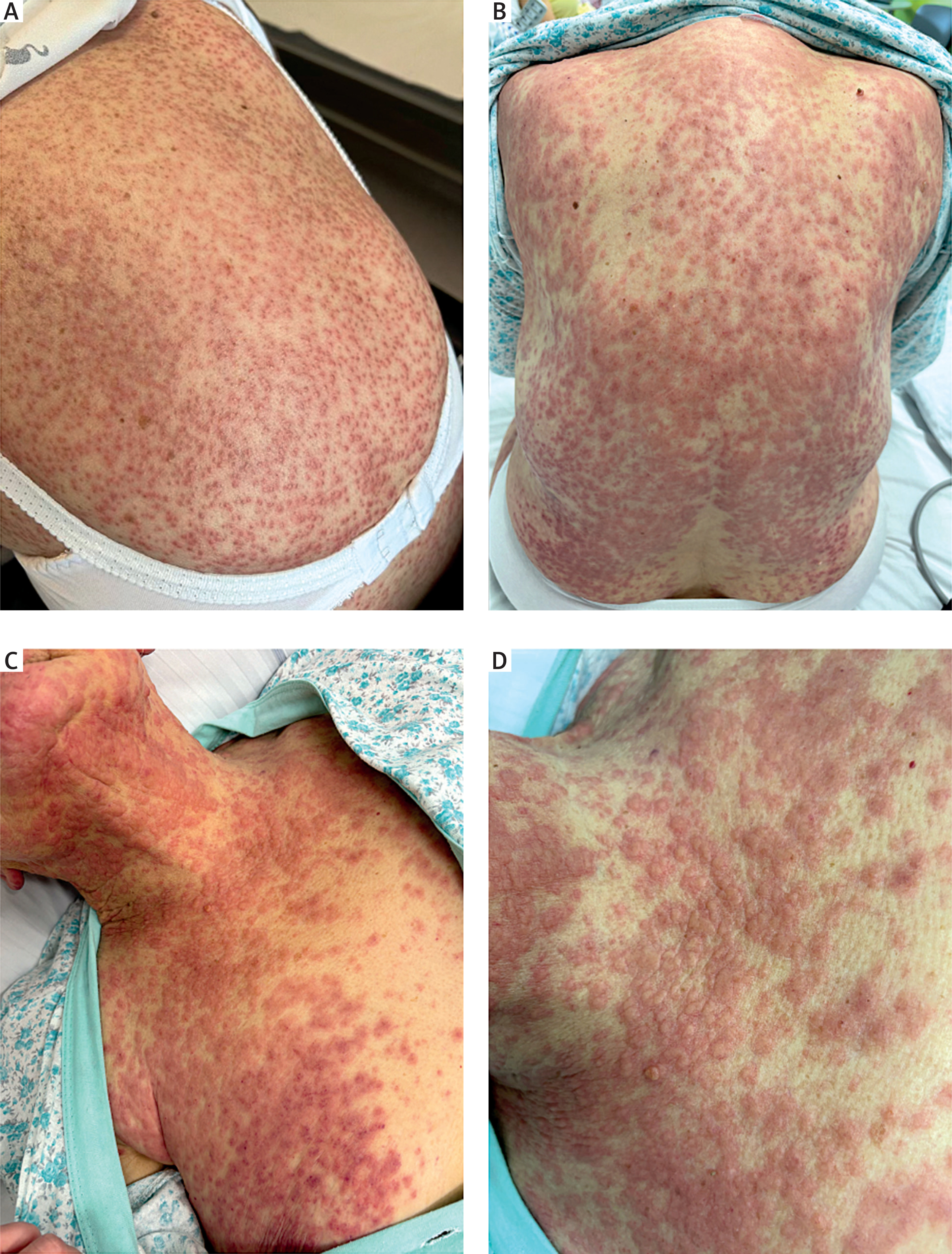
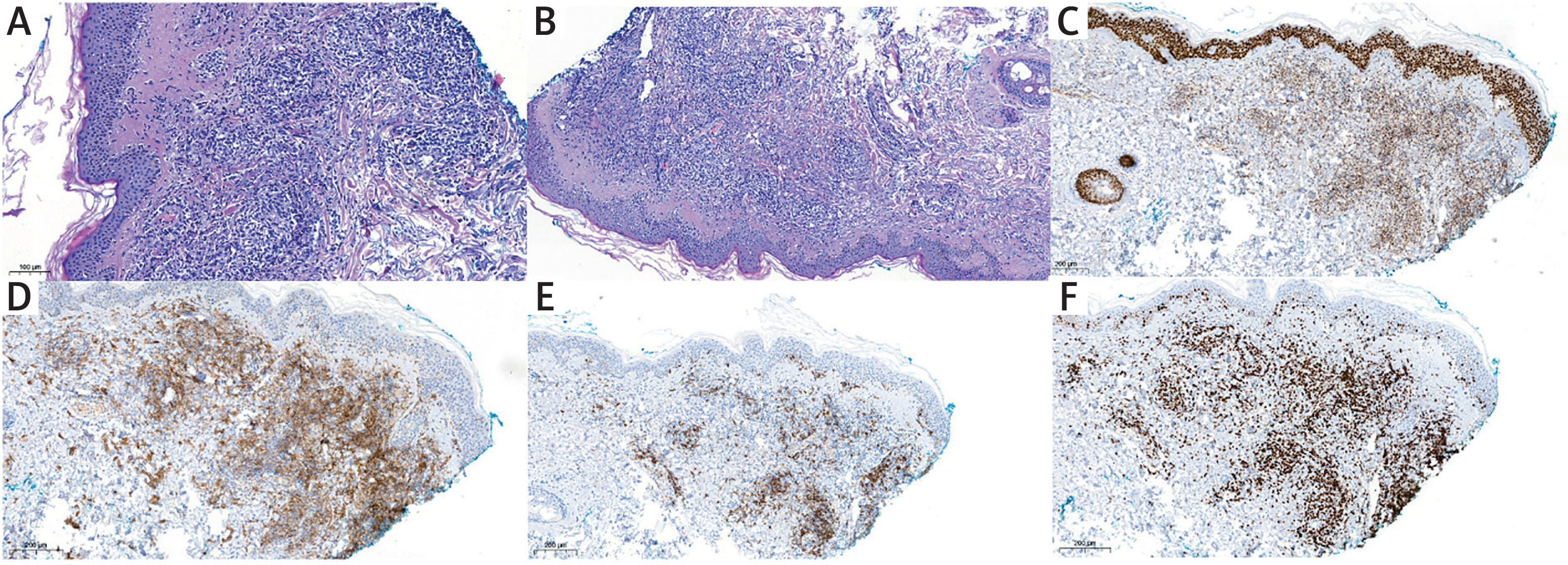
Clinical manifestation of skin lesions and the initial Registry of Severe Cutaneous Adverse Reactions (RegiSCAR) score of 3 (possible case) suggested the drug reaction with eosinophilia and systemic symptoms (DRESS), including skin eruption involving > 50% of body surface area, rash suggestive of DRESS, and hepatic impairment. Laboratory tests revealed leukocytosis without eosinophilia, thrombocytopenia, and elevated levels of C-reactive protein (CRP), lactate dehydrogenase (LDH), alkaline phosphatase (ALP), γ-glutamyl transpeptidase (GGTP) and bilirubin. Creatinine levels remained normal. Treatment with intravenous methylprednisolone (1 mg/kg/day for 7 days) resulted in improvement of the skin rash. However, a chest X-ray revealed densities in the right lung, and crackles were noted on auscultation and CRP levels continued to rise. The control leukogram showed 69% abnormal lymphocytes, no eosinophils. Due to severe thrombocytopenia, the patient required multiple transfusions of platelet concentrates. Due to the confirmed HHV-6 viremia, ganciclovir was also included in the treatment. On day 13, hemorrhagic changes developed on the lower limbs, resembling vasculitis, with subsequent progression of the initial maculopapular rash, involving additional areas: the face, neck and décolleté, with the formation of confluent erythematous-infiltrative changes, and in the following days (Figure 1 B), with the final formation of bluish-purple nodules (Figures 1 C, D). The control RegiSCAR scoring system was 5 points, which corresponded to a diagnosis of DRESS syndrome (probable case), including: skin eruption involving > 50% of body surface area, suggestive of DRESS; presence of atypical lymphocytes in peripheral blood, hepatic impairment, lung manifestations. However, as the skin lesions evolved into infiltrated nodules, the subsequent biopsy evaluation confirmed infiltration by PTCL-NOS (Figure 2).
Figure 2
The skin biopsy sections of skin samples from décolleté (June 2024, 13th day of hospitalization), diffuse dermal infiltrate of mainly medium-sized atypical lymphocytes in hematoxylin and eosin staining (A, B), BCL6 (-/+), GATA3(+) (C), CD4(+) (D), CD30 heterogeneous expression by approx. 20% dermal atypical lymphocytes (E), high proliferative activity, Ki-67 approx. 90% (F). Immunophenotype compatible with immunoprofile PTCL-NOS infiltrates in thigh core biopsy (October 2023) and adenoids, lymph nodes samples (June 2022)
Given the rapid worsening of the underlying disease, treatment with brentuximab vedotin was initiated, which initially resulted in partial clinical improvement; however, within the following days, enlargement and disintegration of the nodular lesions on the back and chest led to ulcer formation. Control peripheral blood flow cytometry revealed 97.8% CD4+ T-lymphoid cells with an aberrant immunophenotype (reduced expression of CD3 and CD45), with CD30 coexpression on 0.2% of cells. Moreover, parainfluenza type-2 virus infection and Epstein-Barr virus (EBV) reactivation occurred, and the patient developed respiratory failure. Despite the escalation of the treatment and the initiation of rescue cisplatin monotherapy (20 mg/m2 for 5 days), the patient succumbed to infectious complications secondary to lymphoma.
DRESS syndrome is a rare, potentially fatal immunological reaction to drugs [1]. Symptoms typically appear 2–6 weeks after initiating the causative agent and include a maculopapular rash covering > 50% of the body, often with facial oedema. Although eosinophilia is characteristic, it is not required for diagnosis. The diagnosis is based on the RegiSCAR criteria, which include fever, lymphadenopathy, eosinophilia, atypical lymphocytes, rash covering > 50% of the body, skin biopsy results, resolution of lesions within ≥ 15 days, organ dysfunction, and exclusion of other possible causes. Scores determine whether the case is possible, probable, or definitive [1]. Reactivation of HHV-6 is a frequently described phenomenon in patients with DRESS, which additionally prompted such a diagnosis in the patient. The causal relationship of this observation is the subject of discussion [2].
PTCL-NOS, the most common T-cell lymphoma, represents 30% of cases in this group. The median diagnosis age is 60 years, with a slight male predominance. Patients typically present with advanced-stage disease, often involving multifocal lymph nodes and extranodal sites such as the gastrointestinal tract, skin, and bone marrow. Laboratory findings may include anemia, thrombocytopenia, haemolysis, elevated β2-microglobulin, and viral reactivations such as EBV or cytomegalovirus (CMV) [3].
In this case, the patient with PTCL-NOS progression developed disseminated skin lesions resembling DRESS syndrome and hemorrhagic changes resembling vasculitis.
However, lesion progression to nodules, control peripheral blood flow cytometry and skin biopsy results confirmed lymphoma progression. Hemorrhagic changes likely resulted from neoplastic vascular destruction exacerbated by thrombocytopenia and hydrostatic pressure. HHV-6 reactivation, a phenomenon frequently associated with DRESS syndrome, further complicated the diagnosis. This case underscores the diagnostic challenges posed by overlapping features of DRESS syndrome and PTCL-NOS. Early differentiation and appropriate treatment strategies are critical in such complex presentations. Early cutaneous manifestations lacked infiltrative features and, together with the RegiSCAR scoring system, were consistent with DRESS; however, recent PET findings showing disease activity, rapid evolution into infiltrated lesions, and peripheral blood flow cytometry results prompted consideration of lymphoma relapse. Similar observations have been reported in the literature, where both lymphomas [4] and drug-induced pseudolymphomas have been described as mimicking DRESS, consistent with our case [5].










