eISSN: 1644-4124
ISSN: 1426-3912
Central European Journal of Immunology
Current issue Archive Manuscripts accepted About the journal Special Issues Editorial board Abstracting and indexing Subscription Contact Instructions for authors
Editorial System
Submit your Manuscript
SCImago Journal & Country Rank
2/2021
vol. 46
 
Share:
Share:
Short communication

Expression of ORAI1 and STIM1 genes in blood of patients with pulmonary tuberculosis

Oksana Kolesova
1
,
Ksenija Kramica
1
,
Aleksandrs Kolesovs
1
,
Jelena Eglite
1

1.
Riga Stradins University, Latvia
Cent Eur J Immunol 2021; 46 (2): 275-282
Online publish date: 2021/06/18
Article file
- Expression of ORAI1.pdf  [0.31 MB]
Get citation
 
PlumX metrics:
 

Introduction

ORAI1 and STIM1 genes are essential for the store-operated Ca2+ entry (SOCE) mechanism used by immune cells to increase intracellular Ca2+ concentrations [1]. Expression of these genes in blood of patients with tuberculosis (TB) became the focus of our exploration. We expected some impairment in this specific regulatory mechanism of Ca2+ entry in patients with TB.

Tuberculosis is an infection characterized by the persistence of mycobacteria in macrophages and dendritic cells due to imperfect phagocytosis. The local secretion of interferon γ (IFN-γ), produced by type 1 T helper cells, and tumor necrosis factor α (TNF-α), primarily produced by macrophages, are the main factors enhancing the maturation of phagolysosomes, production of reactive oxygen (ROS), nitrogen species, and antimicrobial peptides in macrophages [2]. Activation of immune cells, production of cytokines and inflammation-restricting proteins (Fas-ligand, Foxp3), and production of ROS in phagosomes of phagocytes occur after an increase of intracellular Ca2+ concentration [3, 4]. Many studies [5-9] indicate that mycobacteria use different strategies to manipulate Ca2+ signaling in macrophages and dendritic cells to increase their lifetime and escape from the immune response.

There are two main channels providing Ca2+ influx into the immune cells: Ca2+ release-activated Ca2+ (CRAC) channels, which are activated by decreasing intracellular Ca2+ concentration, and voltage-gated Ca2+ channels (VGCC), which are opened by depolarization of the cytoplasmic membrane [5, 10]. The immune cells predominantly use CRAC channels of SOCE [1]. The prototypical CRAC channel is formed by ORAI1 protein (encoded by the ORAI1 gene) located on the cytoplasmic membrane. Its activation is provided by ligation of stromal interaction molecule 1 (STIM1, encoded by the STIM1 gene) located on the membrane of the endoplasmic reticulum [10].

The importance of STIM1 and ORAI1 genes in TB was demonstrated in an experimental model in TB infected mice [3]. A defect of the STIM1 gene led to the death of mice due to pulmonary hyper-inflammation and loss of respiratory function. It suggests that a dysfunction of SOCE may contribute to imperfect mycobacterial phagocytosis and provide a basis for mycobacterial persistence. Simultaneously, there are no data about the expression of ORAI1 and STIM1 genes in humans with TB. Detection of the expression level of ORAI1 and STIM1 genes in blood despite their non-specificity for TB can provide additional information for a TB pathogenesis, prognosis, and monitoring of TB treatment.

This study aimed to detect the level of expression of ORAI1 and STIM1 genes in blood of TB patients at the beginning of anti-TB treatment in association with immunological status, assessed by levels of IFN-γ and interleukin 18 (IL-18) in serum, and to compare the expression of ORAI1 and STIM1 with a healthy control group. IL-18 was selected for the assessment of the immunological status as a pro-inflammatory cytokine, produced predominantly by activated monocytes/macrophages and having broad immunological effects [11], including an increase of production of IFN-γ by T cells and enhancing the protective Th1 immune response against mycobacteria. Simultaneously, increasing activity of IL-18 induces activation of nuclear factor-kB and expression of Fas ligands, which can lead to tissue destruction [11, 12]. An increasing level of IL-18 in serum also has a negative predictive value for HIV-associated TB [13].

Material and methods

The permission of the Ethics Committee of Riga East University Hospital (No. 9-A/17) was obtained for the study. The study included 45 patients, aged 20 to 86 years (mean age = 49.7 years, SD = 16.0, 33% females), with confirmed TB. The patients underwent observation and treatment from October 2017 to December 2019 in the Lung Disease and Tuberculosis Ward of Daugavpils Regional Hospital (Latvia). Inclusion criteria were: age of 18 and older, bacteriologically confirmed pulmonary TB, and drug-sensitive TB. Exclusion criteria were: age < 18, exclusively extrapulmonary TB, pulmonary TB with concurrent extrapulmonary TB involvement, pregnancy, imprisonment, mental disorders, and HIV-positive status. All patients had bilateral drug-sensitive tuberculous pneumonia and used first-line anti-tuberculous drugs according to the standard regimen.

The control group included 35 volunteers, aged from 21 to 73 years (mean age = 36.8 years, SD = 16.0, 30% females), without active TB and human immunodeficiency virus (HIV) infection. For the control group, the inclusion criteria were age of 18 and older, self-reported physical health, and no ongoing infection.

For analysis of ORAI1 and STIM1 expression, 1 ml of peripheral blood with EDTA was collected and stored at –20oC before detection. RNA was extracted from stored blood samples using the innuPREP Blood RNA Life Science Kits & Assays (Analytik Jena Company, Germany) according to the manufacturer’s procedures. The quality and quantity of extracted RNA were detected by spectrophotometry using the Nanofotometr NF80 (Implen GmbH, München, Germany). The concentration of RNA was 40 µg/ml per reaction. For reverse transcription-PCR (RT-qPCR), the Revert First-Strand cDNA synthesis system with an oligo dT primer (QuantiTect Reverse Transcription, an oligo dT primer Invitrogen, Germany) was used following the manufacturer’s instructions. The first-strand cDNA was diluted 1 : 20 with distilled water with the following used as a template (K1+) in RT-qPCR analysis. Specific primers for human STIM1 (Hs STIM1 FAM_1, QF00208159) and ORAI1 (Hs ORAI 1 FAM_1, QF00163611) were detected by one-step qRT-PCR using sequence-specific probes for gene expression analysis (QuantiFast Probe Assay, Invitrogen, Germany). RT-qPCR was performed in a DTlite Cycler (DNA-Technology, Russia) by the fast real-time PCR System using the following amplification conditions: 5 min of initial denaturation at 95°C, then 45 cycles of 95°C for 30 s, 60°C for 30 s. The specificity of RT-qPCR products was confirmed by the analysis of a melting curve. Absolute quantification of STIM1 and ORAI1 gene expression was detected relative to a standard curve, automatically created by serial dilution of the reference gene glyceraldehyde-3-phosphate dehydrogenase (GAPDH). For amplification, the following primers (Invitrogen by Thermo Fischer Scientific) and reference gene (Bioneer Corporation, Republic of Korea) were used: ORAI1, forward: 5'-CGTATCTAGAATGCATCCGGAGCC-3' reverse: 5'-CAGCCACTATGCCTAGGTCGACTAGC-3'; STIM1, forward: 5'-CCTCGGTACCATCCATGTTGTAGCA-3', reverse: 5'-GCGAAAGCTTACGCTAAAATGGTGTCT-3'; GAPDH, forward: 5'-CCACTCCTCCACCTTTGAC-3', reverse: 5'-ACCCTGTTGCTGTAGCCA-3'; Probe, 5'-TTGCCCTCAACGACCACTTTGTC-3'.

Detection of IFN-γ and IL-18 in serum was performed by the sandwich immunoassay method using the commercial kits IFA-BEST INF-γ (detection range: 0-1000 pg/ml, sensitivity: 2.0 pg/ml, normal values: < 15 pg/ml) and IFA-BEST IL-18 (detection range: 0-1000 pg/ml, sensitivity: 2.0 pg/ml, normal values: 90-260 pg/ml) according to the methodology of the manufacturer (Vector-Best, Russia).

Results and discussion

The Shapiro-Wilk test revealed significant deviance from the normal distribution in both groups of participants. Test values varied from 0.19 to 0.91 (p < 0.01 to p < 0.001). Observed non-normality of distributions led to the use of nonparametric statistics for a comparison of the control and TB groups. The results of the Mann-Whitney U-test (Table 1) indicated a significantly higher level of mRNA ORAI1 in the control group.

Table 1

Comparison of markers in tuberculosis (TB) and control groups

MarkersTB group (n = 45) Median (IQR)Control group (n = 35) Median (IQR)Mann-Whitney U-test
mRNA ORAI1 (copies/ml)55.7 (1.7-520.5)1.3E+12 (1.1E+9-2.6E+13)1.0***
mRNA STIM1 (copies/ml)978.0 (7.4-3230.0)383.0 (9.2-21200.0)778.0
IFN-γ (pg/ml)39.3a (17.3-79.2)0.0b (0.0-0.0)10.0***
IL-18 (pg/ml)276.9c (228.4-397.8)225.8b (208.2-253.1)208.0*

[i] IQR – interquartile range, a presented for a subsample n = 34, β presented for a subsample n = 15, c presented for a subsample n = 44, *p < 0.05, ***p < 0.001

Simultaneously, there were no significant differences between groups in the level of mRNA STIM1. The level of IFN-γ and IL-18 were higher in the group of TB patients. Figure 1 presents the comparison between groups.

Fig. 1

Differences between groups in mRNA ORAI1, mRNA STIM1, IFN-γ and IL-18 (medians and their 95% CI are presented on a decimal logarithmic scale)

/f/fulltexts/CEJOI/44411/CEJI-46-44411-g001_min.jpg

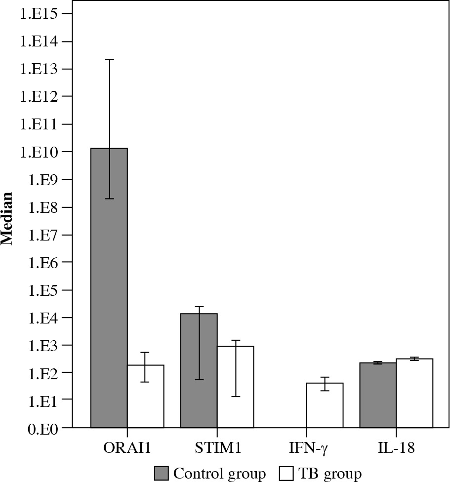
Expression levels of ORAI1 and STIM1 genes in blood dominantly reflected their expression in peripheral blood leukocytes, being immune cells. A more expressed ORAI1 gene in TB patients indicated an impaired SOCE mechanism in these cells.

Taking into account that ORAI1 protein forms a pore in the plasmatic membrane of immune cells for releasing Ca2+ ions into cytoplasm, a lower expression level of ORAI1 in blood can be associated with a lower influx of Ca2+ into cells, leading to lower cytokine production. In contrast, our findings showed that the production of IFN-γ and IL-18 is not disrupted in patients with TB and is at a higher level than in the control group. As described in a model of TB infected mice [3], impaired SOCE is associated with the reduced expression of inflammation-restricting proteins, leading to hyper-inflammation.

The Spearman correlation coefficients indicated no significant relationships among the markers in both the control and TB groups (Table 2). It indicates the relative independence of the level of expression of genes and the level of inflammation.

Table 2

Spearman correlations among markers in control and tuberculosis (TB) group

MarkersControl groupTB group
ORAI1STIM1IFN-γORAI1STIM1IFN-γ
STIM10.07a0.12c
IFN-γ–0.18b–0.38b–0.10d0.16d
IL-18–0.05b0.06b0.13b0.15e–0.03e–0.06d

a presented for a subsample n = 35, β presented for a subsample n = 15, c presented for a subsample n = 45,d presented for a subsample n = 34, e presented for a subsample n = 44

Simultaneously, non-pronounced differences in STIM1 gene expression indicate a low association of its expression with TB. Ca2+ influx into immune cells is also provided by other Ca2+ channels [14], for example, VGCC [6]. In this case, Ca2+ influx through VGCC leads to suppression of the protective immune response [6, 7].

At a more generalized level, observed differences in expression of the ORAI1 gene have at least two explanations. One the one hand, low expression of the ORAI1 gene in patients with TB can be a result of mycobacterial infection. Previous studies [15-18] show that mycobacteria can change Ca2+ signaling and protect survival in macrophages by regulation of expression of microRNAs (endogenous regulators of gene expression) in TB patients. It is possible that microRNAs block ORAI1 expression. The relationship between ORAI1 expression and microRNA specific for TB can be the further direction of the investigation. On the other hand, we have not assessed the dynamics of ORAI1 expression. A low level of expression can be observed before the infection and affect the development of TB. A prospective study can present the dynamics of ORAI1 after the treatment. In addition, low expression of ORAI1 can be a result of genetic polymorphisms of the ORAI1 gene [19, 20].

Focusing on ORAI1 and STIM1 expression constitutes a limitation of the study because regulators of other Ca2+ channels and the intracellular level of Ca2+ were not included in the investigation. We assessed the expression of ORAI1 and STIM1 genes but did not directly address the level of expression of ORAI1 and STIM1 proteins. Moreover, assessing the expression of ORAI1 and STIM1 genes and proteins in blood should be performed in a larger group of patients with different severity of TB and should include monitoring of TB dynamics. Further studies should also include a broader range of Ca2+ channels and their regulators to investigate their complex functioning in TB patients.

Conclusions

It can be concluded that patients with TB have a lower level of ORAI1 expression in blood than individuals without TB. It indicates some impairment in the SOCE mechanism of immune cells, which is associated with TB. Despite the lack of significant correlations among ORAI1 and STIM1 gene expression levels and levels of two inflammatory cytokines, we have demonstrated that a low level of the ORAI1 gene and high levels of markers of inflammation are presented simultaneously in blood of TB patients.

Acknowledgements

This study was supported by Rl-ga Stradin¸š University, Latvia, Grant No. 21-1/1/2019-4.

Notes

[3] Conflicts of interest The authors declare no conflict of interest.

References

1 

Feske S (2009): ORAI1 and STIM1 deficiency in human and mice: roles of store-operated Ca2+ entry in the immune system and beyond. Immunol Rev 231: 189-209.

2 

Dheda K, Schwander SK, Zhu B, et al. (2010): The immunology of tuberculosis: from bench to bedside. Respirology 15: 433-450.

3 

Desvignes L, Weidinger C, Shaw P, et al. (2015): STIM1 controls T cell–mediated immune regulation and inflammation in chronic infection. J Clin Invest 125: 2347-2362.

4 

Demaurex N, Nunes P (2016). The role of STIM and ORAI proteins in phagocytic immune cells. Am J Physiol Cell Physiol 310: C496-C508.

5 

Jayachandran R, Sundaramurthy V, Combaluzier B, et al. (2007): Survival of mycobacteria in macrophages is mediated by coronin 1-dependent activation of calcineurin. Cell 130: 37-50.

6 

Gupta S, Salam N, Srivastava V, et al. (2009): Voltage gated calcium channels negatively regulate protective immunity to Mycobacterium tuberculosis. PLoS One 4: e5305.

7 

Gupta D, Sharma S, Singhal J, et al. (2010): Suppression of TLR2-induced IL-12, reactive oxygen species, and inducible nitric oxide synthase expression by Mycobacterium tuberculosis antigens expressed inside macrophages during the course of infection. J Immunol 184: 5444-5455.

8 

Sharma D, Tiwari BK, Mehto S, et al. (2016): Suppression of protective responses upon activation of L-type voltage gated calcium channel in macrophages during Mycobacterium bovis BCG infection. PLoS One 11: e0163845.

9 

Sharma S, Meena LS (2017): Potential of Ca2+ in Mycobacterium tuberculosis H37Rv pathogenesis and survival. Appl Biochem Biotechnol 181: 762-771.

10 

Feske S, Wulff H, Skolnik EY (2015): Ion channels in innate and adaptive immunity. Annu Rev Immunol 33: 291-353.

11 

Samarani S, Allam O, Sagala P, et al. (2016): Imbalanced production of IL-18 and its antagonist in human diseases, and its implications for HIV-1 infection. Cytokine 82: 38-51.

12 

Keyel PA (2014): How is inflammation initiated? Individual influences of IL-1, IL-18 and HMGB1. Cytokine 69: 136-145.

13 

Tan HY, Yong YK, Andrade BB, et al. (2015): Plasma interleukin-18 levels are a biomarker of innate immune responses that predict and characterize tuberculosis-associated immune reconstitution inflammatory syndrome. AIDS 29: 421-431.

14 

Feske S, Picard C, Fischer A (2010): Immunodeficiency due to mutations in ORAI1 and STIM1. Clin Immunol 35: 169-182.

15 

Zhang X, Guo J, Fan S, et al. (2013): Screening and identification of six serum microRNAs as novel potential combination biomarkers for pulmonary tuberculosis diagnosis. PLoS One 8: e81076.

16 

Kumar R, Sahu SK, Kumar M, et al. (2016): MicroRNA17-5p regulates autophagy in Mycobacterium tuberculosis-infected macrophages by targeting Mcl-1 and STAT3. Cell Microbiol 18: 679-691.

17 

Ouimet M, Koster S, Sakowski E, et al. (2016): Mycobacterium tuberculosis induces the miR-33 locus to reprogram autophagy and host lipid metabolism. Nat Immunol 17: 677-686.

18 

Liu F, Chen J, Wang P, et al. (2018): MicroRNA-27a controls the intracellular survival of Mycobacterium tuberculosis by regulating calcium-associated autophagy. Nat Commun 9: 4295.

19 

Chang WC, Lee CH, Hirota T, et al. (2012): ORAI1 genetic polymorphisms associated with the susceptibility of atopic dermatitis in Japanese and Taiwanese populations. PLoS One 7: e29387.

20 

McCarl CA, Picard C, Khalil S, et al. (2009): ORAI1 deficiency and lack of store-operated Ca2+ entry cause immunodeficiency, myopathy and ectodermal dysplasia. J Allergy Clin Immunol 124: 1311-1318.

INDEX OF PAPERS

Volume 46, Issue 1-2, 2021

Billert H, Bednarek E, Kusza K, Ponichter M, Kurpisz M: Effect of acute isooxic hypercapnia on oxidative activity of systemic neutrophils in endotoxemic rabbits 47

Bogucka-Fedorczuk A, Czyż A, Szuba A, Machnicki MM, Pępek M, Płoski R, Stokłosa T, Wróbel T: Co- occurrence of unclassified myeloproliferative neoplasm and giant cell arteritis in a patient treated with allogeneic hematopoietic stem cell transplantation: a case report and literature review 121

Ding J, Zhang X, Xue J, Fang L, Ban C, Song B, Wu L: CircNPM1 strengthens Adriamycin resistance in acute myeloid leukemia by mediating the miR-345-5p/FZD5 pathway 162

Džopalić T, Božić-Nedeljković B, Jurišić V: The role of vitamin A and vitamin D in modulation of the immune response with a focus on innate lymphoid cells 264

Fasshauer M, Schuermann G, Gebert N, von Bernuth H, Goldacker S, Krueger R, Manzey P, Notheis G, Ritterbusch H, Schauer U, Schulze I, Umlauf V, Widmann S, Baumann U: A structured patient empowerment programme for primary immunodeficiency significantly improves general and health-related quality of life 244

Ferreira S, Masi J, Giménez V, Carpinelli MM, Late-rza O, Hermoso M, Ortiz-Villalba J, Chamorro ME, Lang-jahr P: Effect of gluten-free diet on levels of soluble CD14 and lipopolysaccharide-binding protein in adult patients with celiac disease 225

Glushkov A, Polenok E, Gordeeva L, Mun S, Kostyanko M, Antonov A, Verzhbitskaya N, Vafin I: Immuno-hormonal network in postmenopausal women: disturbance in breast cancer patients 68

Gowin E, Bąbol-Pokora K, Januszkiewicz-Lewandowska D: Mutation in the proline-serine-threonine phosphatase- interacting protein 1 (PSTPIP1) gene in a patient with acute lymphoblastic leukemia 270

Hyla-Klekot L, Wolny A, Janas-Kozik M, Koszutski T: Anorexia nervosa and juvenile lupus erythematosus in a 16-year-old female patient – common disease origin or random coincidence? 127

Jałowska MD, Gornowicz-Porowska J, Seraszek-Jaros A, Bowszyc-Dmochowska M, Kaczmarek E, Dmochowski M: Conceptualization and validation of an innovative direct immunofluorescence technique utilizing fluorescein conjugate against IgG + IgG4 for routinely diagnosing auto-immune bullous dermatoses 183

Kaźmierczyk-Winciorek M, Nędzi-Góra M, Słotwiń-ska SM: The immunomodulating role of probiotics in the prevention and treatment of oral diseases 99

Kolesova O, Kramica K, Kolesovs A, Eglite J: Expression of ORAI1 and STIM1 genes in blood of patients with pulmonary tuberculosis 275

Kołtan S, Kołtan A, Soszyńska K, Matiakowska K, Morgut-Klimkowska M, Grześk E, Grześk G, Dąbrowska A, Urbańczyk A, Konieczek J, Styczyński J, Haus O, Wysocki M: Killer-cell immunoglobulin-like receptor genotype and haplotype combinations in children treated for acute lymphoblastic leukemia 210

Kostic M, Dzopalic T, Marjanovic G, Urosevic I, Milosevic I: Immunomodulatory effects of galectin-1 in patients with chronic lymphocytic leukemia 54

Kuźmicka W, Moskalik A, Manda-Handzlik A, Demkow U, Wachowska M, Ciepiela O: Influence of iron- and zinc-chelating agents on neutrophil extracellular trap formation 135

Lu Y, Wang G, Li C: Expression of peripheral monocytic programmed death ligand-1 in severe sepsis combined with HBV-related cirrhosis. A pilot observational study 217

Marcinkiewicz J, Witkowski JM, Olszanecki R: The dual role of the immune system in the course of COVID-19. The fatal impact of the aging immune system 1

Mizerska-Wasiak M, Gajewski Ł, Cichoń-Kawa K, Siejko A, Małdyk J, Spława-Neyman A, Zachwieja J, Firszt-Adamczyk A, Stankiewicz R, Drożyńska-Duklas M, Żurowska A, Bieniaś B, Sikora P, Pukajło-Marczyk A, Zwolińska D, Szczepańska M, Pawlak-Bratkowska M, Tkaczyk M, Stelmaszczyk-Emmel A, Pańczyk-Tomaszewska M: Relationship between Gd-IgA1 and TNFR1 in IgA nephropathy and IgA vasculitis nephritis in children – multicenter study 199

Nędzi-Góra M, Górska R, Górski B: The utility of gingival crevicular fluid matrix metalloproteinase-8 provides site-specific diagnostic value for periodontal grading 236

Pawlik-Gwozdecka D, Górska-Ponikowska M, Adam-kiewicz-Drożyńska E, Niedźwiecki M: Serum heat shock protein 90 as a future predictive biomarker in childhood acute lymphoblastic leukemia 63

Pérez-Soto E, Oros-Pantoja R, Fernández-Martínez E, Carbonell-Campos JM, Sánchez Monroy V: Seminal pro-inflammatory cytokines and pH are affected by Chlamydia infection in asymptomatic patients with teratozoospermia 76

Samelska K, Zaleska-Żmijewska A, Bałan B, Grąbczewski A, Szaflik JP, Kubiak AJ, Skopiński P: Immunological and molecular basics of the primary open angle glaucoma pathomechanism 111

Shoji S, Uchida K, Inoue G, Takata K, Mukai M, Aikawa J, Iwase D, Takano S, Sekiguchi H, Takaso M: Increase in CD5L expression in the synovial membrane of knee osteoarthritis patients with obesity 231

Skopiński P, Radomska-Leśniewska DM, Izdebska J, Kamińska A, Kupis M, Kubiak AJ, Samelska K: New perspectives of immunomodulation and neuroprotection in glaucoma 105

Słotwiński R, Lech G, Słotwińska SM: Molecular aspects of pancreatic cancer: focus on reprogrammed metabolism in a nutrient-deficient environment and potential therapeutic targets 258

Stelmasiak M, Bałan BJ, Mikaszewska-Sokolewicz M, Niewiński G, Kosałka K, Szczepanowska E, Słotwiński R: The relationship between the degree of malnutrition and changes in selected parameters of the immune response in critically ill patients 82

Sun M, Wu J, Liu W: Profiling changes in microRNAs of immature dendritic cells differentiated from human monocytes 10

Švajger U, Rožman PJ: Mixed cultures of allogeneic dendritic cells are phenotypically and functionally stable – a potential for primary cell-based “off the shelf” product generation 152

Volokha A, Bondarenko A, Chernyshova L, Hilfanova A, Stepanovskiy Y, Boyarchuk O, Kostyuchenko L: Impact of the J Project on progress of primary immunodeficiency care in Ukraine 250

Wielińska J, Tarassi K, Iwaszko M, Kościńska K, Wysoczańska B, Mole E, Kitsiou V, Świerkot J, Kolossa K, Kouniaki D, Athanassiades T, Tsirogianni A, Bogunia- Kubik K: Shared epitope and polymorphism of MICA and NKG2D encoding genes in Greek and Polish patients with rheumatoid arthritis 92

Xu W, Li S, Chang X: E2F2 stimulates CCR4 expression and activates synovial fibroblast-like cells in rheumatoid arthritis 27

Xue X, Liu Q, Xu W, Yuan J, Zhou H, Zou X, Han S, Meng X, Wang X: Imbalanced Th17/Treg in peripheral blood of adult patients with immunoglobulin A vasculitis nephritis 191

Zdanowicz K, Daniluk U, Jewsiejenko E, Krasnodębska M, Motkowski R, Lebensztejn DM: Diagnosis of autoimmune neutropenia in a 10-month-old boy – a case report 118

Zhang B, Zhang Y, Li R, Li Y, Yan W: Knockdown of circular RNA hsa_circ_0003204 inhibits oxidative stress and apoptosis through the miR-330-5p/Nod2 axis to ameliorate endothelial cell injury induced by low-density lipoprotein 140

Zhang X, Zhang J, Li F, Luo Y, Jiang S: PDCD4-mediated downregulation of Listeria monocytogenes burden in macrophages 38

Zhang Y, Xie L, Lu W, Lv J, Li Y, Shao Y, Sun J: LncRNA MIAT enhances systemic lupus erythematosus by upregulating CFHR5 expression via miR-222 degradation 17

INDEX OF AUTHORS

Volume 46, Issue 1-2, 2021

Adamkiewicz-Drożyńska E 63

Aikawa J 231

Antonov A 68

Athanassiades T 92

Bałan BJ 82, 111

Ban C 162

Baumann U 244

Bąbol-Pokora K 270

Bednarek E 47

Bieniaś B 199

Billert H 47

Bogucka-Fedorczuk A 121

Bogunia-Kubik 92

Bondarenko A 250

Bowszyc-Dmochowska M 183

Boyarchuk O 250

Božić-Nedeljković B 264

Carbonell-Campos JM 76

Carpinelli MM 225

Chamorro ME 225

Chang X 27

Chernyshova L 250

Cichoń-Kawa K 199

Ciepiela O 135

Czyż A 121

Daniluk U 118

Dąbrowska A 210

Demkow U 135

Ding J 162

Dmochowski M 183

Drożyńska-Duklas M 199

Džopalić T 54, 264

Eglite J 275

Fang L 162

Fasshauer M 244

Fernández-Martínez E 76

Ferreira S 225

Firszt-Adamczyk A 199

Gajewski Ł 199

Gebert N 244

Giménez V 225

Glushkov A 68

Goldacker S 244

Gordeeva L 68

Gornowicz-Porowska J 183

Gowin E 270

Górska R 236

Górska-Ponikowska M 63

Górski B 236

Grąbczewski A 111

Grześk E 210

Grześk G 210

Han S 191

Haus O 210

Hermoso M 225

Hilfanova A 250

Hyla-Klekot L 127

Inoue G 231

Iwase D 231

Iwaszko M 92

Izdebska J 105

Jałowska MD 183

Janas-Kozik M 127

Januszkiewicz-Lewandowska D 270

Jewsiejenko E 118

Jiang S 38

Jurišić V 264

Kaczmarek E 183

Kamińska A 105

Kaźmierczyk-Winciorek M 99

Kitsiou V 92

Kolesova O 275

Kolesovs A 275

Kolossa K 92

Kołtan A 210

Kołtan S 210

Konieczek J 210

Kosałka K 82

Kostic M 54

Kostyanko M 68

Kostyuchenko L 250

Koszutski T 127

Kościńska K 92

Kouniaki D 92

Kramica K 275

Krasnodębska M 118

Krueger R 244

Kubiak AJ 105, 111

Kupis M 105

Kurpisz M 47

Kusza K 47

Kuźmicka W 135

Langjahr P 225

Laterza O 225

Lebensztejn DM 118

Lech G 258

Li C 217

Li F 38

Li R 140

Li S 27

Li Y 140

Li Y 17

Liu Q 191

Liu W 10

Lu W 17

Lu Y 217

Luo Y 38

Lv J 17

Machnicki MM 121

Małdyk J 199

Manda-Handzlik A 135

Manzey P 244

Marcinkiewicz J 1

Marjanovic G 54

Masi J 225

Matiakowska K 210

Meng X 191

Mikaszewska-Sokolewicz M 82

Milosevic I 54

Mizerska-Wasiak M 199

Mole E 92

Morgut-Klimkowska M 210

Moskalik A 135

Motkowski 118

Mukai M 231

Mun S 68

Nędzi-Góra M 99, 236

Niedźwiecki M 63

Niewiński G 82

Notheis G 244

Olszanecki R 1

Oros-Pantoja R 76

Ortiz-Villalba J 225

Pańczyk-Tomaszewska M 199

Pawlak-Bratkowska M 199

Pawlik-Gwozdecka D 63

Pérez-Soto E 76

Pępek M 121

Płoski R 121

Polenok E 68

Ponichter M 47

Pukajło-Marczyk A 199

Radomska-Leśniewska DM 105

Ritterbusch H 244

Rožman PJ 152

Samelska K 105, 111

Sánchez Monroy V 76

Schauer U 244

Schuermann G 244

Schulze I 244

Sekiguchi H 231

Seraszek-Jaros A 183

Shao Y 17

Shoji S 231

Siejko A 199

Sikora P 199

Skopiński P 105, 111

Słotwińska SM 99, 258

Słotwiński R 82, 258

Song B 162

Soszyńska K 210

Spława-Neyman A 199

Stankiewicz R 199

Stelmasiak M 82

Stelmaszczyk-Emmel A 199

Stepanovskiy Y 250

Stokłosa T 121

Styczyński J 210

Sun J 17

Sun M 10

Švajger U 152

Szaflik JP 111

Szczepanowska E 82

Szczepańska M 199

Szuba A 121

Świerkot J 92

Takano S 231

Takaso M 231

Takata K 231

Tarassi K 92

Tkaczyk M 199

Tsirogianni A 92

Uchida K 231

Umlauf V 244

Urbańczyk A 210

Urosevic I 54

Vafin I 68

Verzhbitskaya N 68

Volokha A 250

von Bernuth H 244

Wachowska M 135

Wang G 217

Wang X 191

Widmann S 244

Wielińska J 92

Witkowski JM 1

Wolny A 127

Wróbel T 121

Wu J 10

Wu L 162

Wysocki M 210

Wysoczańska B 92

Xie L 17

Xu W[encheng] 191

Xu W[anju] 27

Xue J 162

Xue X 191

Yan W 140

Yuan J 191

Zachwieja J 199

Zaleska-Żmijewska A 111

Zdanowicz K 118

Zhang B 140

Zhang J 38

Zhang X[iaochun] 162

Zhang X[ingju] 38

Zhang Y 140

Zhang Y 17

Zhou H 191

Zou X 191

Zwolińska D 199

Żurowska A 199

Copyright: © 2021 Polish Society of Experimental and Clinical Immunology This is an Open Access article distributed under the terms of the Creative Commons Attribution-NonCommercial-ShareAlike 4.0 International (CC BY-NC-SA 4.0) License (http://creativecommons.org/licenses/by-nc-sa/4.0/), allowing third parties to copy and redistribute the material in any medium or format and to remix, transform, and build upon the material, provided the original work is properly cited and states its license.
 
Quick links
© 2024 Termedia Sp. z o.o.
Developed by Bentus.