


Dear editor,
First described in 1973 [1], Frank’s sign is strongly predictive of a myriad of cardiovascular diseases such as coronary, cerebrovascular, and peripheral vascular diseases [2] and is more useful diagnostically in persons younger than 60 years of age [3]. A recent study showed a significant association between the sign and cardiovascular events with a sensitivity and specificity of 43% and 70% [4]. Despite its importance, this sign is poorly used in anesthesiological practice.
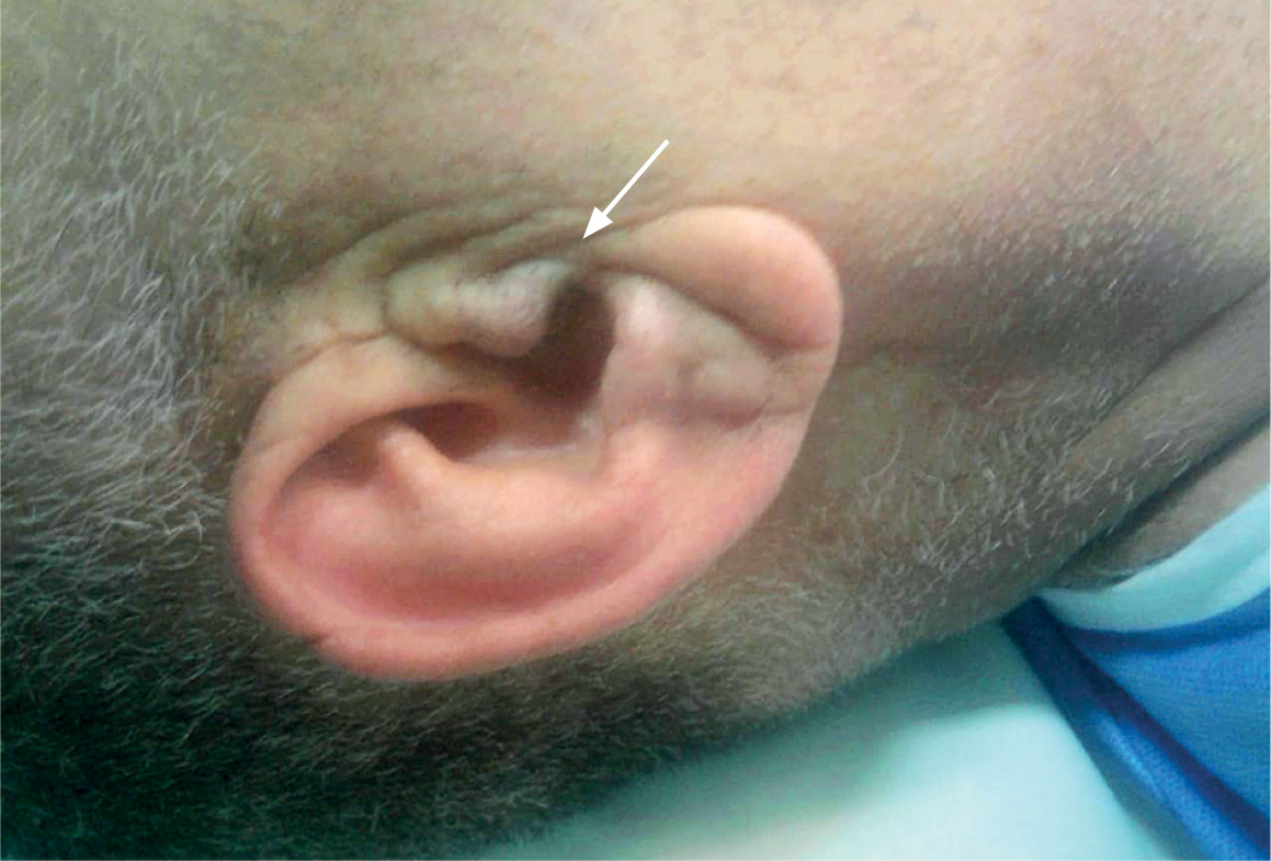
Written informed consent was obtained from the patient for the publication of this report. A 52-year-old man with no past medical or familial history, American Society of Anesthesiologists physical status 2, was scheduled for laparoscopic radical prostatectomy. Prior to presentation, he was a non-smoker and physically active with a metabolic equivalent of > 4. The rest of the examination was unremarkable. The pre-operative hemoglobin level was 13 γ dL-1 and the biochemical profile was normal. The attending anesthesiologist did not deem any cardiac exploration necessary prior to surgery and echocardiography and electrocardiogram (ECG) were not put into practice. One hour after initiation of pneumoperitoneum, the patient became tachycardic with hypotension. End-tidal CO2 and SpO2 did not change. Concomitantly, monitoring of the ST segment showed significant ST depression in lead II of 2 mm, which in the next minutes progressed to 3 mm with reciprocal elevation in lead aVL. The ECG pattern changed to normal with heart rate dropping after giving intravenous ephedrine boluses (total of 6 mg) along with optimizing the vascular filling and reducing the pneumoperitoneal insufflation pressure (from 15 to 12 mm Hg). This was not accompanied by a raised cardiac enzymes profile. The patient was noted to have bilateral Frank’s sign (Figure 1), a diagonal crease that runs across the earlobe at a 45-degree angle from the tragus to the auricle. Later after surgery, the findings from the coronarographic examination revealed 70% occlusion of the right coronary artery and elective coronary stenting was done subsequently.
The pathophysiology of Frank’s sign is not fully understood. Several explanations have postulated that a loss of elastic fibers due to increased circulating free radical oxidative stress and increases in intima-median thickness of blood vessels with diminished blood supply in the absence of collateral vessels in the ear lobe may be a potential explanation of the dermatological changes [5], reflecting a similar pathology in weakened coronary arteries. Shortened telomere length in Japanese patients with metabolic syndrome was also reported as a general pro-ageing and atherosclerotic mechanism [6].
The presence of this sign has a grading system that has been linked to incidence of cardiovascular events based on length, depth, bilateralism, and inclination. Bilateral deep cleft across whole earlobe was reported as the most severe form [7].
Identification of preoperative signs and symptoms that may predict the outcome of surgery is important for the development of adequate interventions. Frank’s sign may be a useful marker in the preanesthetic assessment of patients for coronary artery disease when there may be little or no history available from the patient.