

INTRODUCTION
Generalized pustular psoriasis (GPP) is a severe, potentially life-threatening disease characterized by the acute or recurrent appearance of sterile pustules on erythematous skin, frequently accompanied by systemic symptoms. The most common of these include fever, chills, malaise, leukocytosis, and elevated inflammatory markers, distinguishing GPP from plaque psoriasis and other forms of the disease [1, 2]. In some patients, the disease course may be fulminant, with flares leading to electrolyte imbalances, organ failure, sepsis, and, in extreme cases, death, particularly in elderly individuals or those with comorbidities [3].
GPP is classified as a rare (orphan) disease. Its prevalence is low compared with that of plaque psoriasis and varies substantially across geographical regions and ethnic groups. The rarity of this condition poses major diagnostic and therapeutic challenges and limits the feasibility of conducting large randomized clinical trials. Consequently, for many years, knowledge regarding the optimal therapeutic management of GPP was based primarily on case reports, small case series, and expert experience [4].
A further distinguishing feature of GPP is its specific pathogenetic background, which differs from that of classic psoriasis. In a subset of patients, mutations have been identified in genes involved in the regulation of the innate immune response, specifically interleukin-36 receptor antagonist (IL36RN), as well as CARD14 and AP1S3. These discoveries have contributed to a better understanding of disease mechanisms and enabled the development of targeted therapies, particularly interleukin-36 (IL-36) pathway inhibitors, which have transformed the approach to GPP treatment in recent years [5, 6]. Despite advances in the understanding of GPP pathogenesis and the availability of new therapies, consistent European or national management guidelines are still lacking. Aside from a few expert papers, such as the Asia-Pacific consensus recommendations on the management of GPP [7], clinicians are required to make therapeutic decisions based on limited data and their own clinical experience.
The purpose of this study is to develop up to date, evidence-based, and expert consensus-driven therapeutic recommendations for the treatment of GPP, intended for dermatologists in Poland. This document aims to consolidate current knowledge on GPP, support therapeutic decision-making, and enhance the safety and effectiveness of treatment for this rare yet exceptionally severe dermatosis.
EPIDEMIOLOGY
Generalized pustular psoriasis is a rare disease entity with specific epidemiological characteristics that differ significantly from those of plaque psoriasis. Due to its low prevalence, epidemiological data regarding GPP are limited, and their quality and scope vary substantially across different regions of the world. The highest incidence rates are observed in Japan and other Asian countries, whereas in Europe, including Poland, GPP remains an ultra-rare condition. These epidemiological differences, particularly in the genetic context, are highly relevant to diagnosis, prognosis, and treatment selection and should be considered when formulating therapeutic recommendations. In recent years, new data based on national registries, population analyses, and genetic studies have been published, allowing for a more precise estimation of disease prevalence.
The estimated prevalence of GPP in the general population is low. In most analyses, it ranges from 0.18 to 18 cases per million individuals, depending on the geographical region and study methodology [2, 4]. These differences result, among other factors, from variations in the genetic structure of populations, the availability of registry systems for rare diseases, and historical diagnostic discrepancies. European data are derived primarily from analyses of national registries and large health insurance databases. The prevalence of GPP in European countries is estimated at 0.6–7.5 cases per million inhabitants, with higher rates reported in Western European nations [5]. Globally, GPP accounts for less than 1% of all psoriasis cases. However, its clinical significance is disproportionately high relative to its prevalence due to its severe course, frequent hospitalizations, and substantial risk of systemic complications [3].
In a study analyzing populations from Germany, France, and the United Kingdom, the mean age of onset was 45–55 years, with a slightly higher incidence in women. A history of plaque psoriasis is reported in approximately 30–50% of patients, while in the remaining cases GPP represents the first, and often the only, manifestation of psoriasis (table 1) [1, 2, 4, 8].
Table 1
Estimated prevalence of GPP in selected European countries
| Country | Prevalence (per 1 million population) | Source |
|---|---|---|
| Germany | 1.5–3.5 | Twiss et al., 2019 [8] |
| France | 0.9–2.3 | Bachelez, 2020 [4] |
| UK | 1.8–4.0 | Gooderham et al., 2022 [2] |
| Nordic countries | 0.6–1.2 | Navarini et al., 2017 [1] |
The most detailed epidemiological data on GPP come from Japan, where the disease is subject to a dedicated registration system as a condition of high clinical significance. The prevalence of GPP in Japan is estimated at 7–18 cases per million inhabitants, representing one of the highest rates reported worldwide [7]. Several characteristic features have been noted in the Japanese population, including a lower proportion of patients with prior plaque psoriasis (approximately 20–30%), a strong association with IL36RN mutations, and more frequent onset in early adulthood. Similar, although less comprehensive, data are available from South Korea, China, and Taiwan, where the prevalence of GPP also exceeds that observed in Europe [9].
In Poland, no registry dedicated to patients with GPP is currently available. Existing data are derived primarily from hospitalizations reported to the National Health Fund (NFZ), retrospective analyses from individual centers, and extrapolations from European datasets. Based on these sources, the prevalence of GPP in Poland is estimated at 0.8–2.5 cases per million inhabitants, corresponding to a nationwide total of approximately 30–100 patients. These estimates should be interpreted with caution, although they are consistent with observations from other Central and Eastern European countries [10].
GPP can occur at any age, although the peak incidence is in the fifth and sixth decades of life. The disease is slightly more common in women (F : M ratio ≈ 1.2–1.5 : 1), and distinct ethnic differences correlate with the frequency of specific genetic mutations [6]. In approximately 40–60% of patients, GPP develops against a background of prior or coexisting plaque psoriasis. In the remaining patients, GPP constitutes the initial manifestation of psoriasis. Notably, the absence of prior psoriasis does not exclude a diagnosis of GPP, and mutations in the IL36RN gene encoding the interleukin-36 receptor antagonist are more frequently found in patients with no history of psoriasis [3]. The prevalence of IL36RN mutations in patients with GPP ranges from 30% to 50% in Japan and from 5% to 20% in Europe, reaching up to 60% in individuals with earlyonset disease [6]. More detailed information on genetic abnormalities in GPP is provided below.
DEFINITION AND DIAGNOSTIC CRITERIA
Currently, GPP is recognized as a distinct phenotype of psoriasis, both in terms of its clinical presentation and pathogenic mechanisms, particularly the dominant role of the IL-36 axis [11, 12]. Despite these characteristic features, a unified, universally accepted definition and consistent diagnostic criteria were lacking for many years, which hindered diagnostic standardization, the conduct of clinical trials, and the comparison of epidemiological data [1, 13].
Generalized pustular psoriasis is an autoinflammatory disease characterized by the occurrence of sterile, macroscopically visible pustules on a background of diffuse cutaneous erythema. The condition often presents with systemic symptoms such as fever, malaise, leukocytosis, or elevated inflammatory markers [1, 3, 13]. In recent years, significant international initiatives have been undertaken to standardize the definition and diagnostic criteria for GPP, most notably the work of the International Psoriasis Council (IPC) and the European/International Rare and Severe Psoriasis Expert Network (ERASPEN) [1, 13]. In 2024, the International Psoriasis Council published an international consensus statement on the definition and diagnostic criteria for GPP, developed using the Delphi method with the participation of experts from various regions of the world [13]. According to this consensus, “generalized pustular psoriasis is a systemic inflammatory disease characterized by cutaneous erythema and macroscopically visible sterile pustules” [13].
The IPC definition thus emphasizes the systemic nature of the disease, the presence of diffuse cutaneous erythema, and the occurrence of macroscopically visible sterile pustules. A key element of the IPC definition is the departure from restrictive criteria regarding lesion location, duration, or minimum affected body surface area. According to the IPC consensus, GPP may coexist with other forms of psoriasis or occur independently, and the presence or absence of systemic symptoms is not used to confirm or exclude the diagnosis [13]. The diagnostic criteria proposed by the IPC comprise the following elements [13]:
Macroscopically visible sterile pustules: clinically visible regardless of their size or location; their sterile nature should be confirmed clinically, or, in doubtful cases, microbiologically.
Erythematous background of skin lesions: pustules occur on a background of generalized or diffuse erythema, reflecting an active cutaneous inflammatory process.
No location restrictions: lesions may involve the skin of the trunk, extremities, and acral areas, provided that the other clinical criteria for GPP are met.
The IPC consensus does not define a minimum symptom duration or a threshold for body surface area involvement, aiming to support early diagnosis and prompt initiation of treatment during the acute phase [13].
Previously, for many years, the benchmark was the definition proposed by ERASPEN, which characterized GPP as the occurrence of primary, sterile, macroscopically visible pustules located on non-acral skin and not exclusively related to psoriatic plaques [1]. ERASPEN also proposed a classification of GPP into subtypes based on the presence or absence of systemic symptoms, the presence or absence of coexisting plaque psoriasis, and a relapsing or chronic course [1]. The persistence of lesions for ≥ 3 months was also highlighted as a supportive criterion in the classification of the chronic form of the disease [1]. Table 2 compares the diagnostic criteria for GPP proposed by the IPC and ERASPEN.
Table 2
Comparison of IPC and ERASPEN diagnostic criteria
| Criterion element | IPC (Choon et al., 2024) [13] | ERASPEN (Navarini et al., 2017) [1] |
|---|---|---|
| Sterile pustules | Yes | Yes |
| Cutaneous erythema | Required | Expected |
| Location of skin lesions | No restrictions | Non-acral skin only |
| Concomitant plaque psoriasis | Acceptable | Acceptable |
| Systemic symptoms | May be present | May be present |
| Minimum duration | Unspecified (diagnosis possible at the initial pustular flare) | ≥ 3 months (for the chronic form) |
| Purpose of the definition | Early diagnosis | Clinical staging |
Defining a flare or exacerbation in the course of GPP remains a separate issue. Currently, no single, universally accepted definition of a GPP flare exists. In general, a flare is understood as a sudden or rapid increase in disease activity, manifested by the appearance of new pustules or their rapid progression, worsening erythema, and frequently accompanying systemic symptoms [11, 13]. In clinical trials and patient registries, operational definitions are used, incorporating, among other elements, lesion severity assessed with the GPP Physician Global Assessment (GPPGA), an increase in the number or extent of pustules, and the need for hospitalization or intensification of systemic treatment [13, 14].
PATHOGENESIS AND HISTOLOGICAL FEATURES OF GENERALIZED PUSTULAR PSORIASIS
In contrast to plaque psoriasis, in which the adaptive immune response and activation of the IL-23/IL-17 axis play a key role, the pathogenesis of GPP is dominated by the innate immune response, with an essential role of neutrophils and the IL-1/IL-36 axis [15, 16]. The clinical presentation of GPP, characterized by the acute appearance of sterile pustules on an erythematous base, reflects the rapid activation of inflammatory mechanisms leading to a massive recruitment of neutrophils into the epidermis. Significantly elevated expression of neutrophil chemokines, including CXCL1, CXCL2, and CXCL8, which are responsible for neutrophil chemotaxis and activation, is demonstrated in the skin of affected patients [15, 17].
The histopathological appearance of generalized pustular psoriasis is relatively characteristic, though nonspecific, and depends on the phase of the disease. It reflects a rapid neutrophilic inflammatory reaction in the skin, with predominant involvement of the epidermis and secondary changes in the dermis. Although it is not pathognomonic, when interpreted in conjunction with the clinical presentation, it constitutes an important diagnostic aid, particularly in equivocal cases or those requiring differentiation from other pustular dermatoses [1, 18]. As noted above, the histological features of GPP may vary depending on the stage of the disease (early, active, or resolving); however, certain findings are consistently observed in the majority of patients.
The most characteristic element of the histopathological picture of GPP is the presence of intraepidermal neutrophilic pustules, referred to as spongiform pustules of Kogoj. These are aggregates of neutrophils located within the stratum spinosum and stratum granulosum of the epidermis, accompanied by extracellular edema corresponding to spongiosis [11, 15]. Further epidermal alterations may include parakeratosis, acanthosis of varying extent, and neutrophilic exocytosis in the absence of keratinocyte necrosis [19]. During the active phase of the disease, an increased epidermal exocytosis of neutrophils is observed, correlating with the clinical eruption of pustules. At the dermal-epidermal junction, enhanced migration of neutrophils from the dermal capillaries into the epidermis is visible. This process is mediated by neutrophil chemokines induced by cytokines associated with the IL-36 pathway (CXCL1, CXCL2, CXCL8), which show significantly increased expression in the skin of patients with GPP [19, 20].
Typical features of interface dermatitis and keratinocyte damage are absent, which distinguishes GPP from certain drug-induced cutaneous reactions. However, the dermis shows dilation and congestion of blood vessels, edema of the papillary dermis, and inflammatory infiltrate predominantly composed of neutrophils, with sparse accompanying lymphocytes and macrophages.
The pathophysiology of GPP is strongly influenced by genetic abnormalities, although the disease remains heterogeneous in this regard. To date, several genes predisposing to the development of the disease have been identified. Most of them are functionally linked to the regulation of the neutrophilic inflammatory response and the activation of IL-36.
The most important genes associated with the development of GPP include:
IL36RN encoding the IL-36 receptor antagonist (IL-36Ra);
CARD14 regulating NF-κB activation in keratinocytes;
AP1S3 associated with intracellular transport and regulation of the innate immune response;
MPO, SERPINA1, SERPINA3 affecting the activity of neutrophil proteases;
TNIP1 regulating the NF-κB pathway.
Mutations in the IL36RN gene are the best characterized and occur more frequently in patients with isolated GPP without coexisting plaque psoriasis. Their presence is associated with earlier disease onset, a more severe clinical course, and a higher number of flares, particularly in the case of biallelic mutations [21, 22].
The pathophysiology of GPP is driven primarily by activation of the IL-36 pathway, which represents the central disease mechanism and is reflected in the histological background. IL-36 cytokines (IL-36α, IL-36β, IL-36γ) belong to the IL-1 family and act as potent mediators of cutaneous inflammation. Under physiological conditions, their activity is tightly regulated by the IL-36 receptor antagonist (IL-36Ra), encoded by the IL36RN gene. In GPP, uncontrolled activation of the IL-36 pathway occurs due to mutations in the IL36RN gene, overactivity of neutrophil proteases (e.g., in myeloperoxidase deficiency), and increased expression of IL-36 agonists in keratinocytes. Activation of the IL-36 receptor leads to an inflammatory cascade involving NF-κB and MAPK, the induction of neutrophil chemokines, and massive recruitment of neutrophils into the epidermis, which clinically manifests as the formation of pustules [19, 20]. The significance of this pathway is confirmed by clinical trials using IL-36R inhibitors, such as spesolimab and imsidolimab, which demonstrate rapid and profound inhibition of disease activity in the majority of patients with an acute GPP flare [23–25].
ASSESSMENT OF CLINICAL SEVERITY OF GENERALIZED PUSTULAR PSORIASIS
GPP is a disease with a variable, dynamic, and potentially fulminant course, in which the rapid progression of skin lesions can lead to severe systemic complications, hospitalization, and, in extreme cases, life-threatening conditions [1, 11]. Consequently, clinical severity assessment is a key element of diagnostic and therapeutic management. Given the characteristics and clinical course of GPP, disease activity assessment tools used in clinical practice should facilitate rapid evaluation, demonstrate sensitivity to shortterm fluctuations, and support the monitoring of treatment response.
Standardized disease severity assessment is important for several reasons: it enables the objectification of disease activity, allows for the recognition of exacerbations, facilitates therapeutic decision-making regarding the urgency and intensity of treatment, and enables the monitoring of treatment response. Its use is also essential in clinical trials and patient registries [14, 26].
Classical skin lesion severity assessment tools used in plaque psoriasis, such as the Psoriasis Area and Severity Index (PASI) are insufficient for GPP because of the disease’s distinct dynamics, the predominance of pustular lesions, and the frequent presence of systemic symptoms. For this reason, several scales dedicated to GPP have been developed. However, no single universal tool has yet been established for use across all clinical scenarios. In both clinical practice and clinical trials, the scale should be selected according to the purpose of the assessment and the clinical context [14, 26].
The most frequently used tools are GPPASI, GPPGA, and JDA-GPPSI. These scales differ in the scope of parameters assessed, their degree of complexity, and their utility in daily practice.
The GPPASI (Generalized Pustular Psoriasis Area and Severity Index) scale was proposed as a modification of the classical PASI scale, expanded to include the assessment of pustule severity and to reflect lesion dynamic to a greater extent [27]. The final score is determined based on the affected body surface area, erythema severity, and pustule severity. The utility of the GPPASI scale has been demonstrated in clinical trials and long-term observation, although it has proven less intuitive for assessing disease flares [26].
The GPPGA (Generalized Pustular Psoriasis Physician Global Assessment) scale was originally developed for clinical trials but has been widely adopted in routine clinical practice since 2019. It relies on an overall physician assessment covering two core components: pustule severity and erythema severity. Each component is scored from 0 to 4, with the final score reflecting global disease activity [14, 27]. A GPPGA score of ≥ 3 is frequently used in clinical trials as a threshold for active disease or a flare, indicating the need to initiate or escalate systemic therapy.
In 2018, the Japanese Dermatological Association proposed its own scale, the JDA-GPPSI (Japanese Dermatological Association Generalized Pustular Psoriasis Severity Index), which subsequently gained widespread use throughout the Asian region [28]. This scale accounts for the severity of skin lesions, systemic symptoms, laboratory findings, including C-reactive protein (CRP) and white blood cell count, and the presence of metabolic disturbances. While the JDA-GPPSI is a comprehensive tool, its use in daily clinical practice outside Japan remains limited, primarily due to the need to score laboratory parameters and systemic symptoms, which makes rapid assessment in an outpatient setting more difficult, as well as the fact that the scale was developed and validated mainly in a Japanese population [26, 28].
Patient-reported outcomes (PROs), in which patients assess disease severity based on their own experienced of symptoms, are equally important. The PSS (Psoriasis Symptom Scale) serves this purpose by allowing patients to evaluate subjective symptoms such as pain, burning, itching, and general discomfort. Although it is not a formal clinical severity scale for GPP, it is a valuable adjunct for monitoring the course of the disease, particularly with regard to quality of life and treatment response [26]. Table 3 provides a comparison of the key features of the various scales used to evaluate the clinical severity of GPP.
Table 3
Comparison of clinical scales used to assess the severity of GPP
[i] GPPGA – Generalized Pustular Psoriasis Physician Global Assessment, GPPASI – Generalized Pustular Psoriasis Area and Severity Index, JDA-GPPSI – Japanese Dermatological Association – Generalized Pustular Psoriasis Severity Index, CRP – C-reactive protein, PSS (PROs) – Psoriasis Symptom Scale (patient-reported outcomes).
Currently, the GPPGA scale remains the most practical and frequently recommended tool for assessing GPP activity in both daily clinical practice and clinical trials. Quantitative scales and PROs should be viewed as complementary tools, and the choice of scale should be tailored to the specific purpose of the evaluation, whether assessment of an acute flare or long-term monitoring.
When an active GPP flare is suspected, including in patients presenting to the emergency department or requiring hospitalization, the use of the GPPGA scale is recommended. The rationale is that it allows for a rapid, intuitive assessment focused on the hallmark features of GPP, namely pustules and erythema, and has been validated as an inclusion criterion and endpoint in clinical trials [13, 14]. The authors note that, during an acute GPP flare, a GPPGA score of ≥ 3 should be regarded as evidence of active disease requiring urgent systemic treatment, regardless of the affected body surface area. This approach is reflected in both clinical trials and current expert guidelines [13, 14, 26].
For short-term monitoring of treatment response, over days to weeks, the use of a combination of the GPPGA and PSS scales is advisable. A reduction in the number of pustules may precede an improvement in the patient’s overall well-being, or vice versa. Additionally, the PSS captures clinically significant symptoms, such as pain and burning, that are not visible during a standard physical examination [26].
For long-term monitoring and clinical trials, quantitative scales capable of tracking changes over time, such as the GPPASI and GPPGA, are recommended. While the GPPASI can be used as a supplementary tool in clinical trials and registries, it should not replace the GPPGA in routine clinical practice. In specialized centers, particularly for patients with severe disease courses and laboratory abnormalities, the JDA-GPPSI may be used for a more comprehensive assessment [29]. Table 4 summarizes the recommendations for the preferred GPP severity assessment scales across different clinical scenarios.
Table 4
Recommendations for the use of scales in GPP
| Clinical status | Recommended scale | Role | Source |
|---|---|---|---|
| Suspected GPP flare | GPPGA | Primary | Bissonnette et al. [14], 2021; Burden et al., 2023 [27]; Choon et al., 2024 [13] |
| Therapeutic decision | GPPGA | Decisive | Burden et al., 2023; Strober et al., 2022 [26] |
| Early response | GPPGA + PSS | Monitoring | Burden et al., 2023 [27]; Strober et al., 2022 [26] |
| Long-term follow-up | GPPASI | Complementary | Strober et al., 2022 [26] |
| Reference centers | JDA-GPPSI | Additional | Fujita H. et al., 2018 [28]; Rivera-Diaz et al., 2023 [30] |
[i] GPP – generalized pustular psoriasis, GPPGA – Generalized Pustular Psoriasis Physician Global Assessment, PSS – Psoriasis Symptom Scale, GPPASI – Generalized Pustular Psoriasis Area and Severity Index, JDA-GPPSI – Japanese Dermatological Association – Generalized Pustular Psoriasis Severity Index.
CLINICAL PRESENTATION OF GENERALIZED PUSTULAR PSORIASIS
The typical clinical presentation of GPP includes:
numerous, small, superficial pustules, often coalescing into “lakes of pus”;
intense skin erythema;
skin tenderness, burning, and a sensation of skin tightness;
systemic symptom, including fever, chills, malaise, and fatigue.
In a multicenter analysis of 156 patients with GPP conducted in Turkey, acute, recurrent flares were the predominant clinical presentation, with fever and weakness being the most common systemic symptoms [31]. Approximately 60% of patients had a prior history of psoriasis, and metabolic and cardiovascular comorbidities were frequently observed. These findings were supported by another large study, which reported that 72.2% of patients with GPP had comorbidities, most commonly arterial hypertension (43.3%), but also other cardiovascular diseases, diabetes mellitus, and hyperlipidemia [32]. In an analysis of 102 adult patients from Malaysia, Choon et al. found that GPP is characterized by rapid onset, a high frequency of systemic symptoms, and the frequent co-occurrence of infections and electrolyte disturbances during severe flares [33].
Observations from Japan and other Asian countries further indicate a higher frequency of mutations in genes associated with the IL-36 pathway, a higher prevalence of severe, recurrent forms of the disease, and the significant impact of systemic symptoms on the assessment of disease severity and appropriate therapeutic decisions-making [26]. American authors highlight the pronounced clinical heterogeneity of GPP, which encompasses both acute and chronically relapsing forms, a high rate of hospitalizations due to disease exacerbations, and a substantial impact on quality of life and the risk of complications [34]. Similar observations have been reported in European populations [26, 35].
For many years, data on GPP in the Polish population remained very limited. In 2024, the results of a multicenter retrospective analysis conducted by the Polish GPP Working Group were published. The analysis included patients hospitalized at 14 specialized dermatology centers across Poland between 2013 and 2023 [32]. A total of 90 patients meeting the current diagnostic criteria for GPP were included. The median age at the time of analysis was 59 years (range: 5–85 years), with a clear female predominance (68.9%; female-to-male ratio approximately 2 : 1) [32]. Analysis of the age distribution confirmed that GPP can occur across a broad age range, from childhood to advanced age, although nearly half of the patients were over 60 years, which has important clinical implications, particularly regarding comorbidities and therapeutic options.
In some patients, GPP develops against a background of pre-existing plaque psoriasis. However, in a substantial proportion of individuals, it represents the primary manifestation of the disease [33, 34]. Available data indicate that individuals with GPP carry a considerable burden of comorbidities, particularly metabolic and cardiovascular disorders. Depending on the study population, hypertension is reported in approximately 29–43% of patients, diabetes in 18–26%, and clinically overt cardiovascular disease in 13–19% [31–34]. Similar observations have been reported in the Polish population. In a multicenter retrospective study comprising 90 patients with GPP, plaque psoriasis coexisted in 59.1% of patients, and the most common comorbidity was hypertension (43.3%), followed by cardiovascular diseases, diabetes, and hyperlipidemia (approximately 20% each) [32]. The high prevalence of comorbidities among patients with GPP underscores the need for a comprehensive internal medicine evaluation, consideration of comorbidities when selecting systemic treatment, and close collaboration between the dermatologists and the primary care physicians, cardiologists, and diabetologists. Table 5 presents data regarding the prevalence of various comorbidities in patients with GPP.
Table 5
Most common comorbidities in patients with GPP
| Study/country | Number of patients | Pre-existing plaque psoriasis | Arterial hypertension | Diabetes mellitus | Dyslipidemia | Cardiovascular diseases | Obesity/metabolic syndrome |
|---|---|---|---|---|---|---|---|
| Kara Polat et al., 2022 (Turkey) [31] | 156 | ~60% | 32% | 18% | 21% | 15% | 27% |
| Choon et al., 2014 (Malaysia) [33] | 102 | 67% | 29% | 24% | nd | 13% | nd |
| Noe et al., 2021 (USA) [34] | 201 | 56% | 38% | 26% | 30% | 19% | 35% |
| Kołt-Kamińska et al., 2024 (Poland) [32] | 90 | 59.1% | 43.3% | 22.2% | 25.6% | 17.8% | 31.1% |
Essential components of the clinical presentation of GPP include systemic symptoms and laboratory abnormalities. Elevated CRP levels were observed in 85.6% of patients, leukocytosis in 62.2%, hypoalbuminemia in one-third of cases, and elevated transaminase levels in approximately 25%. In 33.3% of patients, flares were accompanied by fever, and nearly 9% exhibited involvement of other organs, confirming the systemic nature of GPP [32].
GPP coexisted with plaque psoriasis in 59.1% of patients. These individuals were significantly younger (median age: 48 years) than those with isolated GPP (median age: 66 years). They also had a higher prevalence of other psoriasis phenotypes, such as nail or scalp involvement. Conversely, patients with isolated GPP experienced a greater number of flares and a higher burden of cardiometabolic comorbidities [32].
GPP flares are characterized by extensive skin lesions involving, on average, 43% to 70% of the body surface area, depending on the severity of the episode. Clinical severity assessed using the GPPGA scale is typically high in these cases: more than 75% of patients with flares classified as severe have a GPPGA score ≥ 3. The median duration of a flare is approximately 4 weeks, with a median hospitalization time ranging from 1.6 to 2.3 weeks, reflecting the abrupt onset and severe clinical course of the disease. GPP flares are frequently accompanied by systemic symptoms, including a fever, profound weakness, and features of generalized inflammation. Laboratory findings in the majority of patients include elevated CRP levels and leukocytosis. In more severe cases, electrolyte imbalances, hypoalbuminemia, and abnormal liver function tests are also observed [35].
Published analyses demonstrate a significant correlation between the severity of systemic symptoms, the total GPPGA score, and the length of hospital stay. This confirms the close relationship between cutaneous inflammatory activity and the systemic response in GPP [35, 36]. A subset of patients experienced severe organ complications, including acute kidney injury, respiratory failure, and septic episodes, underscoring the potentially life-threatening nature of severe GPP flares [5, 23]. More than 90% of patients with flares classified as severe required hospitalization, and approximately 11% required treatment in an intensive care unit [35]. These data are consistent with reports indicating that GPP flares must be treated as medical emergencies requiring urgent diagnosis and intensive systemic therapy [1, 36].
DIAGNOSIS OF GENERALIZED PUSTULAR PSORIASIS
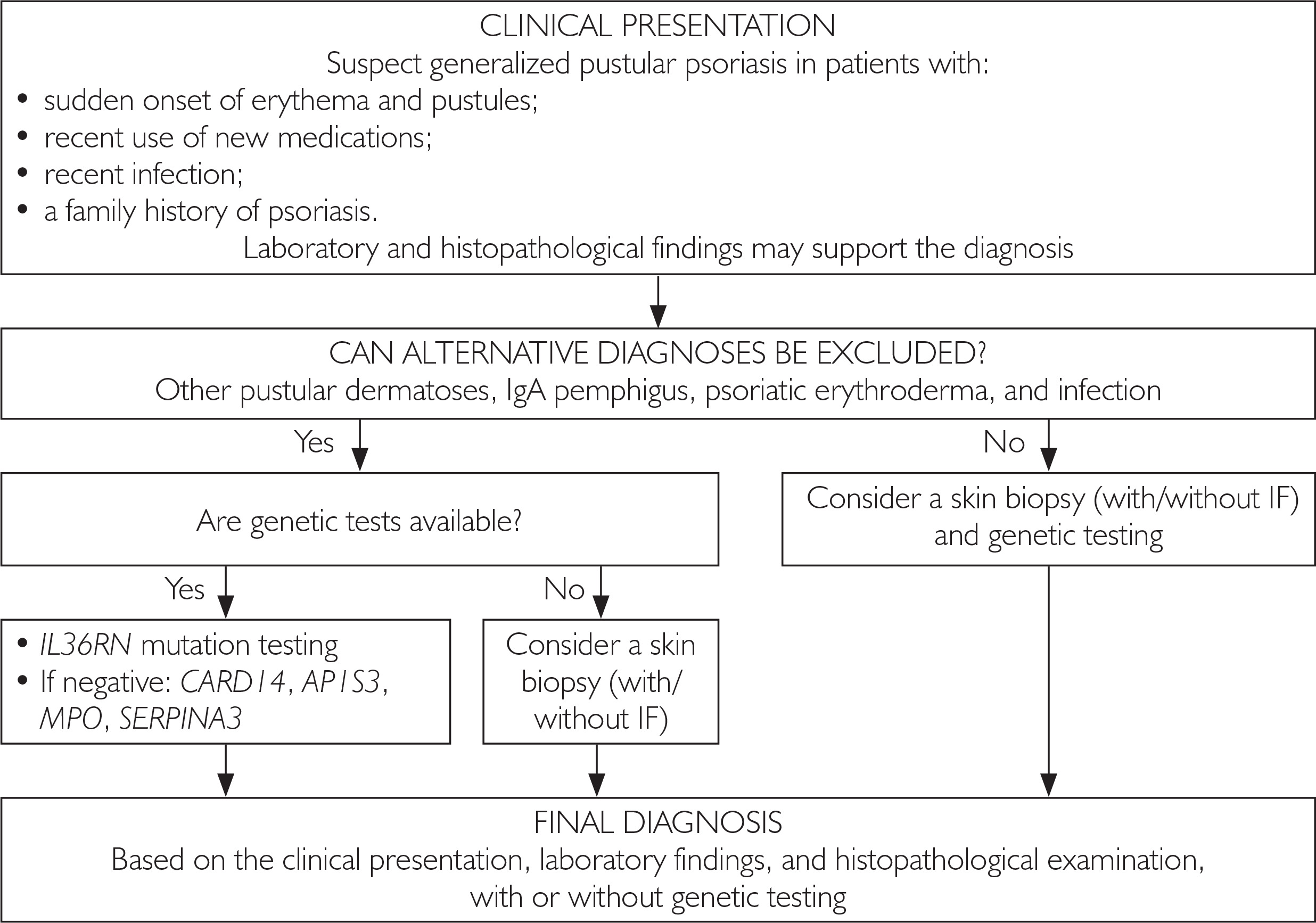
The primary objective when evaluating patients with suspected GPP is to confirm the diagnosis, followed by assessment of disease severity, establishment of a differential diagnosis, and identification of potential complications or comorbidities.
Recommended baseline laboratory evaluations include a complete blood count with differential, CRP and erythrocyte sedimentation rate (ESR), plasma electrolytes (Na+, K+, Ca²+), renal and hepatic function tests, glucose, total protein and albumin levels, and a lipid profile. Additional diagnostic evaluations, guided by the clinical presentation, may include cultures to rule out infection and virological testing if a triggering viral infection is suspected [31, 34].
Histopathological examination of a skin biopsy is strongly recommended during the initial episode of the disease, in cases with an atypical clinical presentation, and to support the differential diagnosis. The typical histological features of GPP are outlined above [18].
Genetic testing, for example for IL36RN mutations, is not mandatory as part of the routine diagnostic workup, but it may be considered in patients with a severe, recurrent disease course or an early-onset form of the condition [26]. A practical diagnostic algorithm is presented in figure 1.
TREATMENT OF PATIENTS WITH GENERALIZED PUSTULAR PSORIASIS
A precise understanding of the pathogenesis of GPP, specifically the role of the IL-36 pathway and its links to other inflammatory mediators, such as IL-1, IL-17, IL-23, and TNF-α, has led to a fundamental shift in the therapeutic approach to this disease. GPP is not merely a skin condition but a severe systemic inflammatory syndrome that can cause organ complications and pose an immediate threat to life. Consequently, treatment goals extend far beyond the management of cutaneous symptoms alone (table 6).
Table 6
General principles and treatment goals for patients with GPP
The primary, overarching goal in the treatment of patients with GPP is the rapid suppression of new pustule formation, which represents the clinical manifestation of activation of the IL-36–neutrophil–keratinocyte axis. This goal is most effectively achieved through therapies with a rapid onset of action, enabling direct modulation of the innate immune response. Another crucial treatment goal is the control of cutaneous inflammatory symptoms, including erythema, edema, and tenderness of the lesions. These symptoms not only significantly impair patients’ quality of life but also serve as clinical indicators of ongoing inflammatory activity. Reducing the severity of erythema and edema reflects effective modulation of the cytokine cascade and limitation of epidermal barrier damage.
Systemic symptoms, such as fever, malaise, weakness, chills, and tachycardia, signal generalized inflammatory activation and are key factors determining GPP severity. These symptoms are a direct consequence of systemic inflammation and carry a risk of serious complications, including electrolyte imbalances, sepsis, or multiple organ dysfunction syndrome. Their rapid control is essential to prevent organ damage. Cases of acute respiratory failure, renal failure, and exacerbations of cardiovascular disease have been reported in patients during severe flares. Consequently, treatment should aim not only to control cutaneous symptoms but also to mitigate the systemic consequences of chronic and fulminant inflammation.
The outlined therapeutic goals can be achieved with various classes of medications, each characterized by distinct response kinetics:
IL-36 receptor inhibitors:
demonstrate the ability to rapidly reduce inflammatory markers and resolve systemic symptoms;
provide the fastest clinical effect, frequently leading to a significant reduction in pustule formation, often within a few days. This therapy directly blocks the key causative pathogenic mechanism of GPP;
effectively reduce skin inflammation by limiting neutrophil recruitment and keratinocyte activation.
TNF inhibitors, including infliximab:
infliximab has a rapid onset of action and is effective during disease flares. However, it acts less selectively and does not directly target the IL-36 pathway;
although TNF inhibitors can effectively reduce systemic symptoms, their action is less specific and less documented in the available literature.
Traditional systemic agents, including acitretin, cyclosporine, and methotrexate:
can suppress pustule formation, but their effects are typically slower and less predictable during severe flares;
acitretin affects keratinocyte proliferation and can be effective during the chronic phase, but its anti-inflammatory effect is indirect;
cyclosporine provides rapid immunosuppression, but its use is limited by its toxicity profile and the risk of a rebound effect if discontinued prematurely;
often require supportive care and hospitalization when used during acute flares.
Detailed information regarding specific therapies used in patients with GPP is provided later in this paper.
In GPP therapy, pain management is of paramount importance. Pain in GPP can be intense, and therapeutic interventions are frequently inadequate. Pain associated with extensive skin involvement, epidermal cracking, and severe inflammation significantly impairs patients’ daily functioning and can hinder treatment success. Appropriate anti-inflammatory therapy, and, when necessary, symptomatic treatment should be integral components of standard management. The greatest improvement in pain is observed with therapies that lead to a rapid reduction in pustules, such as IL-36 inhibitors and infliximab. PRO scales, such as the PSS, are most commonly used to monitor treatment efficacy in this regard.
The long-term goal of treating patients with GPP is to maintain disease remission and prevent relapses. The recurrent nature of GPP, often marked by abrupt flares, demands a therapeutic strategy focused not only on managing the acute phase but also on stabilizing the disease course over time. This involves the careful selection of maintenance therapy and monitoring for triggers that may precipitate subsequent flares. Of note, IL-23 and IL-17 inhibitors may play a role in maintenance therapy, particularly in patients with a concomitant plaque psoriasis. In selected cases, acitretin, or less frequently methotrexate, is used for maintenance treatment. A treat-to-target strategy based on GPPGA scores allows for a rational assessment of long-term treatment efficacy.
Increasing emphasis is also being placed on managing comorbidities, such as metabolic disturbances, cardiovascular disease, chronic arthritis, and liver disease. Effective GPP management should consider the patient’s overall clinical picture and minimize the long-term consequences of chronic inflammation.
For many years, therapeutic management of GPP relied primarily on medications used for plaque psoriasis, despite significant pathogenic differences between these two disease entities [37]. The lack of randomized clinical trials, combined with the low prevalence of GPP, has resulted in a scarcity of robust data. Consequently, treatment decisions have relied largely on case series, retrospective studies, and expert consensus statements [28, 38]. In recent years, major advances in understanding GPP pathogenesis, particularly the pivotal role of the IL-36 axis, have led to the development of targeted therapies that are transforming the treatment paradigm for this disease [24, 25, 37].
NON-BIOLOGIC TREATMENT: TRADITIONAL SYSTEMIC AGENTS
Retinoids – acitretin
Acitretin is one of the most frequently used first-line agents for the treatment of GPP in adults. Its mechanism of action involves the normalization of keratinocyte proliferation and differentiation and modulation of the immune response, including effects on Th17 cells [28]. In a multicenter study conducted in Japan, treatment with etretinate was effective in 84.1% of 188 patients with GPP, although the definition of clinical response was not clearly specified [39]. Other retrospective studies and case series confirm the relatively rapid onset of action of acitretin in GPP, often within 1–2 weeks, which distinguishes it from the response observed in plaque psoriasis [40]. The use of acitretin is limited by its teratogenicity, hepatotoxicity, and the risk of lipid abnormalities, necessitating close laboratory monitoring [41].
Methotrexate
Methotrexate is a classic immunomodulatory drug used in numerous inflammatory diseases. However, evidence regarding its efficacy in GPP remains limited. It has been postulated that methotrexate may modulate the immune response by affecting regulatory T cells and the mTOR pathway [42].
In retrospective analyses, its effectiveness in GPP has been variable, with clinical improvement reported in approximately 30–75% of patients, typically after several months of therapy [39, 42]. Methotrexate is particularly considered in patients with concomitant psoriatic arthritis, although its slow onset of action limits its utility in acute, severe episodes.
Cyclosporine
Cyclosporine is a calcineurin inhibitor that inhibits T-cell activation by suppressing IL-2 synthesis. It is characterized by a rapid onset of action, making it a useful therapeutic option for acute episodes of GPP [43]. Available data are derived mainly from case reports and small patient series, in which clinical improvement was typically observed within 2–4 weeks of treatment at doses of 2–5 mg/kg body weight/day [44]. However, the long-term use of cyclosporine is limited by the risk of nephrotoxicity and arterial hypertension.
Other non-biologic drugs
Isolated cases of the successful use of other medications, such as mycophenolate mofetil, hydroxyurea, apremilast, or colchicine, have been described in the literature. However, these data remain anecdotal and are insufficient to establish definitive clinical recommendations [45, 46].
BIOLOGICAL TREATMENT
TNF inhibitors
Anti-TNF agents, such as infliximab, adalimumab, and etanercept, were among the first biologic therapies used in GPP. Infliximab is characterized by a rapid onset of action and is occasionally used in severe, refractory cases. However, clinical responses are often short-lived, and the risk of disease relapse remains substantial [47, 48].
IL-17 and IL-23 inhibitors
Agents blocking IL-17, such as secukinumab and ixekizumab, and IL-23, such as guselkumab and risankizumab, have demonstrated high therapeutic efficacy in plaque psoriasis. Their use in GPP is supported by a growing body of case reports and observational studies [46]. These therapies are considered particularly in patients with concomitant plaque psoriasis.
THERAPY TARGETING THE IL-36 PATHWAY AS A NEW STANDARD OF TREATMENT FOR GENERALIZED PUSTULAR PSORIASIS
In view of the pathophysiology of GPP, direct blockade of the IL-36R has emerged as a compelling therapeutic target. Monoclonal antibodies directed against IL-36R, such as spesolimab and imsidolimab, inhibit the binding of IL-36 ligands to the receptor, thereby interrupting a pivotal step in the initiation and amplification of neutrophilic inflammation in GPP [26, 37]. This mechanism differs fundamentally from that of traditional biologic therapies used in psoriasis, which primarily target cytokines along the IL-23/IL-17 axis or TNF-α.
The most convincing clinical data are available for spesolimab, the first IL-36R antagonist approved as monotherapy for treating flares in adult patients with GPP. In the randomized, double-blind, phase II EFFISAYIL-1 trial, a single intravenous dose of spesolimab led to complete pustule clearance, defined as a GPPGA pustulation subscore of 0, after 1 week in 54% of patients, compared with 6% in the placebo group [14]. Crucially, clinical improvement was observed very rapidly, often within the first 24–48 hours after administration. Treatment was also associated with a significant reduction in pain, improvement in quality of life, and normalization of inflammatory markers such as CRP and white blood cell count [14, 26].
Further data from the EFFISAYIL-2 trial and long-term analyses suggest that IL-36R-targeted therapy may not only effectively control acute GPP flares but also reduce the risk of disease recurrence [37]. The safety profile of spesolimab was considered favorable, and the incidence of adverse events was comparable to that observed with placebo in the controlled studies [14].
An alternative IL-36R antagonist is imsidolimab, which has also demonstrated high clinical efficacy in phase II and III trials. In open-label clinical studies, rapid pustule clearance and improvement in overall disease activity were observed, with responses sustained for many weeks after administration [25]. Although the evidence base for imsidolimab is currently smaller than that for spesolimab, the available results support a class effect for IL-36R blockade in GPP.
The accumulated clinical data indicate that IL-36-targeted therapies meet the core objectives of GPP management: they enable rapid control of acute flares, effectively eliminate pustules and erythema, resolve systemic symptoms, and mitigate the risk of severe organ complications. Unlike traditional systemic therapies and older biologic agents, IL-36 inhibitors directly target the underlying pathogenesis of the disease, supporting their position as the preferred first-line therapy for acute GPP flares [3, 26].
In summary, anti-IL-36 therapies provide: rapid control of acute flares; reduction of systemic symptoms; a decreased risk of organ complications; the potential prevention of disease recurrence.
IL-36 inhibitors are currently regarded as the most rational and preferred treatment option for patients experiencing severe episodes of GPP [13, 37]. Table 7 summarizes the key information on therapies used in patients with GPP.
Table 7
Summary of therapies used in GPP
An analysis of data from the Polish cohort revealed that all patients with GPP received systemic therapy during disease flares, reflecting the severe clinical course and the necessity for intensive management [48]. The most frequently prescribed medication was acitretin, administered to 68 patients. Within this group, complete clearance of skin lesions was achieved in 25 patients, a good clinical response, defined as a greater than 90% reduction in pustule formation, was observed in 15 patients, and a satisfactory response, defined as a greater than 50% reduction in pustules, was observed in 14 patients, although the time to improvement varied. A lack of efficacy with acitretin was observed in 3 patients, indicating that its effectiveness in the treatment of GPP flares is substantial but not universal [48].
Phototherapy was of limited utility. Narrowband ultraviolet B (NB UVB) was used in 5 patients, yielding a good response in only 1 case, while psoralen and ultraviolet A (PUVA) was administered to 3 patients, with moderate clinical outcomes. These data confirm that phototherapy plays a supportive rather than a primary role in GPP management [48].
Biologic treatment was used less frequently but was associated with distinctly higher efficacy. TNF inhibitors led to a complete response in 10 patients, while IL-36 axis inhibitors administered to 6 patients resulted in complete remission in 5 and a good response in 1 patient. IL-23 and IL-17 inhibitors also demonstrated high efficacy, although the number of treated patients was small [48].
The authors emphasize that the high hospitalization rate, frequent relapses, and limited efficacy of traditional systemic therapies indicate a significant unmet therapeutic need in the Polish population. These findings unequivocally support the expanding role of targeted therapies, particularly IL-36 axis inhibitors, in the management of GPP flares [48].
DISTINCT THERAPEUTIC APPROACHES TO THE MANAGEMENT OF GENERALIZED PUSTULAR PSORIASIS FLARES
Traditional systemic treatment
The therapies most commonly used in patients with GPP flares have included conventional systemic agents, such as acitretin, cyclosporine, methotrexate, and systemic glucocorticosteroids [35]. Their use has been driven primarily by their availability and long-standing clinical experience, despite a limited number of high-quality studies confirming their efficacy in patients with GPP [28, 38]. It should be noted that the use of systemic glucocorticosteroids is associated with a risk of disease recurrence or exacerbation following their discontinuation and therefore requires particular caution [2].
Biologic and targeted therapies
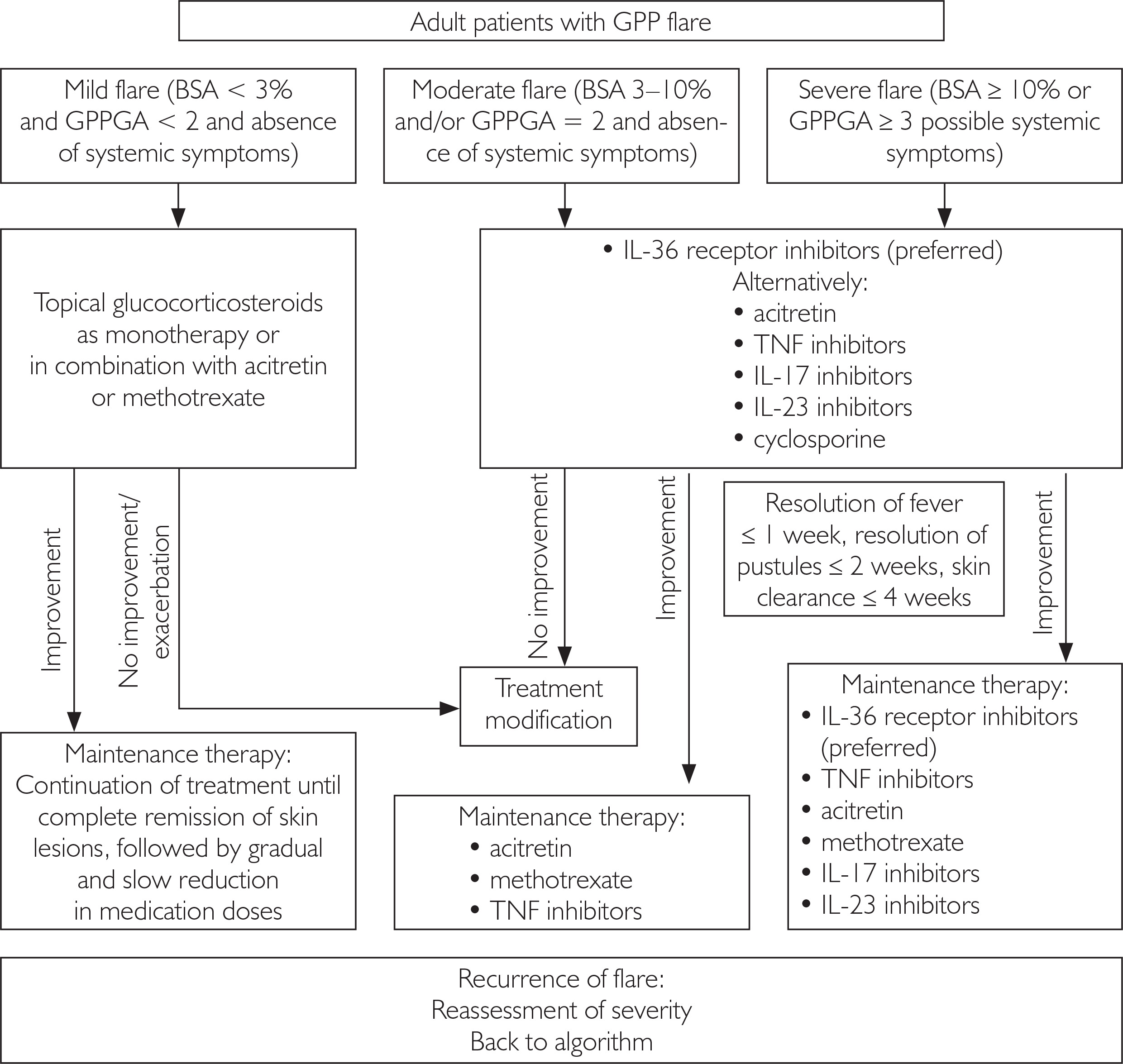
Biologic therapies are playing an increasingly prominent role in the management of GPP flares, particularly agents that target the core pathogenic mechanisms of the disease. The strongest evidence of efficacy is available for spesolimab, a monoclonal antibody targeting the IL-36 receptor. Spesolimab treatment was the first targeted therapy assessed in a randomized clinical trial for acute GPP episodes [23]. The EFFISAYIL-1 study demonstrated rapid and significant clearance of neutrophilic pustules and improvement in systemic symptoms within a few days of drug administration, representing a major advantage over conventional systemic therapies [14, 26]. Other biologic therapies, such as TNF, IL-17, and IL-23 inhibitors, have been used off-label with moderate and variable efficacy, primarily in selected patient populations [13, 46]. Figure 2 presents a proposed treatment algorithm for GPP flares.
Figure 2
Management algorithm for patients with GPP flares; modified from Choon et al., 2024) [13]
GPP – generalized pustular psoriasis, BSA – body surface area, GPPGA – Generalized Pustular Psoriasis Physician Global Assessment, TNF – tumor necrosis factor, IL – interleukin.
SIGNIFICANCE OF THE THERAPEUTIC STRATEGY
Current data indicate that the management of GPP exacerbations should include rapid recognition and severity assessment of the episode, taking systemic symptoms into consideration, together with the prompt initiation of systemic or biologic therapy [35, 36]. In severe and relapsing forms of the disease, targeted therapies, particularly IL-36 axis inhibitors, should be recommended as the most pathogenetically justified therapeutic approach [23, 24].
TREATMENT OF GENERALIZED PUSTULAR PSORIASIS IN CHILDREN
No randomized clinical trials on the treatment of GPP in children are currently available. The existing literature consists of systematic reviews, retrospective studies, and case reports or case series. Treatment options for GPP in children include oral retinoids, cyclosporine, methotrexate, phototherapy, and biologic therapies. A combination of these treatments is often used to achieve clinical improvement. Systemic steroids are not recommended because of disease relapses observed after treatment discontinuation and the risk of complications associated with their use [49, 50].
Methotrexate is used in pediatric rheumatology and is considered a safe therapeutic option in this population [51, 52]. There are also reports on the efficacy of methotrexate at a dose of 0.2–0.4 mg/kg body weight/week in the treatment of GPP in children [53, 54]. This drug should be considered particularly in patients with coexisting psoriatic arthritis.
Cyclosporine is another drug used in GPP therapy. Cyclosporine is characterized by a rapid onset of action, which may be useful in the acute phase of the disease [55]. Potential limitations include adverse effects such as nephrotoxicity and hypertension. Therefore, patients require close monitoring. In most reports, cyclosporine was used at a dose of 1–3 mg/kg body weight/day [56–58].
There are isolated reports on the use of biologic drugs in the treatment of GPP. Robinson et al. [59] reported that etanercept and infliximab were recommended for the management of severe cases of pustular psoriasis in children. Additional reports describe successful treatment in the pediatric population with etanercept, adalimumab, and secukinumab [60–69].
A systematic review by Posso-De Los Rios et al., which evaluated 12 publications encompassing a total of 24 patients with GPP, with a mean age of onset of 6.3 ±4.9 years, showed that acitretin, cyclosporine, and methotrexate were the most frequently used therapies in this group [70]. Although biologic treatment was administered to only a few patients, it was used as a later-line therapy following the failure of prior interventions. Overall, 50% of the patients required more than one treatment modality.
Partial or complete disease control was achieved in 88% of patients treated with acitretin at a dose of 0.8 ±0.2 mg/kg body weight/day, 63% of those receiving cyclosporine at a dose of 3.3 ±3.1 mg/kg body weight/day, and 57% of those treated with methotrexate at a dose of 0.3 ±0.1 mg/kg body weight/week. None of the patients receiving infliximab at a dose of 4.3 ±1.2 mg/kg body weight at weeks 0, 2, and 6, followed by administration every 7 weeks, achieved disease control. Patients receiving etanercept and adalimumab demonstrated a satisfactory response to treatment [70].
DEFINITION OF TREATMENT RESPONSE
The lack of uniform, universally accepted definitions for remission and treatment response in GPP has long hindered the interpretation of clinical trial results. In recent years, operational definitions based mainly on the GPPGA have been proposed and are now the most commonly used [13, 14].
RAPID RESPONSE
Rapid response is defined a:
a reduction in pustulation to a GPPGA pustulation subscore of 0–1 within 7 days of treatment initiation.
This definition is of particular importance in the context of biologic therapies targeting the IL-36 pathway [24].
CLINICAL REMISSION
Clinical remission of GPP is defined as:
A stable clinical condition maintained for ≥ 4 weeks.
SUSTAINED REMISSION
Sustained remission is defined as:
maintenance of clinical remission for a minimum of 3 months,
absence of disease episodes during this period.
Standardization of the definitions of response and remission definitions enables an unambiguous assessment of treatment efficacy, facilitates comparison of study results, and supports decisions on whether to continue, modify, or discontinue therapy.
IMPLICATIONS FOR CLINICAL PRACTICE AND THERAPEUTIC RECOMMENDATIONS IN POLAND
Available epidemiological and clinical data clearly indicate that the population of patients with GPP in Poland carries a high disease burden, characterized by a significant frequency of severe flares and a substantial prevalence of comorbidities, particularly cardiometabolic conditions [48]. In clinical practice, this underscores the need to treat GPP as a systemic disease with a potentially life-threatening course, rather than exclusively as a severe form of skin disease.
The high rate of hospitalizations related to GPP flare in Poland and the frequent co-occurrence of systemic symptoms and laboratory abnormalities, including elevated CRP, leukocytosis, and hypoalbuminemia, provide clear justification for classifying acute GPP flares as a dermatological emergency. Consequently, in the Polish healthcare setting, it is essential to develop clear, practical management algorithms for acute disease episodes, encompassing rapid diagnosis, severity assessment, for example using GPPGA, and the immediate initiation of systemic treatment without unnecessary therapeutic delays [35, 48].
The demographic structure of the Polish GPP population, predominantly comprising older adults with multiple comorbidities, requires particular caution when selecting traditional systemic drugs. The prevalence of hypertension, diabetes, dyslipidemia, and cardiovascular diseases limits the feasibility of long-term treatment with cyclosporine and retinoid in a substantial proportion of patients, while hepatotoxicity and myelosuppression represent significant barriers to methotrexate therapy. Furthermore, data from the Polish cohort confirm that the efficacy of traditional non-biologic therapies is variable and that a significant proportion of patients experience an insufficient or short-lived response [48].
In this context, the role of biologic and targeted therapies becomes particularly prominent. Although biologic treatment has so far been used in a limited number of patients with GPP in Polish clinical practice, the outcomes achieved, especially with IL-36 axis inhibitors, indicate very high efficacy and a rapid clinical response. These observations are consistent with the results of international clinical trials and analyses, which identify IL-36 receptor blockade as the most pathogenetically justified treatment strategy for GPP flares [13, 14, 26]. For the Polish therapeutic recommendations, this underscores the need to explicitly identify IL-36 inhibitors as the preferred option for moderate-to-severe flares, particularly in patients with systemic symptoms or rapidly progressing skin lesions.
Another important implication is the need to personalize treatment according to the disease phenotype. Data from Poland show that approximately 60% of patients with GPP have coexisting plaque psoriasis, whereas the remaining group presents with isolated GPP, often characterized by a later onset and a greater burden of comorbidities. These differences should be considered both when selecting maintenance therapy and when developing flare-prevention strategies. In patients with GPP coexisting with psoriasis vulgaris, the use of agents effective for both phenotypes may be justified, whereas in isolated GPP, the priorities are rapid control of flares and minimization of the risk of subsequent hospitalizations [3, 48].
Finally, Polish data underscore the urgent need to standardize management and systematically monitor GPP patients. The high recurrence rate, variability in the therapeutic regimens used, and substantial use of healthcare resources support the establishment of national GPP registries and the implementation of uniform definitions of disease activity, treatment response, and remission. Such an approach will not only facilitate the comparison of treatment outcomes between centers but also provide a foundation for further optimization of therapeutic recommendations tailored to the realities of the Polish healthcare system [26, 48].
In conclusion, the characteristics of the Polish GPP patient population indicate the need for early, decisive, and personalized therapeutic intervention, with a clear shift in emphasis from purely symptomatic management to targeted therapies, specifically those directed at the IL-36 axis. Incorporating these implications into national recommendations may translate into improved prognosis, fewer hospitalizations, and a better quality of life for patients with GPP in Poland.












