Introduction
Globules are clusters of melanocytes or melanin deposits that may be present in the majority of pigmented lesions. Dermoscopically, they present as small, round or oval pigmented structures, whereas histologically, they correspond to a variety of morphologic features based on their colour [1]. They exhibit a wide colour spectrum (brown, dark-brown, black, blue, grey, red and white) and variable shape, ranging from homogeneous and symmetrical to irregular and asymmetrical structures. Specifically, brown or black globules correspond to melanocytic nests, grey or blue globules correlate with the presence of melanin within the dermis, and red globules may suggest active angiogenesis. White structures represent fibrosis. Uniform and symmetrical globules are characteristic of benign naevi, while asymmetrical and multi-coloured ones suggest a melanoma. Within naevi, globules may be distributed peripherally, centrally or scattered throughout the lesion [2]. They may be observed across all age groups, but their presence in adults, particularly at the periphery of the lesion, is considered an indication for excision, as it signifies dynamic growth of the melanocytic lesion [1]. Videodermoscopy is a fundamental non-invasive diagnostic technique that allows the visualization of the skin surface using a camera with up to 100× magnification. It enables the precise assessment of the morphological features of pigmented lesions, including globules. A definitive diagnosis relies on histopathological examination, which enables the accurate classification of the lesion and, most importantly, the determination of its benign or malignant character.
Aim
The aim of the present study was the retrospective analysis and evaluation of the significance of globules in pigmented lesions as a single factor and the correlation between the dermoscopic features of globules (distribution, colour, and shape) and the histopathological diagnosis of excised skin lesions.
Material and methods
A total of 159 patients (96 women and 63 men) with melanocytic lesions, with a mean age of 39 years (range: 13–90 years) were examined between 2017 and 2025 by routine videodermoscopy (using Foto Finder HD 800 – FotoFinder Systems GmbH, Bad Birnbach, Germany). Images were captured in polarized light at a standard 20–70-fold magnification by two experienced dermatologists. A total of 209 melanocytic lesions were included in the retrospective study.
The inclusion criterion was the presence of globules within the melanocytic lesion, as confirmed by dermoscopy. The following features were evaluated: location of globules (central, peripheral, disseminated), colour (brown, dark brown, black, blue, grey, red, white), symmetry/asymmetry of the globules within the lesion, presence of a reticular pattern, and uniformity of the globule shape and size. The decisions regarding excision were made in accordance with generally accepted guidelines updated and published in 2025 by Garbe et al. [3]. Following excision, all lesions underwent histopathological verification (Table 1).
Table 1
Distribution of histopathological diagnoses by classification
Data analysis relied on Python 3.11, specifically using the Pandas, SciPy, and Statsmodels libraries. Categorical variables, including dermoscopic features (coded as binary: present/absent) and anatomical location (limbs vs. trunk/head), were compared with the histopathological diagnosis (malignant vs. benign) using the χ2 test of independence or Fisher’s exact test, as appropriate.
The distribution of expected counts in contingency tables was used to choose between the χ2 test (applied when all expected frequencies were ≥ 5) and Fisher’s exact test (used otherwise). Binary associations were quantified using odds ratios (OR) along with 95% confidence intervals (95% CI).
The relationship between dermoscopic features and the diagnosis (melanoma vs. benign lesions) was analysed using multivariable logistic regression. Based on Wald statistics, variables achieving a p-value of less than 0.05 were considered independently predictive.
All reported p-values are two-sided, with significance set at p < 0.05.
Results
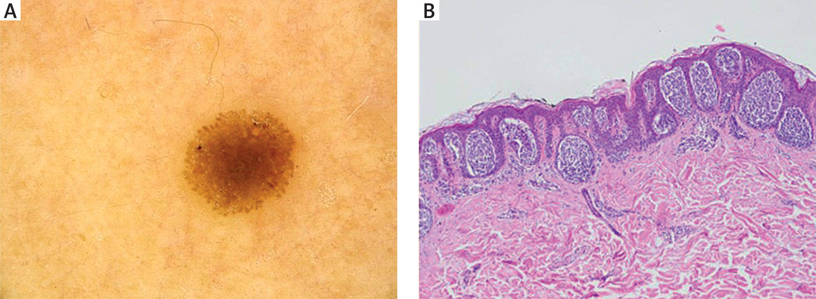
Our study showed that in benign lesions, globules were most common in compound naevi (34.3%), junctional naevi (28.6%), and lentiginous naevi (15.6%). However, in melanomas, they occurred with similar frequency in all histological variants we analysed (Table 1, Figure 1).
Figure 1
Videodermoscopic (A) and histopathological (B) picture of a junctional naevus with multiple globules of similar shape and size at the periphery (magnification 20×)
Melanomas were significantly more common in older patients (median age: 48 vs. 39 years; p = 0.028), however the youngest patient with diagnosed melanoma was 23 years old. There was no statistically significant difference in melanoma incidence between women and men (p = 0.244) (Table 2).
Table 2
Demographic characteristics of the studied patients according to malignancy status
In our study, globules were observed in both benign and malignant lesions. Benign lesions most frequently presented with a diffuse globular pattern (76.6%) of brown (82.3%) or dark brown (70.8%) colour, with preserved general symmetry (72.9%). A statistically significant differentiating feature, observed exclusively in benign lesions, was the presence of globules of one shape and size (24.5% vs. 0.0% in melanoma; p = 0.0149). On the other hand, malignant lesions were associated with a higher prevalence of red globules (11.8%; p = 0.0340) and a significantly lower frequency of symmetry (41.2%; p = 0.0060). However, it is worth emphasising that both melanomas and benign naevi frequently exhibited brown colour (76.5%) and a diffuse globular pattern (70.6%), which may constitute a diagnostic pitfall (Table 3).
Table 3
Descriptive characteristics of dermoscopic features by outcome and univariate association between dermoscopic features and melanoma
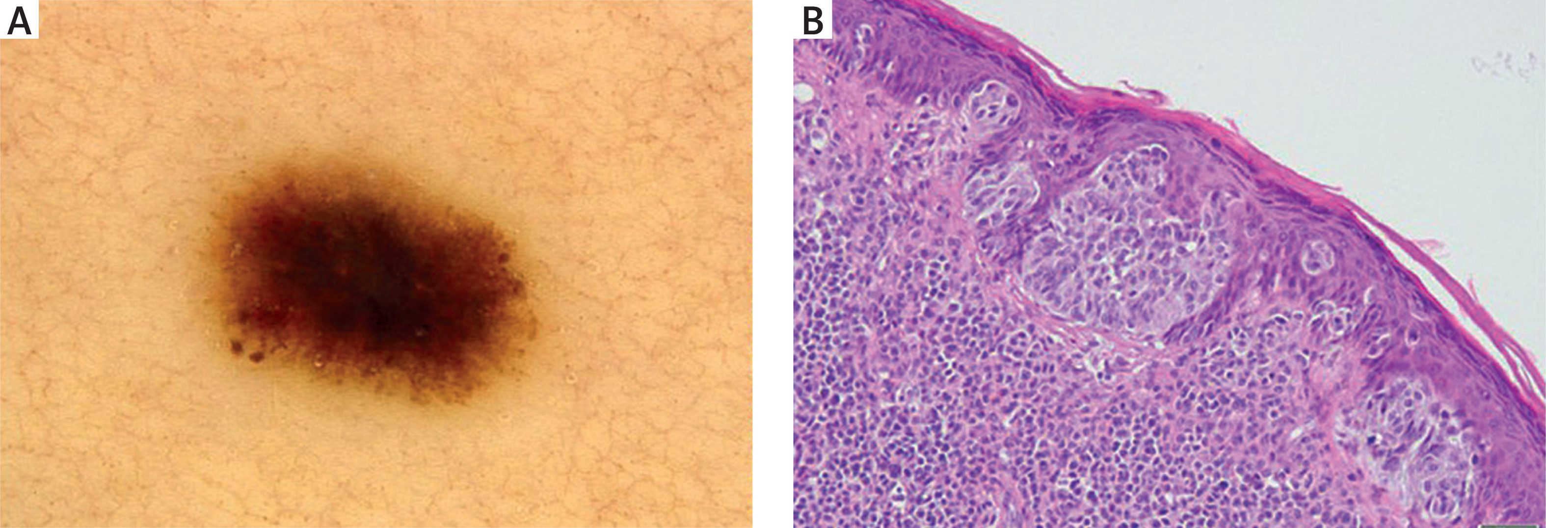
Univariate analysis demonstrated that within our study group, the presence of red globules was the strongest positive predictor of malignancy, associated with a more than 12-fold increase in the risk of melanoma (OR = 12.67; p = 0.0340). In comparison, lesion symmetry (OR = 0.26; p = 0.0060) and the presence of globules of one shape and size (OR = 0.09; p = 0.0149) proved to be statistically significant protective factors, indicating a benign character of the lesion. Our results are consistent with reports by other authors, confirming that while brown colour and a diffuse pattern do not effectively differentiate between lesions, globule uniformity and symmetrical distribution are strong indicators of being benign (Figure 2). However, the absence of these features and the presence of red globules suggest a malignant process (Table 3).
Figure 2
Videodermoscopic (A) and histopathological (B) picture of superficial spreading melanoma (pTis) presenting with single globules of different size at the periphery (magnification 20×)
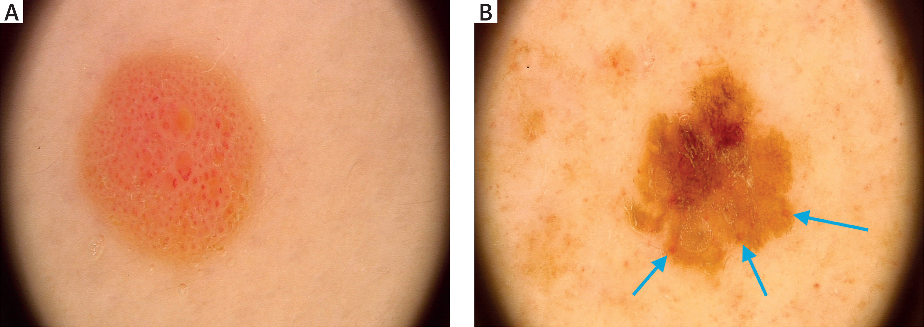
It has been demonstrated that a symmetrical distribution of globules, along with their uniform shape and size, is indicative of a benign lesion, whereas the presence of red colour increases the likelihood of melanoma (Table 3, Figure 3).
Figure 3
Videodermoscopic images of a Spitz naevus with red globules (A) and a lentigo maligna melanoma with isolated single, asymmetric red globules (B) (magnification 20×). Red globules are marked with blue arrows
Most of the benign lesions (74.5%) were located on the trunk or head, while only 25.5% were found on the extremities. A comparison of malignant cases suggests that the frequency of melanomas, which were located on the limbs, was similar to that on the trunk/head within the study group. This finding warrants further investigation with a larger size of the melanoma group, so the statistical significance of this result might be further strengthened (Table 4).
Discussion
The presence of peripheral globules within a naevus is a sign of its active growth [4]. Kittler et al. demonstrated that a symmetrical border of brown globules is a marker of benign and horizontal enlargement of the lesion [5]. This is a dynamic process. Follow-up studies analysing naevus growth confirm that globules appear during the growth phase and progressively disappear upon stabilization, which is a physiological phenomenon. This appearance is observed frequently in the paediatric population. It has been reported that the globular pattern occurs in 40% of naevi in children, and during follow-up, 61.1% of peripheral globules undergo spontaneous involution [6]. Studies suggest that this mechanism does not occur only in children. Ilut et al. observed that the growth rate of naevi with globules was independent of age, as the median age of patients participating in the study was 36 years. This suggests that the presence of peripheral globules in adults may also represent a benign proliferation, while lesion symmetry is preserved [4]. This is not consistent with the results of our study, which showed that there is no correlation between peripherally located globules and the benign character of the naevus. Available data in the literature on the correlation between globule patterns and specific naevus types are limited. There are studies reporting that globules were most frequently observed in dysplastic naevi, which is different from the findings of our study [7, 8]. According to our research, globules were most frequently found in compound naevi.
Despite the physiological nature of globules in many naevi, their presence can constitute a diagnostic trap, particularly in the context of superficial spreading melanoma (SSM) [9]. The key to differentiation depends on the lesion’s morphology. Xu et al. demonstrated that the highest risk of melanoma is associated with diversity in the globule shape (OR = 5.37) and size (OR = 4.72) within a single lesion. Large globules exceeding 0.2 mm are also a concerning feature [10]. Avcı et al. confirmed that asymmetry in globule distribution is an independent predictive factor for early melanomas and high-grade dysplasia, and it increases the risk of malignancy more than threefold [11]. This aligns with the findings of Lallas et al., who identified irregular globules as a key characteristic of SSM, and Reiter et al., who showed that benign lesions are characterized by globule monomorphism and symmetry, whereas polymorphism and asymmetry suggest malignancy [7, 9]. Moreover, previous studies showed that atypical globule colour, such as red, is strongly associated with an increased risk of melanoma, which was confirmed in our study [12]. Lesion location also requires attention. Moraes et al. found that lesions with peripheral globules on the lower extremities carry a higher melanoma risk, particularly when they are accompanied by a disorganised pattern and blotches [8].
Although globules can occur in benign lesions in adults, there is a risk that this presentation masks an underlying oncological process. Lazaridou et al. highlight that peripheral globules are far more concerning in the elderly than in children [13]. These observations were confirmed by Avcı et al., who demonstrated that age significantly increases the risk for high-grade dysplasia in such lesions. Consequently, authors suggest excision of all lesions presenting with peripheral globules in patients who are over 60 years old [14]. This approach is further supported by the potential risk of nested melanoma. Jegou et al. describe this rare melanoma variant, which frequently affects older individuals and is histologically characterised by large nests of melanocytes that dermoscopically mimic the globular pattern typical of benign naevi [15]. However, our study demonstrated that young age did not preclude malignancy. It is worth noting that the youngest patient diagnosed with melanoma was 23 years old. In the analysed material, only two lesions were excised in minors: a Spitz naevus in a 17-year-old patient and a junctional naevus in a 13-year-old patient. In the second case, the decision to excise was made due to the dermoscopic spitzoid pattern of the diagnosed lesion, which on histopathological examination turned out to be a junctional naevus. Therefore, the authors did not include the patients’ age in the analysis in the presented study.
The present findings should be interpreted in the context of current dermoscopic practice, which strongly prioritises the principle of avoiding missed melanoma diagnoses [16]. Although black globules are included among melanoma-associated criteria within established algorithms such as the seven-point checklist, their clinical significance is not uniform and must be evaluated in relation to the patient age and lesion morphology [17, 18]. Previous studies have suggested that the appearance of globules in patients over 60 years may reflect dynamic lesion evolution and justify excision, while spitzoid-pattern lesions developing after the age of 12 years are typically managed surgically due to the well-recognised diagnostic overlap with melanoma in this age group [14, 19]. Our results indicate that, in routine clinical settings, lesions with asymmetric globular structures are frequently perceived as high-risk ones, even in the absence of additional dermoscopic features of malignancy. This tendency appears to be further reinforced by patient-related factors, such as a personal or family history of melanoma. An independent factor could be the patient’s preference for excision over short-term dermoscopic monitoring. While this precautionary management helps to avoid missing melanoma, it may contribute to increased excision rates of biologically benign lesions.