
INTRODUCTION
Halo nevi, also known as Sutton nevi, are nevi surrounded by a depigmented border [1]. They occur in approximately 1% of the population, with no sex or race predominance, and are most commonly observed in young adults, particularly on the trunk [2, 3]. As for the potential underlying pathomechanism, two main theories have been proposed: antibody-mediated mechanisms and cytotoxic T-cell response [2].
OBJECTIVE
To present a case of melanoma associated with multiple Sutton nevi and to discuss its clinical relevance.
CASE REPORT
A 55-year-old man, with a negative personal or family history of dermatoses, except for one dysplastic nevus removed many years ago, presented to dermatologic ambulatory care due to a rough erythematous patch on his cheek. Although initially hesitant, the patient agreed to undergo a full-body dermoscopic examination.
Apart from the lesion he pointed out, which was diagnosed as actinic keratosis, multiple oval hypopigmented macules were observed on the trunk, resembling the end stage of Sutton nevi regression (figs. 1 A, B). The patient confirmed that he previously had multiple nevi, which had gradually disappeared.
Figure 1
Clinical images of the patient’s front (A) and back (B): multiple hypopigmented macules distributed over the trunk in the area of previously existing nevi. In panel B the excised nevus on the chest is shown (marked with a circle)
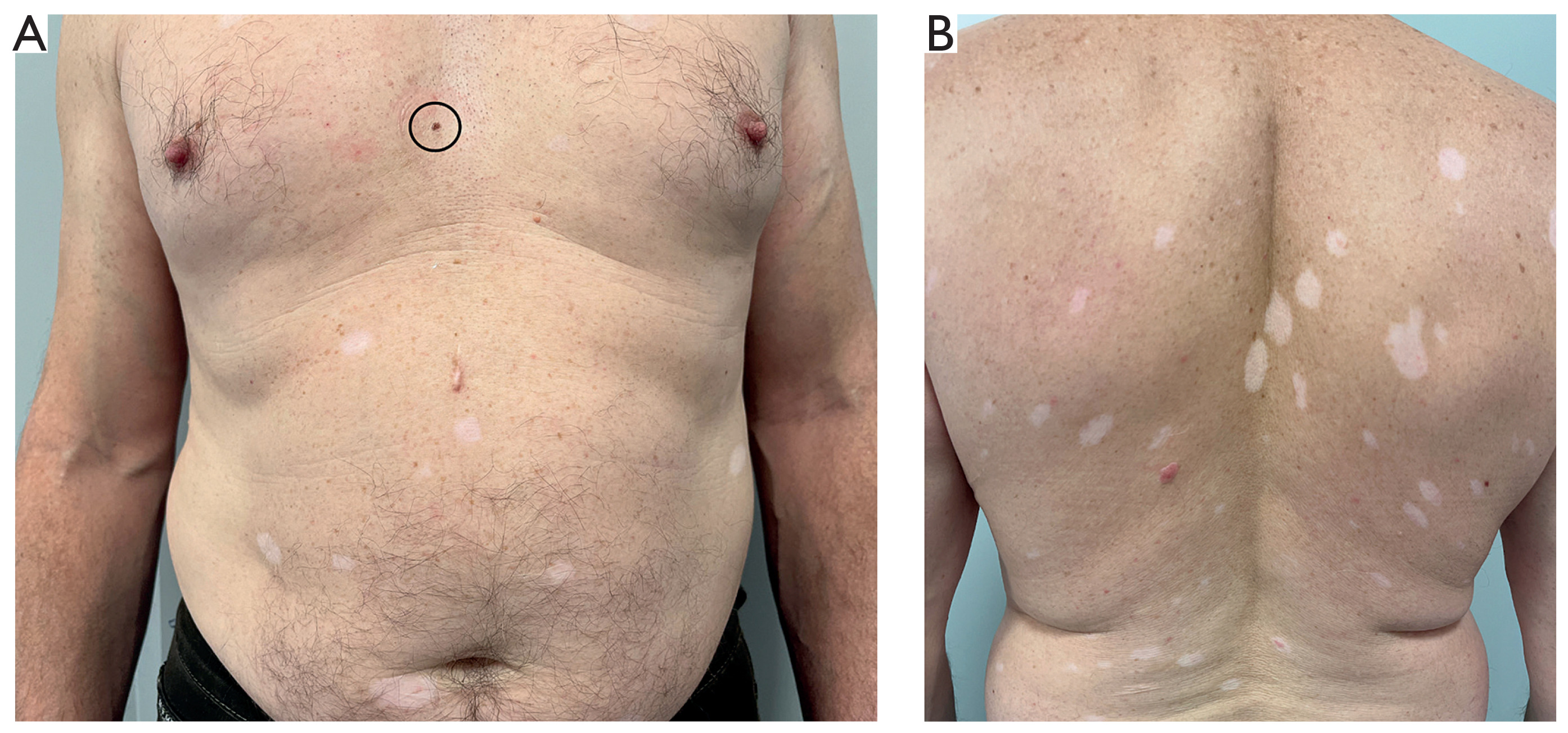
Moreover, one atypical melanocytic lesion was identified on the chest (fig. 1 A, marked with a circle). Dermoscopy revealed asymmetry of structure, color, and border, as well as blue-grey structures and angulated lines (fig. 2).
Figure 2
Dermoscopic image of the excised nevus with features suggestive of melanoma: asymmetry of color, structures, and border, as well as blue-grey structures and angulated lines and peppering
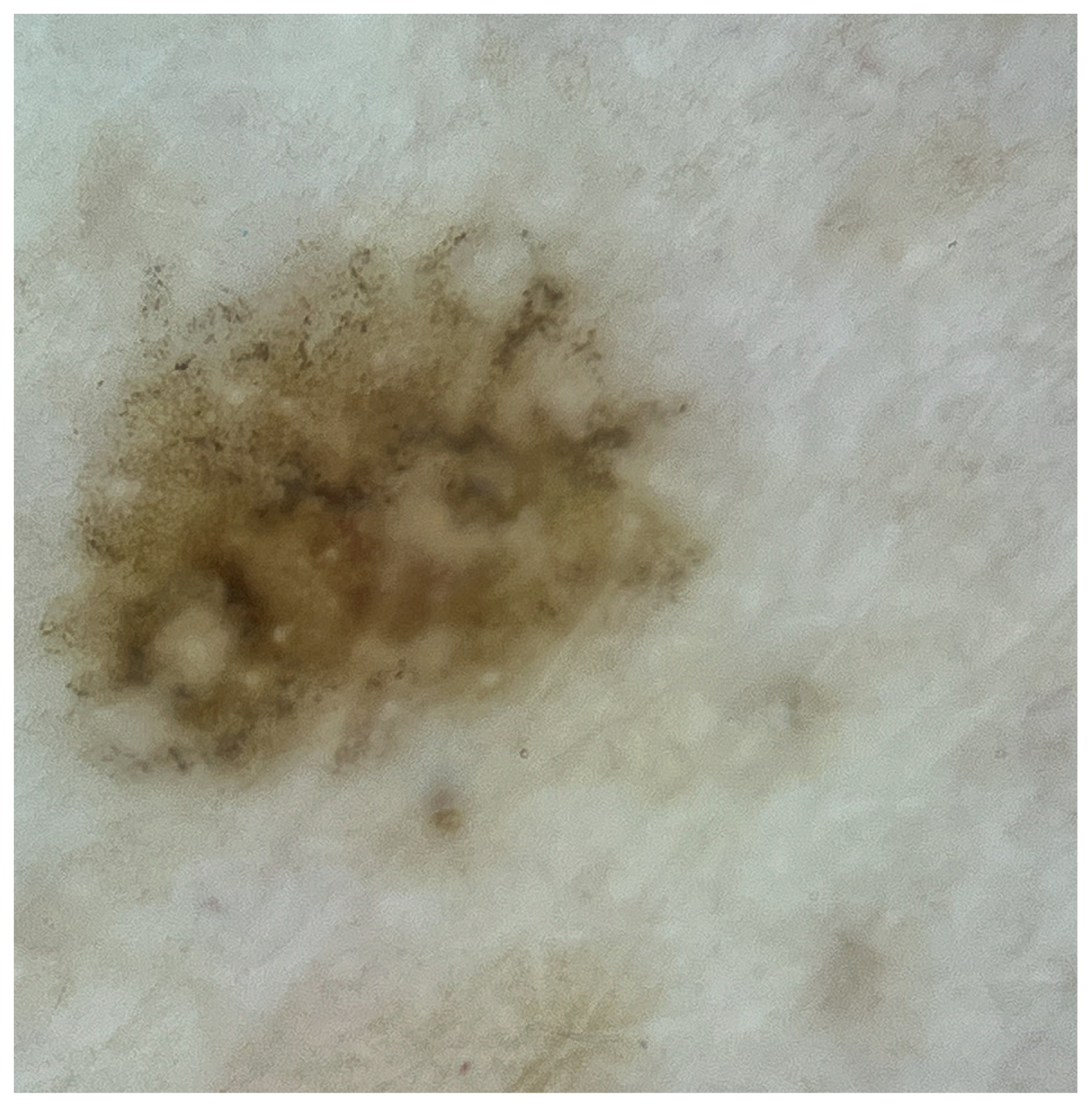
The lesion was excised with 2 mm margins and examined histopathologically, confirming superficial spreading melanoma in situ. The patient was subsequently referred for wide local excision.
DISCUSSION
The presence of Sutton nevi may be associated with several conditions, especially vitiligo, but also thyroid diseases, malignancies, or may occur after immunotherapy [2]. Halo nevi associated with vitiligo usually occur at a younger age and in individuals with a family history of vitiligo [2]. Multiple halo nevi, as opposed to a single lesion, are also more commonly associated with vitiligo [2].
It has been suggested that halo nevi themselves are not associated with an increased risk of melanoma; however, eruptive Sutton nevi have been reported in patients with malignancies [2]. In one of the largest available case series, dermoscopy of a total of 138 halo nevi in 87 patients revealed no melanomas [4, 5]. In another case series, 5 out of 16 patients with eruptive halo nevi had melanoma (including one with a prior history), and some had additional malignancies (not skin-related) [5].
In general, when melanoma and halo nevi coexist, hypopigmentation is typically peri-lesional. In contrast, the presented case is atypical due to the presence of multiple distant halo nevi. Hypopigmentation in melanoma may occur in several forms, including regression of the primary melanoma (and/or its metastases), halo phenomenon, and vitiligo-like melanoma- associated hypopigmentation at distant sites [6]. In older individuals, multiple Sutton nevi may raise suspicion of melanoma [1]. This may reflect an immune-mediated response targeting antigens shared by melanocytes and melanoma cells [6].
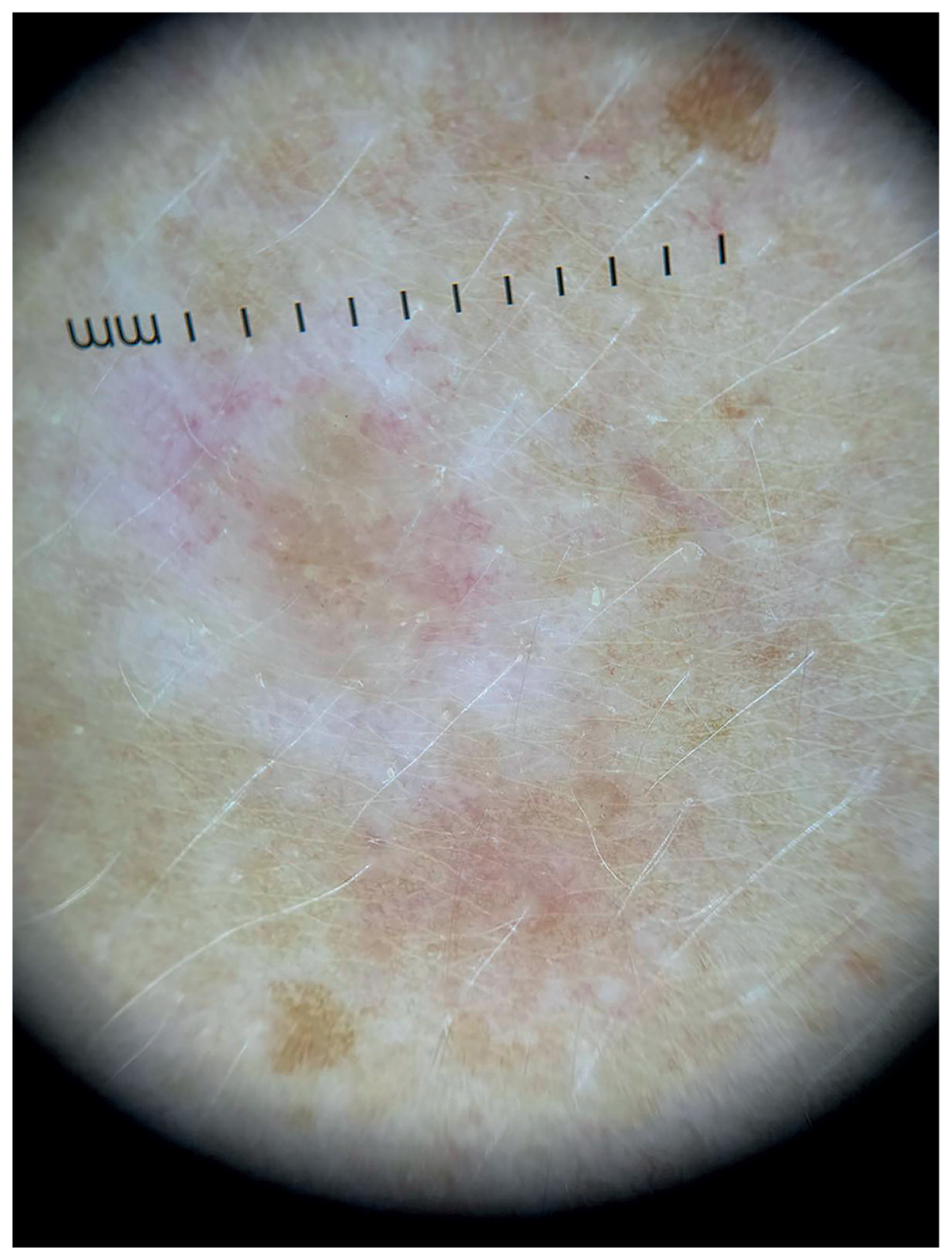
Dermoscopy may help distinguish halo nevi from regressing melanoma. Halo nevi typically show features of a benign melanocytic lesion, including symmetry and a regular depigmented halo [4]. In contrast, melanoma often show chaos of color, structures and borders, blue-white veil, and irregular hypopigmented areas that do not form a true halo [2]. Figure 3 presents an almost fully regressed Sutton nevus.
Figure 3
Dermoscopic image of a representative almost fully regressed Sutton nevus. There is a white halo surrounding a pale, fading brown pigment network in the center
Histopathologically, markers such as HMB-45 may aid in differentiation [2]. While it is relatively specific for melanoma, it is less sensitive than S-100, which is highly sensitive but less specific [7]. In halo nevi, HMB-45 expression is typically limited to superficial dermal layers, whereas in melanoma it may be diffuse, irregular, or absent [2].
Another marker, PRAME, is expressed in melanomas of various origins, although it is not entirely specific, as it can also be detected in other neoplasms [8]. Its increased expression detected in melanoma transformed melanocytes [8].
The described patient developed melanoma in a lesion without a halo, while multiple distant halo nevi appeared simultaneously. The patient had no family history of vitiligo. Melanoma-associated depigmentation occurs in approximately 2–16% of patients and is more commonly observed after treatment, often correlating with a favorable prognosis [1].
Patients presenting with Sutton nevi should be approached with caution. However, no clear recommendations exist. The authors suggest careful clinical history-taking (including personal and family history of melanoma, autoimmune diseases, and other skin conditions), followed by thorough clinical and dermoscopic examination. Additional laboratory tests, such as TSH and fasting glucose, may be considered. In the absence of abnormalities, regular follow-up is recommended. If a lesion shows features suggestive of melanoma, it should be excised.












