
Fixed drug eruption (FDE) is a cutaneous adverse drug reaction characterised by the recurrence of well-demarcated erythematous or violaceous plaques at the same anatomical location after repeated exposure to the causative drug. Nonsteroidal anti-inflammatory drugs (NSAIDs), including ibuprofen, are inducers of FDE, particularly in children, in whom the frequent use of antipyretics during febrile illnesses can obscure the diagnosis of FDE. The clinical presentation ranges from solitary lesions to extensive bullous eruptions. Diagnostic confirmation is crucial for preventing recurrence and guiding safe pharmacological therapy. Patch testing applied to residual lesions is a useful, non-invasive method for identifying the offending agent, particularly in paediatric patients, in whom oral provocation is difficult and may not be ethically justifiable.
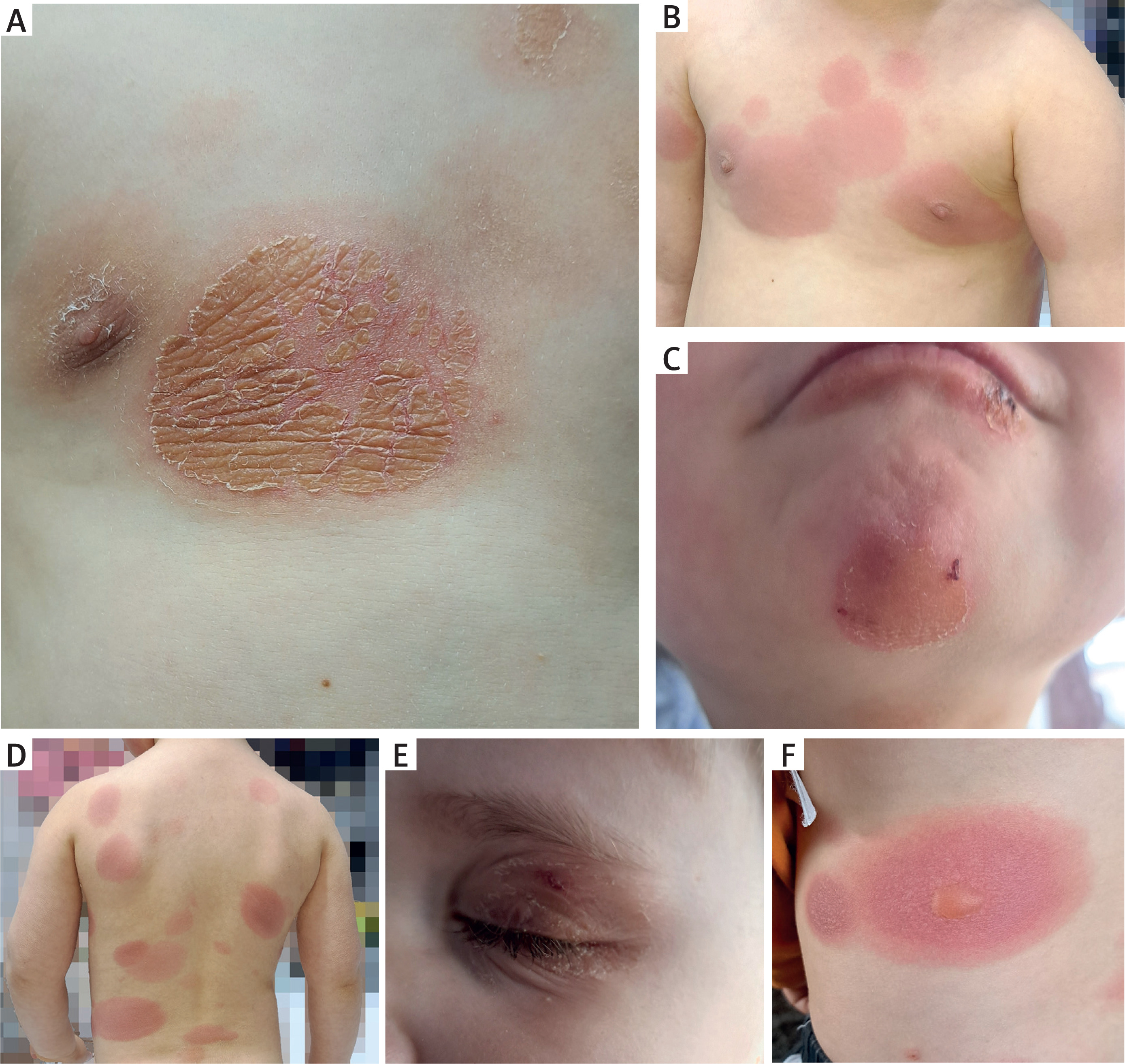
A 3-year-9-month-old boy was admitted for evaluation of recurrent cutaneous eruptions associated with febrile episodes. The first episode occurred at 20 months of age, presenting as erythematous changes on the left upper eyelid and was initially misdiagnosed as viral conjunctivitis with secondary irritation. During subsequent febrile illnesses, approximately eight documented episodes, the child developed sudden oval erythematous plaques on the trunk, face, and neck, often accompanied by a low-grade fever (up to 38.5°C) (Figures 1 A–F). Although lesions consistently recurred at the same sites, new plaques occasionally appeared in later episodes. Each episode was preceded by upper respiratory or gastrointestinal infection symptoms.
In January 2024, the child was hospitalised for a similar episode, presenting with bullous lesions. Laboratory studies revealed elevated total IgE and D-dimer levels; however, allergy screening (including skin prick testing for aeroallergens) was negative. Bullous impetigo was suspected initially.
During the current/latest admission in April 2025, physical examination revealed multiple sharply demarcated erythematous plaques on the trunk, neck, and face, with areas of post-inflammatory hyperpigmentation. No mucosal involvement or systemic symptoms were observed. Laboratory evaluations, including serum tryptase levels, were within normal limits.
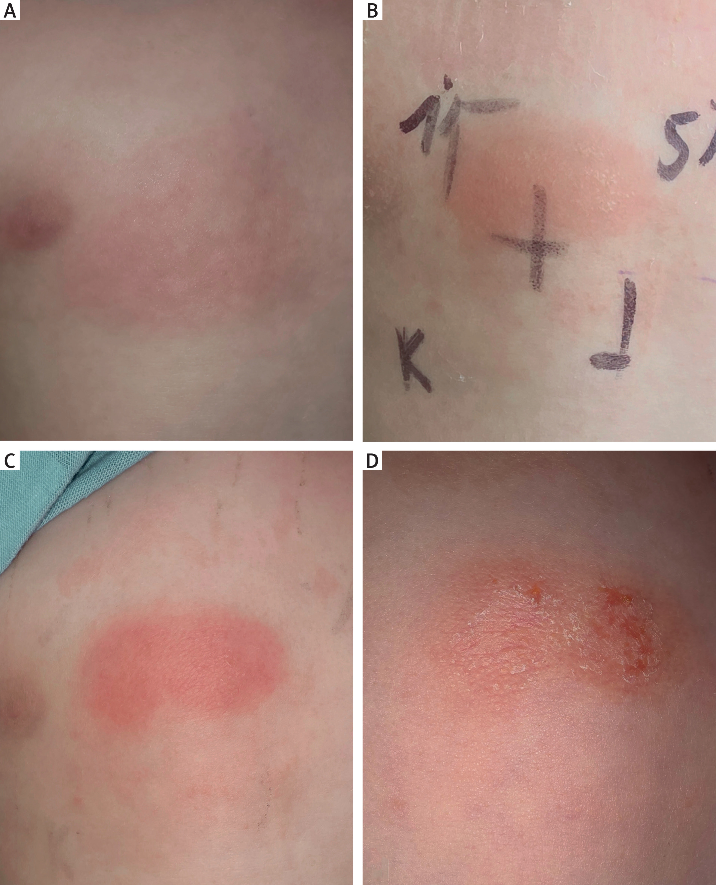
A detailed drug history revealed ibuprofen administration prior to each eruption. Based on the temporal relationship and lesion recurrence pattern, an FDE was strongly suspected. Patch testing with 1% and 5% ibuprofen in white petrolatum was performed on both lesional and non-lesional skin. Positive reactions, manifested as erythema and infiltration, were observed at 48, 72, and 96 h exclusively on the lesional skin for both concentrations, confirming a delayed hypersensitivity response (Figure 2).
Figure 2
Skin patch testing with ibuprofen: A – the place where the test was applied – erythematous lesion, B – after 48 h, C – after 72 h, D – after 96 h
The final diagnosis was multiple fixed drug eruptions induced by ibuprofen. The patient was discharged in good condition, instructed to avoid ibuprofen-containing products, provided with an allergy warning card, and advised to use paracetamol as a safe alternative to ibuprofen.
FDE is a T cell-mediated cutaneous hypersensitivity reaction that poses diagnostic challenges in paediatric populations, particularly when multiple lesions, blistering, or atypical locations obscure the classical presentation. The hallmark feature of FDE is the recurrence of identical skin lesions at the same anatomical sites following re-exposure to a specific drug. In our case, the consistent emergence of well-circumscribed erythematous plaques in response to ibuprofen, combined with post-inflammatory hyperpigmentation and a favourable response to the withdrawal of the drug, supported the diagnosis.
NSAIDs are among the most frequently implicated drugs in paediatric FDE, likely because of their widespread use as antipyretic and analgesic agents. While antibiotics such as trimethoprim-sulfamethoxazole, tetracyclines, and anticonvulsants have traditionally been the causative agents, ibuprofen-induced FDE is increasingly recognised in clinical practice [1, 2]. Importantly, early lesions may be misattributed to viral infections or impetigo, delaying diagnosis and exposing children to further risk through repeated administration of the offending drug.
Histologically, FDE is characterised by basal cell hydropic degeneration, necrotic keratinocytes, pigmentary incontinence, and dermal lymphocytic infiltration. These findings are not unique to FDE and may overlap with other drug-induced or autoimmune dermatoses, reinforcing the importance of clinical correlation and drug-specific testing [3]. In our patient, the evolution of lesions to a bullous form prompted the exclusion of more severe conditions, such as Stevens-Johnson syndrome (SJS) or major erythema multiforme. However, the absence of mucosal involvement and the sharply demarcated lesion pattern were consistent with multifocal bullous FDE rather than a systemic blistering disease.
The pathophysiology of FDE is driven by skin-resident memory CD8+ T cells (TRM) that remain at previously affected skin sites long after clinical resolution. These TRM cells respond rapidly to re-exposure by releasing IFN-γ and cytotoxic mediators, including granzyme B and perforin, resulting in localised keratinocyte apoptosis [4, 5]. Keratinocyte-derived IL-15 plays a pivotal role in maintaining memory T-cell populations over time, making reactivation at the same anatomical site a consistent and reproducible event [6]. This immunological mechanism explains the “fixed” nature of the eruption and the relatively short latency between drug exposure and lesion reactivation, often less than 48 h.
Patch testing is a critical diagnostic modality in the evaluation of FDE, especially in children, where oral drug challenges may be ethically and clinically inappropriate due to the risk of triggering extensive reactions. Positive results are more commonly obtained when the test is applied to lesional skin rather than uninvolved skin, as confirmed in this case [7]. The higher diagnostic yield is thought to result from the persistence of drug-specific T cells at previously affected sites.
Despite its diagnostic value, patch testing is not universally standardised, and false-negative results can occur, particularly with non-immunogenic drugs or in cases with long drug-free intervals. In a cohort of 52 patients, 40.4% demonstrated positive results on patch testing, predominantly to NSAIDs such as nimesulide, piroxicam, and etoricoxib, with 1 case involving cetirizine [7]. In a separate study involving 34 patients, 27 (79.4%) individuals showed patch test positivity, primarily to NSAIDs (accounting for 63% of all positive results) and paracetamol (14.8%) [8].
Importantly, paediatricians and dermatologists must remain vigilant for FDE, especially in toddlers and preschool-aged children with recurrent skin lesions that exhibit a site predilection or residual pigmentation. The potential to confuse early bullous FDE with evolving SJS or other immune-mediated dermatoses necessitates careful clinical assessment, a thorough drug history, and timely dermatological input. Although the prognosis of FDE is favourable, prevention of re-exposure is paramount. Repeat episodes not only increase the lesion number and intensity but may also predispose patients to more severe variants, such as generalised bullous fixed drug eruption (GBFDE), which has been associated with systemic involvement and complications [9].
This case contributes to the growing recognition of NSAID-induced FDE in the paediatric population and underscores the need for greater awareness of its immunopathogenesis, clinical variability, and diagnostic strategies in the future. A multidisciplinary approach involving paediatricians, allergists, and dermatologists can facilitate early recognition and prevent unnecessary morbidity.










