
Introduction
Latex allergy remains one of the significant problems in modern medicine, combining clinical, immunological, and hygienic aspects. The widespread use of latex-containing products in surgery, anaesthesiology, dentistry, gynaecology, and other medical fields poses a high risk of contact with potentially sensitizing materials for both patients and medical personnel. Despite known attempts to standardize diagnostic approaches and introduce latex-free protocols, practice shows that episodes of allergic reactions continue to be reported. Given the high burden on the healthcare system and the intensified use of disposable medical devices, especially in the context of pandemics, there is a significant need for a comprehensive understanding of latex allergy as an interdisciplinary phenomenon.
According to a review by Gracz-Bernaciak et al. [1], the latex plant in general is a biologically active emulsion containing up to 5% morphine (in poppies) and up to 20% alkaloids (in celandine), as well as many proteins, proteases, lectins, and oxidases. The pathophysiology of natural rubber latex allergy (NRLA) is primarily associated with protein allergens derived from Hevea brasiliensis, rather than with alkaloids or morphine-like compounds found in other plant species. Despite centuries of use of latex-bearing plants, many of their molecules remain insufficiently studied, which highlights the need for further research into their allergenic potential and immune response mechanisms in clinical practice. From a clinical point of view, the introduction of latex-free protocols in surgical practice can significantly reduce the risk of allergic reactions. Thus, in a study by Stinkens et al. [2], when switching to powder-free gloves or synthetic analogues in operating rooms, no cases of severe immunoglobulin E (IgE)-mediated reactions were reported among patients at high risk of latex allergy. Only 3.1% had mild forms of contact dermatitis, which highlights the effectiveness of abandoning powdered latex products. Similar trends are confirmed in outpatient dental practice. Critchley and Pemberton [3] found that 90% of dentists completely abandoned latex gloves in favour of alternatives, mainly nitrile ones. This shift toward hypoallergenic materials reflects a high level of concern about latex sensitization and a desire to reduce occupational risks for medical personnel.
Nevertheless, cases of severe sensitization persist. Ngamchokwathana and Chaiear [4] described anaphylaxis in a 39-year-old female medical worker, with two episodes of systemic reaction after contact with latex, accompanied by pronounced sensitization (specific IgE = 10.20 kUA/l). The clinical picture included urticaria, vesiculopapular rash, and anaphylaxis, which highlights the high risk of even brief contact with latex in sensitized individuals.
Latex-fruit syndrome (LFS) deserves special attention, in which sensitization to latex is combined with a cross-reaction to food products. Gromek et al. [5] showed that the prevalence of LFS varies from 4% to 88% depending on the diagnostic methods used. The most common allergens in such patients are bananas, avocados, kiwis, and papayas, and the most accurate diagnosis is considered to be a combination of clinical history and laboratory tests.
A review by Mwanga and Shebe [6] showed that in African countries, avoiding contact with latex remains the only available prevention strategy. At the same time, reactions to latex are divided into IgE-mediated (type I), cell-mediated (type IV), and nonspecific irritation. The authors emphasize the need to provide healthcare workers with systemic reactions with access to adrenaline auto-injectors. Data on the frequency of skin manifestations also deserve attention. According to a study by Knežević Krajina and Knežević [7], almost half of healthcare workers who use latex gloves experience skin symptoms, with 18% suffering from contact urticaria. The risk increases significantly with prolonged glove wear (more than 5 h/day). Most symptoms disappear during vacation, indicating a direct link to occupational exposure. As emphasized in the review by Jiménez-Carrillo et al. [8], complete avoidance of latex remains the primary means of preventing allergic reactions. At the same time, the prevalence of latex allergy worldwide is 4.3%, and in 20% of cases of surgical interventions involving sensitized patients, anaphylaxis develops. Despite the potential of immunotherapy, the lack of standardized extracts and protocols currently limits its use in clinical practice.
Aim
The aim of this study was to analyse the etiopathogenic mechanisms, clinical manifestations, diagnostic approaches, and assessment of sensitization factors among healthcare workers. The study addressed the following objectives: to identify key pathophysiological mechanisms of latex sensitization; to examine the prevalence of allergy across different risk groups; to evaluate the effectiveness of component-resolved and molecular diagnostics; and to summarize approaches to primary and secondary prevention in healthcare settings. Additionally, it considered issues of latex-fruit cross-sensitization and the role of occupational history in shaping individual risk.
Methods
This study is a systematic review based on 42 peer-reviewed publications, including systematic reviews, clinical and epidemiological studies, and case reports related to latex allergy.
Sources were searched in authoritative international databases, including PubMed, ResearchGate, PMC, Asthma Allergy Immunology, JAPM, Elsevier, and Springer. The time range of publications covered the period from 2013 to 2025, with an emphasis on research from the last 10 years. The following keywords and their logical combinations were used in the search: “latex allergy”, “natural rubber latex”, “Hevea brasiliensis”, “Hev b allergens”, “IgE sensitization”, “occupational exposure”, “latex-fruit syndrome”, “component-resolved diagnostics”, “powdered gloves”, “cross-reactivity”, and “spina bifida and latex allergy”.
The publications were screened in stages. In the first stage, the titles and abstracts of the works were analysed in order to exclude duplicates, irrelevant publications, materials that had not undergone expert review, as well as studies that did not contain a description of the methodology or data on the allergenic characteristics of latex. Next, the selected publications were subjected to a full-text analysis, during which the structure of the study, the characteristics of the sample, the diagnostic methods used, and the interpretation of the results were evaluated.
The criteria for inclusion in the review were publications containing empirically confirmed data of scientific and clinical value in the context of studying latex allergy. The analysis included original clinical studies of various designs – cross-sectional, cohort, and retrospective – as well as systematic reviews published in peer-reviewed scientific journals. Clinical data on the frequency and structure of allergic manifestations were quantitatively synthesized, including information on respiratory and systemic reactions. Particular attention was given to the classification and severity of clinical symptoms, with reference to recommended therapeutic interventions, such as the intramuscular administration of epinephrine in cases of anaphylaxis. In addition, experimental studies aimed at the molecular characterization of latex allergens, including component analysis of Hevea brasiliensis allergen (Hev b) 1–15 proteins, recognized as key in sensitization mechanisms, were taken into account [9–12]. Considerable attention was also paid to case reports highlighting the clinical features of NRLA in non-standard or severe cases [4, 13]. Publications were excluded if they did not align with the study objectives, lacked adequate scientific rigour or peer review, failed to present clinically interpreted or quantitative data, relied solely on animal models without clinical validation, or were published before 2013 without subsequent corroboration in later studies.
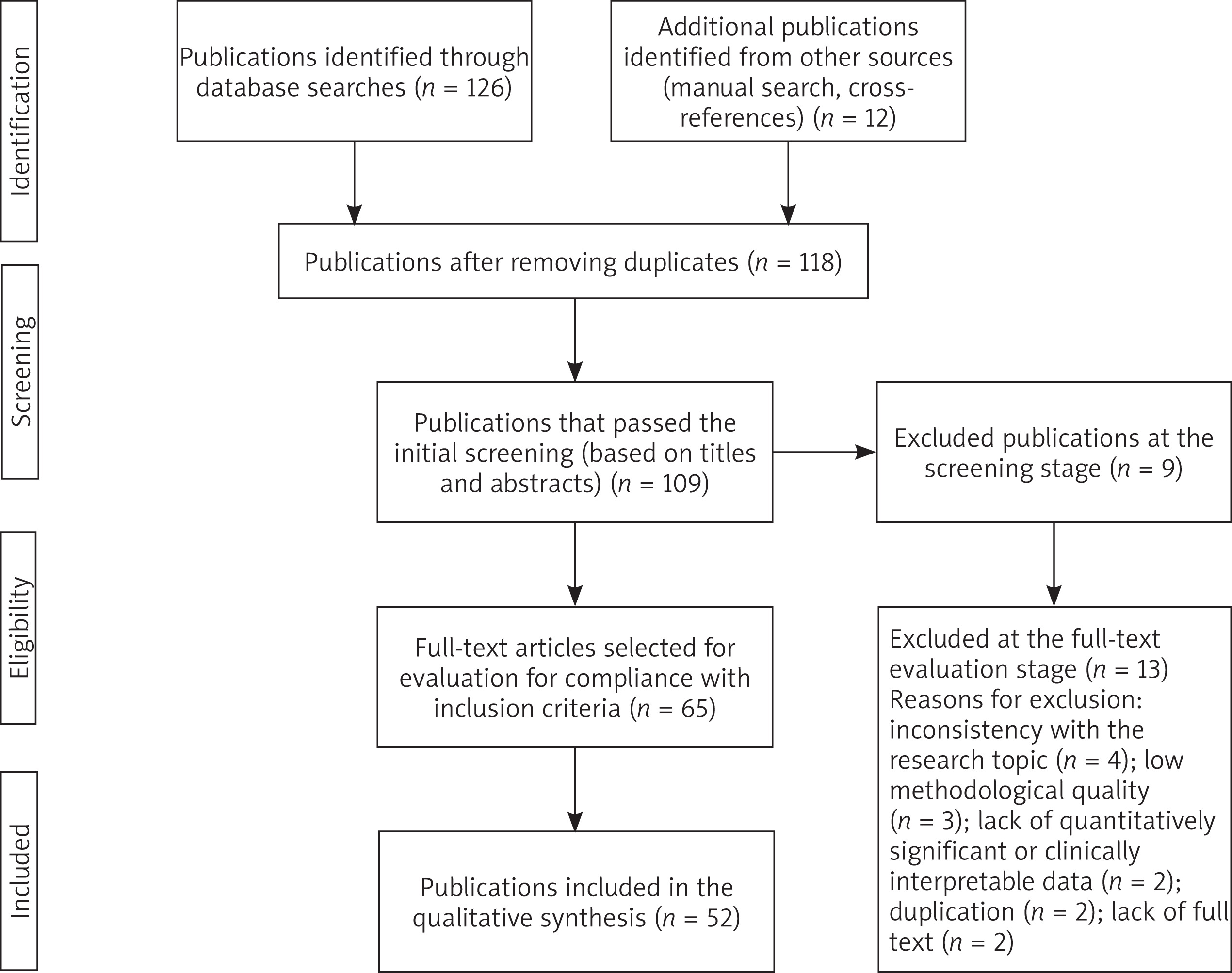
As a result of the literature search and selection based on the specified criteria, 52 publications corresponding to the research topic and scientific quality requirements were included in the analysis. Data extraction from the selected sources was performed manually using a structured approach, including study characteristics (type, design, sample size), allergen characteristics (structure, MW, immunogenicity), data on the frequency and clinical manifestations of NRLA, diagnostic algorithms, preventive strategies, as well as case descriptions and regional epidemiological differences. Particular attention was also paid to non-standard routes of exposure, including inhalation of aerosolized latex particles and alimentary contact through contaminated food or pharmaceutical products, as these factors influence the clinical variability of latex allergy and are especially relevant in vulnerable populations such as patients with spina bifida and healthcare workers.
Data synthesis was carried out using a thematic approach, which allowed the information to be structured according to key research areas. In particular, the following analytical clusters were identified: molecular and pathophysiological aspects of latex sensitization, including a description of the structure and biological activity of Hev b allergens; the prevalence and clinical specificity of NRLA in vulnerable groups, such as patients with myelomeningocele and healthcare workers; issues of differential diagnosis with an emphasis on the use of component-resolved analysis and the use of recombinant allergens; LFS and mechanisms of cross-sensitization with food and pollen allergens; as well as strategies for primary and secondary prevention of latex allergy in clinical practice. This approach made it possible to generalize heterogeneous data and identify both consistent patterns and current contradictions.
To ensure the quality of the analysis and the reliability of the summary, the principles of a systematic approach and transparency in the selection of sources were used. The assessment of the reliability of the evidence took into account the characteristics of the sample, the diagnostic methodology (SPT, sIgE, ImmunoCAP, molecular panels), and the publication source. In evaluating the diagnostic methods, the use of specific brands and equipment was also considered. For skin prick test (SPT), standardized commercial extracts of natural non-ammoniated latex and food allergens from ALK-Abelló A/S (Hørsholm, Denmark) were applied, with procedures conducted using the MedBlue 10® device (Asistan Medikal). The technique also included prick-to-prick testing with fresh food items, histamine dihydrochloride as a positive control, and saline solution as a negative control. For specific immunoglobulin E (sIgE) detection, ImmunoCAP and the 3gAllergy system (Alastat) were employed as established laboratory platforms. Molecular diagnostics involved component-resolved approaches for identifying specific IgE antibodies to latex allergens (e.g., Hev b 1-15), primarily using ImmunoCAP technology. Patch testing was carried out using IQ Chambers (Chemotechnique MB Diagnostics AB), with standard panels including rubber additives, additional haptens, and samples of powdered and non-powdered latex gloves. The study used approaches that comply with international quality standards for review studies, including GRADE (Grading of Recommendations Assessment, Development and Evaluation) recommendations [14], especially when interpreting heterogeneous data on prevalence and diagnostic methods.
In addition to the core diagnostic and analytical parameters, several supplementary aspects were taken into account to enhance the comprehensiveness of the review. The analysis included data on the allergenic protein content in finished latex products (e.g., gloves), as well as the localization of specific allergenic components on the inner and outer surfaces, which may influence the sensitization pathways in various professional groups. The biochemical composition of natural latex was also examined in relation to technological processing, particularly the distribution of proteins across different fractions of centrifuged latex. Clinical conditions such as occupational asthma were considered, with attention to the role of specific latex allergens associated with respiratory manifestations. The analysis also included a detailed assessment of the prevalence of latex-related allergic symptoms among different categories of healthcare workers, including nurses, physicians, technicians, laboratory personnel, and patient care staff. Differences in symptom frequency among atopic and non-atopic individuals were reviewed to clarify risk stratification. The importance of thorough medical history taking was emphasized as a central element in the diagnostic process for latex allergy, especially in the context of distinguishing between immediate and delayed hypersensitivity reactions.
The process of identifying, screening, and including publications was carried out in accordance with the international PRISMA (Preferred Reporting Items for Systematic Reviews and Meta-Analyses) recommendations [15], which ensured transparency, reproducibility, and validity of selection decisions. Figure 1 shows the corresponding PRISMA flowchart, reflecting the stages of searching and selecting the literature included in the review.
Figure 1
Scheme for identifying, screening, and including publications in the review in accordance with PRISMA. Source: compiled by the authors
In cases of discrepancies between sources, a comparative and critical analysis was conducted to identify possible causes (e.g., differences in diagnostic criteria, allergen composition, or population characteristics). This allowed for a more accurate delineation of areas of consensus and gaps in existing knowledge about NRLA, which formed the basis for recommendations on further research directions and clinical prevention measures.
Results and discussion
Aetiology, pathogenesis, and classification of latex allergy
Previous studies have shown that NRLA is a hypersensitivity reaction to latex proteins or chemicals used in production [16]. According to the results of a comprehensive analysis, natural latex obtained from the rubber tree Hevea brasiliensis contains 250 different polypeptides. Of these, 60 are capable of binding to human IgE antibodies, and 15 of them are officially classified as allergens (Hev b 1-15). The most important in terms of allergenicity are Hev b 1, 2, 3, 4, 5, 6.02, 7.01, and 13 [17]. Other reviews have reported that NRLA is caused by IgE-mediated sensitization to latex proteins, which account for only 1–2% of the fresh mass of rubber tree sap [18]. According to Kahn et al. [16], the aetiology of NRLA is associated with proteins (comprising 2% of the “milky” sap of the rubber tree Hevea brasiliensis), as well as with chemical additives such as thioureas and antioxidants used in the rubber production process.
Among the more than 240 polypeptides detected in latex, 60 bind IgE, and to date, 26 Hev b 1-15, including isoforms, have been included in the WHO/IUIS list [10, 17]. The allergenic potential of latex products can be significant: in 2016, 50% of commercial latex gloves contained between 215 and 1308 μg/g of allergenic latex protein [3, 18, 19]. Hev b 5 and Hev b 6.01 (especially the Hev b 6.02 hevein domain) are recognized as the main allergens in healthcare workers [20]. In patients with spina bifida, IgE reactivity to Hev b 1, Hev b 3, and Hev b 5 predominates [21, 22]. As reported in the literature, the different localization of allergens, for example, Hev b 5 and Hev b 6.02 on the inside of gloves, and Hev b 1 and Hev b 3 on the outside, explains the different routes of sensitization in different risk groups and the subsequent selectivity of the IgE response [10, 23]. The main characteristics of the allergenic components of Hevea brasiliensis latex are presented in Table 1.
Table 1
Allergenic components of Hevea brasiliensis latex: MW and presumed physiological functions. Source: compiled by the authors based on [10, 12, 17]
As shown in Table 1, most latex allergens perform functions related to plant protection (e.g., Hev b 2, b 7, b 11-15), while others are involved in rubber synthesis (Hev b 1 and b 3) or structural stabilization of latex (Hev b 5, b 6). Of particular importance is Hev b 6, which includes hevein, a lectin-like protein involved in latex coagulation and recognized as a potent allergen. The low-molecular-weight allergen, Hev b 15 (7.5 kDa), recently identified as a serine protease inhibitor, also demonstrates high IgE-binding capacity and likely plays a role in the pathogenesis of late allergic reactions. Thus, the molecular diversity and functional heterogeneity of allergens may determine the different immunogenicity of latex in representatives of individual risk groups.
As described by Kitabayashi [24], when centrifuged, latex is divided into three fractions: the “rubber phase” (upper) contains about 27% of total protein (including Hev b 1 and Hev b 3), the lower fraction (B-serum, or lutoids) contains about 25% of total protein, and the intermediate yellowish C-serum contains about 48% of total protein. Previous clinical reviews have shown that the pathogenesis of latex allergy includes various routes of exposure, such as skin, percutaneous, mucous membranes, or parenteral, which determines a wide range of clinical manifestations [25, 26]. The risk of developing latex allergy is directly dependent on the degree and duration of exposure, especially in patients with spina bifida and healthcare workers, who are considered the most vulnerable groups [21, 23]. In addition to direct contact, exposure can occur through inhalation of aerosol particles (dust from gloves or tires), as well as through contamination of food and medicinal forms, which requires consideration of non-standard routes of sensitization in clinical practice, as reported in previous reviews [16, 26]. The mechanisms of allergic reaction development include immediate-type reactions (IgE-dependent) and delayed-type reactions [18, 25].
In the late 1980s and 1990s, NRLA reached epidemic proportions among healthcare workers and other professions that use protective gloves (e.g., cleaners, hairdressers). This was due to the aerosol spread of latex proteins adsorbed on glove powder [18, 23]. In patients with spina bifida or who have undergone multiple surgeries, sensitization occurs through direct contact of latex products with body fluids and mucous membranes [21, 22]. The reduction or ban on the use of powdered latex gloves in developed countries has led to a significant decrease in the number of NRLA cases [23, 27]. According to Raulf [23], 25% of sensitized healthcare workers lost skin reactivity after powdered latex gloves were eliminated from their work environment.
In adult healthcare workers, sensitization to Hev b 5 and Hev b 6 is generally most common, while in children with spina bifida, sensitization to Hev b 1 and Hev b 3 is more common [20, 22]. A study assessing the predictive value of molecular markers in occupational asthma demonstrated that specific IgE reactivity to rHev b 5, rHev b 6.01, and rHev b 6.02 was significantly more frequent among individuals with a confirmed bronchial response to natural rubber latex. These allergens were identified as the most efficient molecular predictors of clinically relevant sensitization and respiratory involvement in healthcare workers [20]. Up to 40–50% of people with latex allergies may suffer from LFS caused by cross-reactivity with food allergens such as bananas, avocados, chestnuts, kiwis, and peppers [5, 18]. This is due to cross-reactivity between defensive and structural proteins in latex and proteins in fruits, vegetables, nuts, and grains [26, 28]. Although Hev b 15 is unique to latex, according to experimental research data, serine protease inhibitors are present in other biological sources (e.g., pollen, fruits), which paves the way for cross-sensitization and may explain LFS [12].
Sensitization to Hev b 8 (profilin) usually indicates clinically irrelevant cross-reactivity, which is confirmed by its significantly higher frequency in the control group with concomitant pollen allergy but without symptoms of latex allergy [10, 11]. The presence of both proteins and chemicals in latex causes multiple sensitization mechanisms, which requires a comprehensive understanding of the pathogenetic pathways of the disease [16, 25]. In the context of latex allergy, two main types of allergic reactions are distinguished: type I hypersensitivity (immediate type) and type IV hypersensitivity (delayed type), as well as non-immunological mechanisms [16, 18].
Type I hypersensitivity (immediate type, IgE-mediated) manifests itself as immunological contact urticaria, respiratory symptoms (rhinitis, conjunctivitis, cough, asthma), and systemic reactions, including life-threatening anaphylaxis [16, 18, 25]. Clinical manifestations range from localized urticaria to life-threatening anaphylaxis, depending on the route of contact (direct contact with the skin, mucous membranes, inhalation of glove dust, or through the vascular system) [26]. The classification of reactions by severity ranges from localized urticaria (stage I) to anaphylactic shock (stage IV) [16]. Hypersensitivity to latex can develop through both immunological and non-immunological mechanisms. Immunological reactions include both immediate-type reactions (type I) – IgE-mediated, manifested by urticaria, rhinoconjunctivitis, bronchospasm, and anaphylaxis – as well as delayed-type reactions (type IV), in which allergic contact dermatitis develops in response to chemical additives in latex [18, 25]. The pathogenesis of allergy depends on the route of allergen exposure (cutaneous, mucosal, inhalation, or parenteral), which determines the nature of the clinical picture: from local skin manifestations to systemic reactions. At the same time, there are also non-immunological forms of damage, in particular irritant contact dermatitis, which occurs as a result of the direct toxic effect of latex additives, which does not involve the immune system [16].
A large study by Vandenplas et al. [20] involving 129 individuals with suspected latex-induced occupational asthma demonstrated that specific IgE antibodies against the recombinant allergens Hev b 5, Hev b 6.01, and Hev b 6.02 were significantly more prevalent in patients with a confirmed diagnosis via specific inhalation challenge (SIC), as compared to those with negative SIC results. These allergens were recognized as the dominant sensitizers among healthcare workers, and their combined IgE scores yielded high diagnostic accuracy, with a PPV of up to 96%. In contrast, sensitization to Hev b8 – a profilin associated with cross-reactivity to pollen – was observed in a minority of patients and was not predictive of a positive SIC. These findings clarify the differential diagnostic value of individual latex components: Hev b 5 and Hev b 6 are reliable markers of true occupational sensitization, while Hev b 8 is often associated with false-positive IgE. These findings partially correlate with the data of Ebo et al. [10], which emphasize that different fractions of latex contain different groups of proteins that cause sensitization in different categories of patients. Sensitization to Hev b 5 and Hev b 6 is more often associated with occupational allergies in adults, while children with spina bifida are more sensitized to Hev b 1 and Hev b 3. Thus, component-resolved diagnostics significantly enhance the accuracy of latex allergy assessment by distinguishing true sensitization from cross-reactivity.
Data from Raulf [23] complement the existing picture by providing an expanded list of allergens (Hev b 1-15) and confirming the dominance of Hev b 5 and Hev b 6.01, including the Hev b 6.02 domain, in healthcare workers, and Hev b 1 and Hev 3 in patients with spina bifida. Thus, the differentiation of sensitization profiles depending on the clinical group is confirmed, which is consistent with the results of previous studies. De Amici et al. [9] draw attention to the need to consider latex allergy in the differential diagnosis of skin lesions in patients with COVID-19. The use of component resolved diagnostics (CRD) with recombinant allergens provides a more accurate diagnosis, reflecting current trends in clinical practice and emphasizing the importance of molecular methods, which have already been discussed in other studies.
Thus, the combination of the presented data demonstrates consistency in the identification of key allergens Hev b 5, Hev b 6, and Hev b 1-3 in various clinical contexts and confirms the role of cross-reactivity, especially associated with Hev b 8 and cross-reactive carbohydrate determinant markers, as a factor complicating diagnosis. Data on the use of molecular diagnostics expand the possibilities for accurate detection of latex allergy, which is relevant in complex clinical situations, such as skin manifestations in COVID-19.
Clinical manifestations and diagnosis of latex allergy
As emphasized by Siddiqi and Mallapaty [29] in a chapter of the Board Review in Allergy and Immunology, the clinical manifestations of latex allergy range from mild to life-threatening and depend on the underlying mechanism of hypersensitivity. Type I reactions (IgE-mediated) play a leading role, developing within minutes after contact with the allergen, as confirmed by analysis presented in two review studies [16, 18]. A cross-sectional study found that they can manifest as various symptoms affecting the skin, respiratory system, and in the form of systemic reactions [19]. The most common skin manifestations include urticaria, which is observed in 55.6% of healthcare workers with a positive latex prick test. Redness and itching are also characteristic, especially in areas of direct contact with latex, with skin hyperaemia observed in 100% of sensitized individuals [30]. Angioedema is also possible [4, 6].
Respiratory manifestations include rhinitis (33.3% of cases among sensitized individuals) [19], conjunctivitis, as well as bronchospasm, cough, wheezing, and shortness of breath [25, 26]. The most dangerous are systemic reactions, including anaphylaxis, an acute and potentially fatal form of hypersensitivity that requires immediate administration of epinephrine (0.3–0.5 ml intramuscularly in adults, up to 0.3 ml in children) [18]. According to published reviews, latex anaphylaxis accounts for about 20% of all severe allergic reactions in the perioperative period [25]. A particularly severe course is observed when latex comes into contact with mucous membranes, internal organs, and when it is administered parenterally [19].
These reactions, caused by chemicals added during the latex production process (e.g., thiourea compounds), manifest as allergic contact dermatitis 24-48 h after contact. Symptoms include redness, itching, and peeling of the skin [16]. Cross-reactivity with certain fruits (banana, avocado, kiwi, chestnut, tomato) is characteristic, which increases the severity of symptoms in sensitized patients [5, 26, 28]. LFS has been reported in approximately 30–50% of patients with clinically confirmed latex allergy, particularly in cases associated with contact-related and occupational sensitization [26]. In contrast, broader literature reviews indicate prevalence estimates ranging from 4% to 88%, largely depending on the diagnostic criteria and methods used [5]. An experimental study found that homologues of S-adenosyl-L-homocysteine hydrolase have more than 92% amino acid sequence identity and are a new cross-reactive food allergen that should be considered in the diagnosis of pollen-LFS [25].
Clinical study data showed that nurses reported allergic symptoms after contact with latex medical devices significantly more often (59.7%) than other groups of healthcare workers (physicians 17.6%, technicians 5.7%, laboratory technicians 6.9%, patient care workers 10.1%; p = 0.001). Latex-related symptoms were significantly more common in atopic healthcare workers (52.3%) compared to non-atopic workers (19.4%; p = 0.001) [31]. A detailed medical history is key to diagnosis [16]. The sensitivity of medical history taking is 87–89% [25]. Self-reported latex allergy was found in 9.7% of healthcare workers in a cross-sectional study [32]. A history of personal atopy and the use of latex gloves are independent predictors of latex allergy [33].
Skin tests play a key role in the diagnosis of latex allergy, especially when type I and IV reactions are suspected. The most sensitive method is SPT, which has a sensitivity of 89% and a specificity of over 92% [25]. The sensitivity of the immediate reading method (prick contact latex immunoassay – PCLI) reaches 93%, making it the first-line method for diagnosing IgE-mediated reactions [11]. Positive SPTs for latex were found in 81.8% of healthcare workers in an experimental study [34], and in one study, 9.2% of participants had a positive SPT result, all of whom had previously reported symptoms of immediate hypersensitivity [30]. When delayed-type hypersensitivity (type IV) accompanied by allergic contact dermatitis is suspected, a patch test is used to confirm the presence of a delayed-type reaction [16]. The frequency of clinical manifestations and positive SPT results for latex is presented in Table 2.
Table 2
Prevalence of immediate hypersensitivity symptoms and positive SPTs to latex. Source: compiled by the authors based on [31]
As can be seen in Table 2, the greatest differences between the groups with positive and negative SPT are observed in symptoms such as skin hyperaemia (p = 0.003), peeling (p = 0.011), urticaria (p = 0.02), and ophthalmological manifestations – redness of the eyes and lacrimation (p < 0.001). The probability of rhinitis was also higher in SPT-positive subjects (33.3% vs. 4.5%, p = 0.016). The absence of significant differences in other symptoms, such as itching and the presence of at least one symptom, may be due to the high overall frequency of non-specific skin complaints in the population. These results confirm the high diagnostic value of skin tests, especially in the presence of pronounced immediate-type skin and ophthalmological symptoms.
Laboratory methods for diagnosing latex allergy include both standard measurement of specific IgE levels and modern molecular diagnostic approaches. Determination of specific IgE antibodies to latex in blood serum using ImmunoCAP or Alastat 3gAllergy tests allows confirmation of sensitization, with a sensitivity of 70–80% [18, 25]. The level of IgE to latex can vary from 0 to 40.6 kU/l, reflecting the degree of sensitization [16]. More accurate information about sensitization can be obtained through molecular diagnostics (CRD), based on the use of recombinant latex allergens such as Hev b 1, 3, 5, 6.01, 6.02, 8, 9, and 11 [17]. These tests have greater diagnostic specificity compared to latex extract tests, which can give false-positive results in patients with pollen allergy due to cross-reactivity [10]. According to comparative SIC-based analysis, subjects with confirmed latex-induced asthma more frequently showed reactivity to multiple recombinant components simultaneously, whereas mono-sensitization patterns were characteristic of those without clinically significant reactions, which may help refine differential diagnosis in suspected occupational cases. [20]. In addition, the determination of specific IgE to Hev b 6.01, 6.02, 5, and 8 allows differentiation between occupational asthma and asthma associated with the work environment [10, 20].
Patients with spina bifida have an increased risk of developing latex allergy, as concluded in two studies, a review and a clinical study [11]. A clinical study involving children and adolescents with myelomeningocele documented a latex sensitization prevalence of 25% and confirmed latex allergy in 20% of participants. Independent risk factors included current asthma, atopic status, and a history of four or more surgical interventions, while specific IgE to recombinant latex allergens (rHev b 1, b 3, b 5, b 6.01, and b 6.02) was detected in over half of sensitized patients [22]. Screening is extremely important in risk groups for early detection and prevention of further exposure [25].
Issues of clinical manifestations and diagnosis of latex allergy are addressed in a number of studies focusing on both vulnerable patient groups and the use of various diagnostic techniques. A comparative analysis reveals both commonalities and differences in emphasis and approach. According to López et al. [35], patients with spina bifida and children with skin barrier disorders, including atopic dermatitis, are at particular risk of developing latex allergy. This category is characterized by a high predisposition to the production of specific IgE, which is probably due to multiple surgical interventions and regular contact with latex-containing materials. The reported frequency of sensitization varies from 3% to 9.4%, reflecting the heterogeneity of clinical situations.
Another study presented by Kitabayashi [24] emphasizes the importance of a detailed medical history as a key diagnostic step. The diagnosis is confirmed using skin tests (prick test, patch test) and laboratory methods (determination of sIgE to latex), with the suggested differentiated use of tests depending on the presumed type of reaction (immediate or delayed). The clinical significance of LFS, which occurs in 30–50% of patients with latex sensitization, is also emphasized, indicating the need to assess concomitant food reactions when taking a medical history.
The results presented by Vale et al. [36] deepen the understanding of the severity of clinical manifestations, including respiratory symptoms and the risk of anaphylaxis, especially in the context of the perioperative period. High sensitivity of SPTs (up to 89%) and high specificity of laboratory methods (up to 95%) are noted, confirming the reliability of component diagnostics. At the same time, the limited specificity of clinical history (up to 50%) is noted, especially when occupational asthma is suspected, which requires caution when using this criterion as a primary diagnostic tool.
Compared to the above studies, the case report described by Talekar et al. [37] raises the issue of primary prevention and emphasizes the importance of strict latex-exclusion/latex-free protocols even in the absence of confirmed sensitization. Despite negative diagnostic tests, dental intervention in a child with myelomeningocele was performed using alternative latex-replacing materials, demonstrating the importance of a preventive approach to high-risk patients.
Thus, the presented data allow us to conclude that the clinical manifestations of latex allergy are characterized by a wide spectrum, ranging from skin and respiratory reactions to systemic anaphylaxis. Diagnostic approaches combine anamnestic, skin, and serological methods, but in conditions of high risk of sensitization, preference should be given to a strategy of preventing contact with latex, regardless of the test results. The combination of molecular methods, cross-reactivity assessment, and strict preventive measures can be considered the optimal clinical diagnostic algorithm for latex allergy.
Medical aspects and prevention of latex allergy in clinical practice
Published reviews indicate that latex allergy remains an important issue in clinical practice, requiring a systematic approach to prevention and risk management. Despite the introduction of latex-free alternatives, a thorough understanding of risk groups and the development of reliable perioperative protocols are necessary to minimize complications and improve the safety of patients and healthcare personnel [25, 26].
Data from a clinical cross-sectional study indicate that latex products are widely used in medicine, creating risks of sensitization for both patients and healthcare personnel. The overall prevalence of latex sensitization among nurses in Thailand was 2.8% [33]. Another clinical study reported that 7.9% of healthcare workers reported having a latex allergy in a survey, with 2.2% forced to change professions [30]. A study conducted among healthcare workers at Ain Shams University Hospital showed that 22% of these workers suffered from latex allergy [38]. Even when using hypoallergenic latex gloves, according to the results of a cross-sectional study, the prevalence of latex sensitization and allergy was 7.1% and 5.9%, respectively, among exposed healthcare workers [32].
An important factor influencing sensitization risk is the protein content of latex gloves. In a study of nurses, although no significant difference in sensitization levels was found between groups with low (4.1%) and high (2.2%) protein content in gloves, clinical symptoms of latex allergy were significantly less common in the low-protein group (1.2%) compared to the high-protein group (7.2%) [23, 33]. A clinical cross-sectional analysis found that the prevalence of sensitization was also higher in the hospital where gloves with higher protein levels were used (6.6% vs. 2.2%), and the total protein level in powdered examination gloves was 2–3 times higher [32]. Respiratory exposure, in addition to skin exposure, plays an important role in sensitization [18]. These data emphasize the role of the inhalation route of exposure to latex aerosol in addition to the skin route. The relationship between the level of latex antigen aerosol exposure (NRL) and the frequency of sensitization is presented in Table 3.
Table 3
Correlations between the mean NRL value of an aeroallergen and NRL sensitization. Source: compiled by the authors based on [33]
| Exposure to NRL aeroallergen | Sensitized, n (%) | Non-sensitized, n (%) | Unadjusted OR | Adjusted ORa |
|---|---|---|---|---|
| Lowb | 3 (3.0) | 96 (97.0) | 1.00 | 1.00 |
| Moderatec | 4 (3.3) | 119 (96.7) | 3.3 (0.4–30.4) | 3.5 (0.4–32.3) |
| Highd | 5 (4.8) | 99 (95.2) | 7.9 (1.0–65.0) | 6.8 (0.7–68.4) |
| P-trend | 0.03 | 0.08 |
As can be seen in Table 3, the highest frequency of sensitization (4.8%) is observed at the highest concentration of aerosol latex (7.0–9.4 ng/m3), with an adjusted OR of 6.8 (95% CI: 0.7–68.4), and the trend shows a statistically significant direction (p = 0.03 before and p = 0.08 after adjustment). These data indicate a potentially dose-dependent relationship between the level of inhalation exposure and the likelihood of developing sensitization. Thus, controlling the protein level in gloves and reducing latex dust in the air are key measures for the prevention of latex allergy in conditions of increased occupational risk.
Risk factors for the development of latex allergy symptoms include a history of hand dermatitis and changing gloves more than 8 times a day [32, 33]. Female gender and a history of atopy are also associated with symptoms of latex allergy [31]. Sensitization to latex has also been shown to adversely affect the control of allergic diseases, including asthma and allergic rhinitis [39]. Data from a multicentre clinical study indicate that latex sensitization is common in patients with pollinosis and oral allergy syndrome after eating fruit, as well as in patients with oculorhinitis, contact urticaria, and laryngeal oedema after using latex gloves [26].
A retrospective study showed that for patients with non-anaphylactic skin hypersensitivity to latex, the use of latex balloon catheters for right heart catheterization may be safe, while latex catheters have technical advantages, such as the ability to use smaller introducer sheaths and a wider choice of venous access [40–43].
Latex-free gloves, such as nitrile and neoprene gloves, have demonstrated equivalent characteristics in terms of comfort, tactile sensitivity, dexterity, and durability compared to latex gloves, as reflected in data from two comparative clinical studies [27]. They are easy to put on and take off, provide a good grip, and do not restrict fine motor skills. Surgeons with many years of experience working with latex gloves were “pleasantly surprised” by the absence of tears and the excellent feel of latex-free gloves, noting their similarity to latex gloves [41]. The use of powder-free gloves with low protein content (less than 50 μg/g) plays a key role in the prevention of clinical manifestations of latex allergy [23, 44, 45].
Despite the need to replace latex products in conditions of increased risk of sensitization, the selection of alternative materials is associated with certain difficulties. In an experimental comparative study, latex-free intermaxillary elastomers demonstrated significantly poorer resistance to cyclic fatigue and faster degradation of strength compared to latex counterparts, which requires careful selection of substitutes and prior training of personnel in their specific characteristics [22]. Product labelling plays an important role: to create a completely latex-free environment, it is necessary to clearly indicate the absence of latex in the composition of the product [41, 46]. In addition, an important component of prevention is staff training, both in terms of awareness of the sensitizing effect of latex and in the ability to recognize allergy symptoms in a timely manner and provide first aid, including readiness to use antihistamines and adrenaline in emergencies [25, 27].
Prevention of latex allergy requires a comprehensive approach that includes both limiting the use of latex-containing materials and individualized strategies for high-risk groups. One of the key areas is to avoid the unnecessary use of latex products, especially when latex-free alternatives are available [27, 47]. A systematic review found that for patients with congenital conditions such as spina bifida, a lifelong strategy of complete avoidance of latex contact is recommended due to the high likelihood of sensitization as a result of repeated medical interventions and early exposure to the allergen [21]. Even in the absence of confirmed sensitization, patients at risk, such as those undergoing dental procedures, should be treated according to a latex-free protocol [27]. At the same time, it is important to take a rational approach to creating a latex-free environment, focusing on the needs of truly vulnerable patients, which not only increases safety but also reduces medical costs [42, 48, 49]. In cases where complete replacement is not possible, the use of gloves with reduced protein content can be considered as an interim solution [23]. Overall, primary prevention of sensitization through educational measures and reducing the unjustified use of latex remains the basis for combating allergies, as confirmed by the decrease in the number of new cases after the discontinuation of powdered gloves [2, 50–52].
The problem of latex allergy in clinical practice encompasses the need for systematic prevention, especially among patients with allergic diseases and healthcare workers who are regularly exposed to latex-containing materials in their work.
According to the results presented by Romuald et al. [39], latex sensitization has a significant impact on the course of allergic diseases, including bronchial asthma and allergic rhinitis. A direct link has been established between sensitization (both mono- and poly-sensitization) and poor control of the symptoms of these pathologies, which underscores the clinical significance of latex as a factor that can exacerbate the course of existing allergic conditions. Particular attention is paid to the role of latex as a source of cross-reactivity with food allergens, which further complicates the management of such patients.
A study by Phaswana and Naidoo [32], conducted among healthcare workers, demonstrates a high level of work-related allergic symptoms in individuals who come into contact with latex products. The increase in risk is particularly pronounced when using powder-free gloves: the likelihood of sensitization increased more than fourfold, and allergies fivefold. At the same time, it was noted that the length of professional experience has a moderately protective effect. These data emphasize the importance of choosing a specific type of gloves as a factor that directly affects the level of latex sensitization in a clinical setting.
Similar conclusions are confirmed by the results of Ngamchokwathana et al. [19], which revealed a significant association between the protein content in latex gloves and the frequency of clinically expressed allergy symptoms. Despite the absence of a statistically significant difference in the frequency of sensitization between the groups, the use of gloves with a low protein content (< 50 μg/g) was accompanied by a sharp decrease in the frequency of symptoms (from 9.8% to 1.2%), which emphasizes the effectiveness of this preventive approach. It has also been established that a history of contact dermatitis and the intensity of glove use (more than 8 pairs per day) are significant predictors of symptom development, which should be taken into account when assessing occupational risk.
Unlike the above studies, where latex is considered primarily as a risk factor, the data from Houtz et al. [40] allow us to rethink the safety of certain latex products in the context of patients with intolerance not accompanied by anaphylaxis. A retrospective analysis did not reveal any cases of hypersensitivity when using latex catheters for right heart catheterization, including repeat procedures. In addition, latex catheters demonstrated technical advantages: the possibility of using smaller diameter introducer sheaths and expanding vascular access options. The results obtained open up the prospect of safe use of certain latex-containing materials, subject to clear stratification of patients according to their degree of allergic risk.
Thus, the data presented indicate the need for a differentiated approach to the prevention and control of latex allergy. In the presence of sensitization or clinical symptoms, preference should be given to latex-free products with a low content of allergenic proteins and no powdering. At the same time, in cases of non-anaphylactic hypersensitivity, it is possible to safely use certain latex medical devices that have functional advantages.
Conclusions
This review study successfully completed the task of comprehensively analysing and systematizing existing scientific data on the allergenic potential of natural latex, the clinical typology of allergic reactions, diagnostic options, and prevention strategies in the medical environment. The analysis confirmed that latex allergy is predominantly an IgE-mediated immediate-type reaction based on sensitization to latex proteins (Hev b 1-15) and to chemicals used in the manufacture of latex products. It has been established that the key allergens for medical personnel are Hev b 5 and 6.02, while in patients with congenital developmental abnormalities (e.g., myelomeningocele), reactivity to Hev b 1 and 3 predominates, reflecting the routes of contact with the allergen. The clinical picture of allergy varies from limited urticaria and rhinitis to systemic reactions, including anaphylaxis. Diagnostic accuracy is enhanced by the determination of specific IgE to recombinant Hev b components, which avoids false-positive results in cases of cross-reactivity with pollen or food allergens.
It has been shown that in clinical practice, the frequency of sensitization among healthcare workers remains significant and can reach 22%, especially when using gloves with a high protein content and in conditions of increased latex aerosol contamination. A dose-dependent relationship has been found between the level of latex aerosol in the air (7.0–9.4 ng/m3) and the likelihood of sensitization, which emphasizes the importance of the inhalation route of exposure. The most effective preventive measures include: avoiding powdered latex gloves, switching to products with low protein content (< 50 μg/g), using alternative materials (nitrile, neoprene), clearly labelling medical devices, and training staff. Thus, latex allergy remains a relevant interdisciplinary problem requiring a comprehensive approach to diagnosis and prevention, especially in healthcare settings. The limitations of the study are related to the use of secondary data, which excludes the possibility of controlling the methodology of the original works. However, the use of PRISMA criteria and a systematic approach to source selection ensured the representativeness and reliability of the analysis. Promising areas include further research into the molecular mechanisms of sensitization and cross-reactivity, and optimization of molecular diagnostic protocols.










