
Introduction
Wroclaw Medical University in Poland is the depositary of several collections of cultural heritage that survived the turbulent period of World War II and, in accordance with tradition, are exhibited in the museums of individual faculties and/or departments. These collections include wax models that were used as teaching tools for educating future physicians on a daily basis. After establishing a complex of clinics in the 19th century, the university became one of the most modern in Europe. Wax models had several advantages, the most important of which was their durability and legibility, allowing for quick identification. Probably for this reason, it was decided to make a costly investment related to their creation. From 1853, the university employed the outstanding artist Gustavo Zeiller, who made wax models for the Institute of Anatomy and Pathology in Wrocław (formerly: Breslau, Germany) [1]. It is possible that some of the wax models stored in the Anatomical Museum are his masterpieces.
A unique collection of wax models, the so-called dermatological moulages, was created for the purposes of the local dermatology clinic, founded by Heinrich Koebner in 1877. From 1882, this clinic was headed by Albert Neisser, who initiated the creation of the collection of moulages in 1890. At his request, the specimens were created in the building of the clinic by the employed artists: Paul Berliner from 1890 to 1897 and Alfons Kröner from 1897 to 1937. The most intensive production of moulages was 100 pieces per year, making the Breslau collection of 2696 objects one of the largest in Europe at the time. A collection of such a large size necessitated the preparation of a special space for exhibition, so in 1906 the former lecture hall was used as a storage place for the moulages, resulting in the creation of the Moulage Museum, which exists to this day [2].
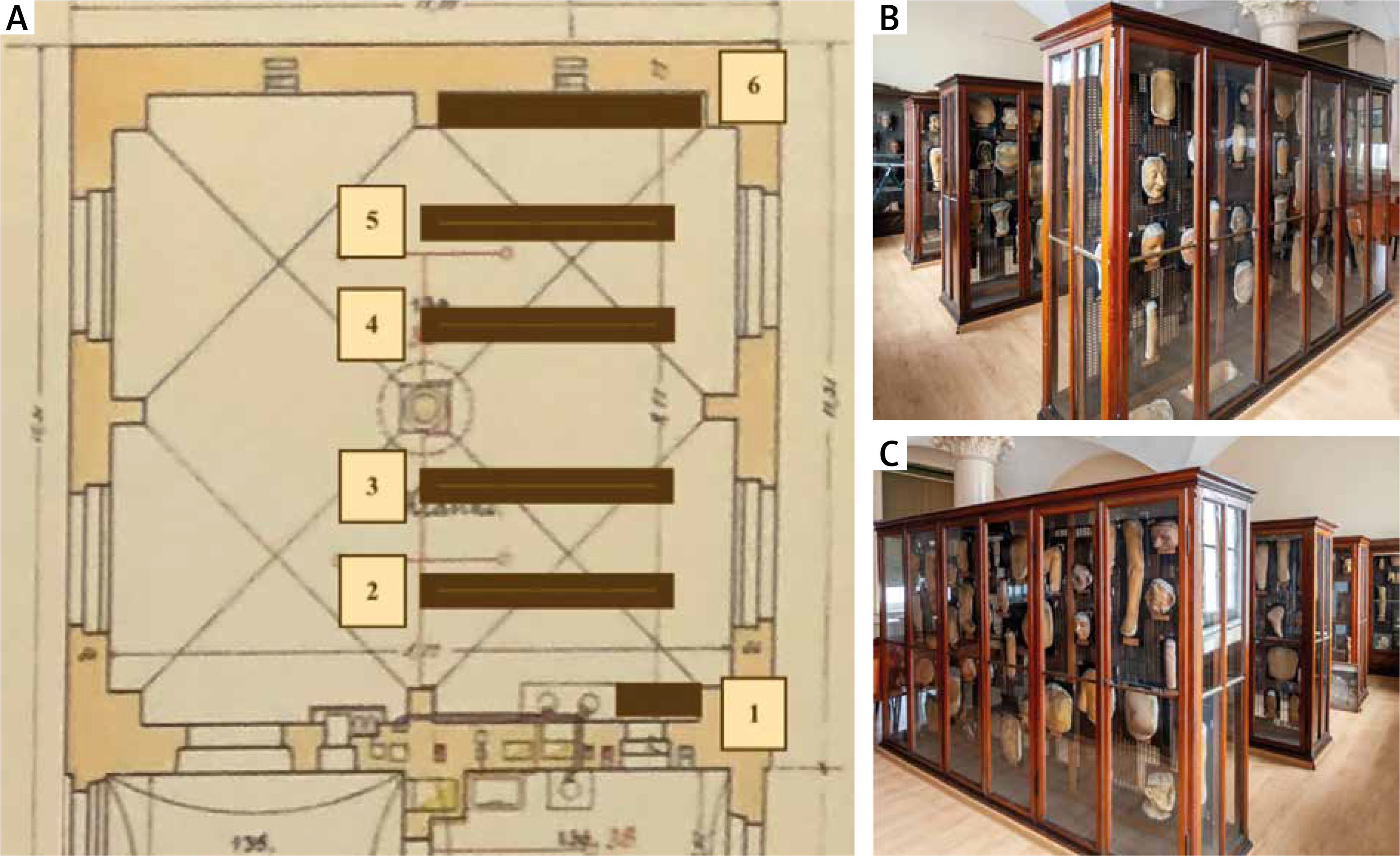
There are 323 original moulages that have survived to the present day, of which 315 are made of wax and 8 of plaster. They are presented in 6 original wooden display cases with glass. The layout of the museum has remained unchanged since its creation (Figure 1).
Figure 1
A – The Moulage Museum is a 9.7 × 9.7 m square with a barrel-vaulted ceiling supported by a 4.88 m high column in the centre of the room. The collections hang on perforated metal panels in 6 wooden display cases with glass. Showcases nos. 1 and 6 are set up against the walls to the right and left: showcase no. 1, with plaster moulages, measuring (width × height × depth) – 1.10 × 2.10 × 0.32 m; showcase 6, facing the lecture hall – 3.70 × 2.00 × 0.32 m (omitted from the study). The centre of the museum is occupied by four display cases (no. 2–5) measuring 3.00 × 1.92 × 0.6 m. B – View of showcases 1 to 4 (C) view of showcases 3 to 6
The protection and maintenance of this unique cultural heritage is a challenge for contemporary times. However, there are not many reports that systematize how to take care of dermatological moulages, although they are part of the collections of many medical universities in Europe [3].
Taking the above into consideration, the current study was undertaken to perform microbiological examinations of dermatological wax models as well as of the neighbouring environment of the museum hall. We aimed to investigate whether the collection of historic dermatological moulages is colonized by specific microorganisms and whether these microorganisms pose a health risk to the environment, including museum visitors. To the best of our knowledge, such studies including dermatologic wax moulages have not been performed so far.
Material and methods
The specimens for microbiological examinations (bacterial and mycological) were swabs obtained from (i) the surfaces of wax moulages, (ii) internal and external surfaces of the cabinets in which they were stored, and (iii) the walls and floor of the museum hall. The air samples taken from the museum’s exhibition room were examined. For technical reasons, showcase 6 and its collections were omitted from the study.
Collection and microbiological proceeding of surface samples
Five museum display cabinets (nos. 1–5) were included in the study. The samples were obtained from at least 5 moulages in each case. A total of 32 specimens, with a variety of textures and morphologies, were selected. In display case no. 1, specimens hanging at each height of the display case were selected. In showcases nos. 2–5, moulages were selected that were hung at different heights of the showcase, on either side of perforated boards.
A sterile swab (Equimed, Poland) moistened with sterile 0.9% sodium chloride solution was used to wipe in 3 different places the waxy surface of a single moulage. Swabs were also taken from the surfaces in the museum space: the upper outer surface of the display cases nos. 2–5 (3 swabs from 3 different display cases), the floor (3 swabs), and the walls (3 swabs taken from a height of 2 m). In addition, material was collected from the top surface of the cabinets nos. 1–5 using Rodac contact plates with Saboraud Dextrose Lab agar (BioMaxima). The imprint plates were opened and the agar side was applied to the top outer surface of each of the 5 cabinets for 10 s. Material from one of the impression plates was transferred to Columbia agar with 5% sheep blood (Becton Dickinson) to assess bacterial growth. All samples were transported to a laboratory within an hour and immediately seeded on microbiological media. From each swab, the material was successively inoculated onto the entire surface of the plate with Columbia agar supplemented with 5% sheep blood (Becton Dickinson) for bacteria and Sabouraud Dextrose Agar supplemented with chloramphenicol (Biomaxima, Lublin, Poland) for fungi. The plates were incubated either at 37°C for up to 3 days (bacteria) or at 28°C for up to 14 days (fungi). Every 24–48 h, the cultures were reviewed and the cultured colonies were identified in accordance with standard microbiological procedures (evaluation of colony morphology and microscopic examination – Gram staining for bacteria and lactophenol slides for fungi). For bacteria species identification was obtained using mass spectrometry (MALDI TOFF MS) and a Bruker MALDI Biotyper IVD instrument (Bruker).
Collection and microbiological proceeding of the air samples
Air was tested using a MicroBio MB1 (220) Bioaerosol Sampler (Cantium Scientific, UK) according to the manufacturer’s instructions. Sampling was carried out from 5 different parts of the museum. At each site, air samples were taken at a hight of 1.5 m sequentially onto an open plate with either Saboraud Dextrose Agar supplemented with chloramphenicol (BioMaxima, Lublin, Poland) for fungi (500 l of air per plate) or Columbia agar with 5% sheep blood (Becton Dickinson) for bacteria (300 l of air per plate), with a flow rate of 100 l/min. Incubation of plates and identification of cultured microorganisms was performed according to the standards described above. The cultures were observed every 24–48 h and the number of grown colonies was counted (each time, the fungal growth sites on the plate were marked to avoid counting secondary colonies). The number of fungi and bacteria in the air was calculated using MicroBio counts. The following formula was used for the calculations: CFU/m3 = 1000 × (nc/Vs), where nc is the corrected number of colonies on the plate, and Vs is the volume of the tested air in litres.
Results
Moulages
Positive moulage swab cultures were obtained from 9/32 (28%) of the tested samples. Microbial growth each time was sparse, from 8 samples grown 1–2 bacterial colonies and from a single mould colony (Table 1). All other cultures were negative. The predominant microorganism identified in the cultures was Micrococcus luteus.
Table 1
Results of the analysis of microbial contamination of the dermatological moulages and internal surfaces of the museum cabinets
| Cabinet number | Moulages | Internal surfaces of cabinets | ||
|---|---|---|---|---|
| Number of positive samples/number of samples tested | Identified microorganisms1 | Number of positive samples/number of samples tested | Identified microorganisms1 | |
| 1 | 3/6 | 2× Micrococcus luteus 1× unidentified fungi (hialohyphomycetes) | 1/1 | 1× Rhodotorula sp. |
| 2 | 3/7 | 2× Micrococcus luteus 1× unidentified Gram-positive bacilli | 4/5 | 2× Micrococcus luteus 1× unidentified Gram-positive cocci (species II) 1× unidentified Gram-positive cocci (species III) |
| 3 | 1/7 | 1× Bacillus pumilus | 2/2 | 1× Penicillium sp. 1× Micrococcus luteus |
| 4 | 1/5 | 1× Bacillus pumilus | 0/1 | – |
| 5 | 1/7 | 1× unidentified Gram-positive bacilli | 0/1 | – |
| Total (Nos. 1–5) | 9/32 (28%) | – | 7/10 (70%) | – |
Interior surfaces of the display cabinets
Microbial growth was obtained from 3/5 cabinets; positive results were obtained from more than half of the swabs taken (70%) (Table 1).
External surfaces of the cabinets
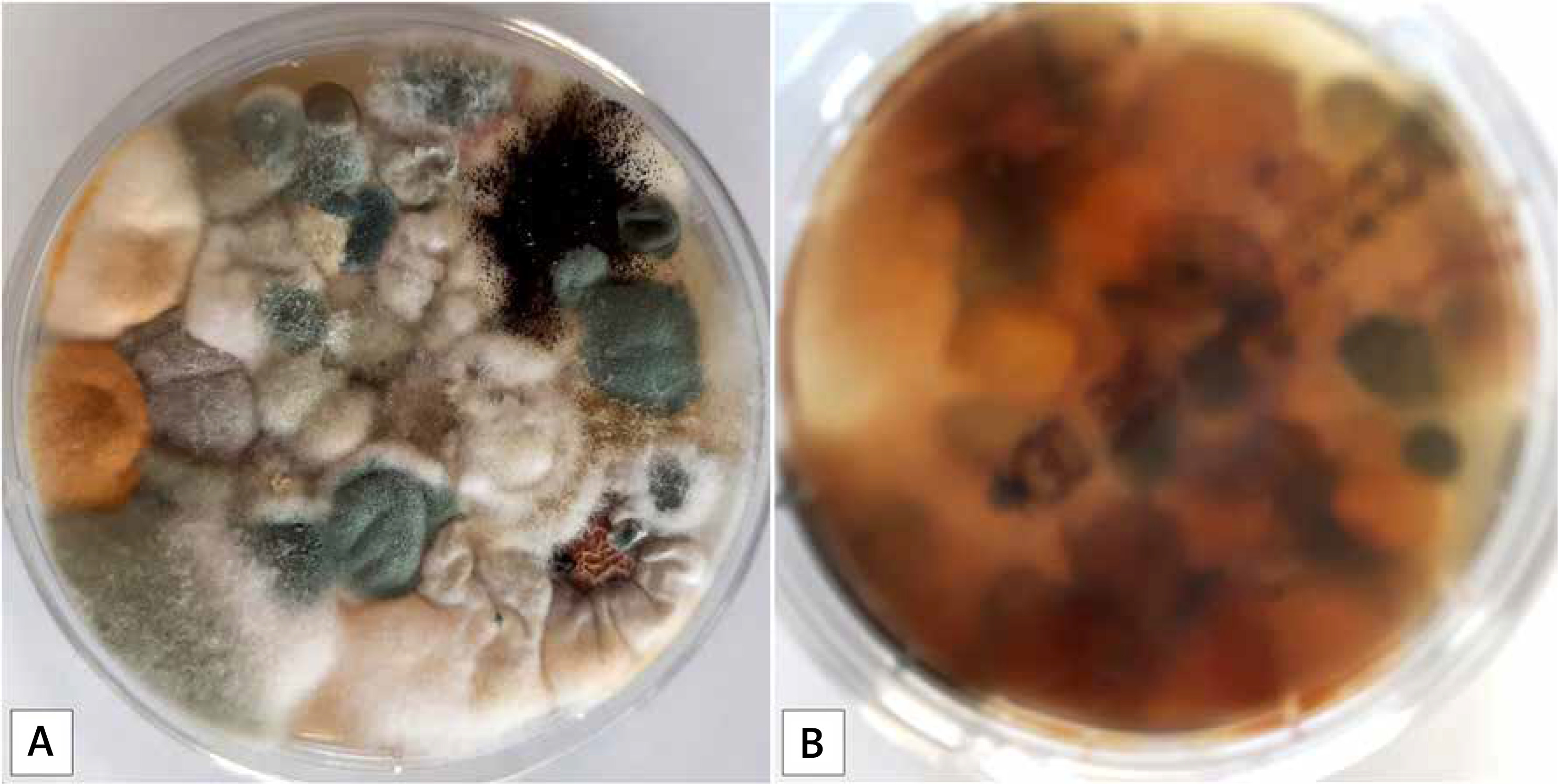
As a result of the examination of the swabs, bacterial growth was obtained from 2/3 and fungal from 3/3 display cases. From each positive sample 2–4 bacterial and 5–10 fungal colonies were obtained. Cultures of samples taken with imprint methods revealed abundant growth of fungi from all 5 showcases tested (at least 50 fungal colonies per 25 cm3 surface) (Table 2, Figure 2). In the culture obtained from the transfer of material to the microbiological medium the growth of numerous bacteria was achieved (Table 2).
Table 2
Results of the analysis of microbial contamination of the upper surfaces of museal cabinets. The results of cultures and direct impressions were included
| Cabinet number | Microorganism cultured from | |
|---|---|---|
| Swab sample | Imprint plate | |
| 1 | Swabs were not taken | Alternaria spp. |
| Cladosporium | ||
| Fusarium spp. | ||
| Mucor spp. | ||
| Penicillium spp. | ||
| Yeast | ||
| 2 | Acremonium spp. | Mucor spp. |
| Alternaria spp. | Other fungi1 | |
| Aspergillus spp. | ||
| Fusarium spp. | ||
| Pheohyphomycetes | ||
| Micrococcus luteus | Micrococcus luteus (strain 1) | |
| Micrococcus luteus (strain 2) | ||
| Paenibacillus lautus | ||
| Pseudomonas plecoglossicida | ||
| Solibacillus silvestris | ||
| Unidentified Gram-positive bacilli (species III) | ||
| Unidentified Gram-positive cocci (species III) | ||
| Unidentified Gram-positive polymorphic bacteria (species I) | ||
| Unidentified Gram-positive polymorphic bacteria (species II) | ||
| 3 | Cladosporium spp. | Mucor spp. |
| Fusarium spp. | Other fungi1 | |
| Penicilium spp. | ||
| Pheohyphomycetes | ||
| No bacterial growth | ||
| 4 | Penicillium spp. | Aspergillus niger |
| Aspergillus fumigatus | Alternaria spp. | |
| Chrysonila spp. | Cladosporium spp. | |
| Cladosporium spp. | Fusarium spp. | |
| Fusarium sp. | Penicilium spp. | |
| Hyalohyphomycetes | ||
| Micrococcus luteus | ||
| Paenibacillus pabuli | ||
| Unidentified Gram-positive bacilli (species II) | ||
| Unidentified Gram-positive cocci (species II) | ||
| 5 | Swabs were not taken | Aspergillus niger |
| Alternaria spp. | ||
| Cladosporium spp. | ||
| Fusarium spp. | ||
| Penicilium spp. | ||
Floor and walls of the museum
Bacterial and fungal growth was obtained in cultures from the floor swabs (Table 3, Figure 3). No bacterial and fungal growth was obtained in culture from swabs from the walls.
Table 3
Results of the analysis of microbial contamination of museal floor
Museum air
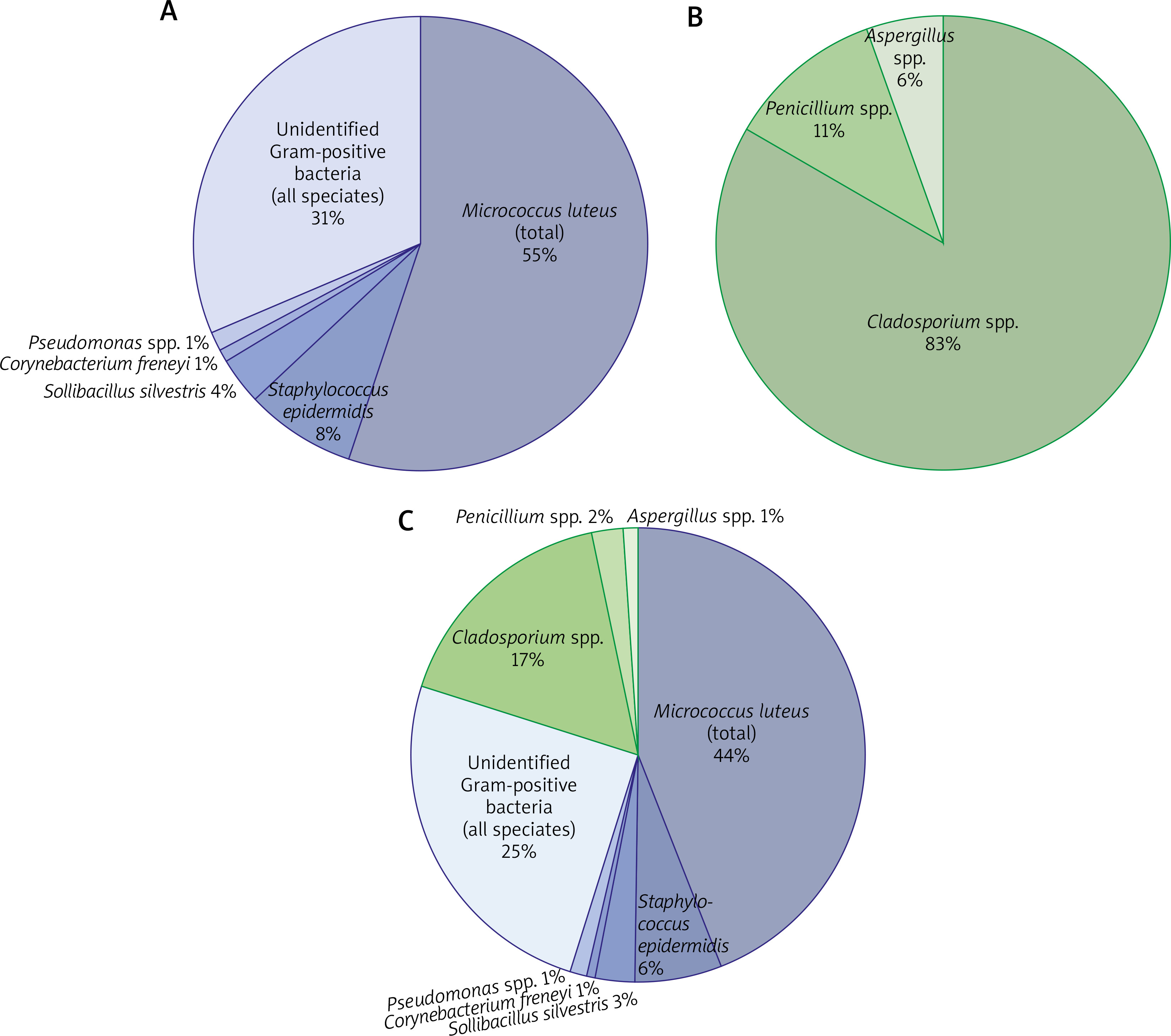
As a result of the analysis, both fungi and bacteria were isolated (Table 4). The mean number of bacteria in a cubic metre of air was 161 CFU/m3, and the number of fungi was 38 CFU/m3. The quantitative proportions of microorganisms in the air are shown in Figure 2.
Table 4
Results of microbial air pollution analysis
| Microorganisms | Number of colonies | Number of CFU/m3 | |||||
|---|---|---|---|---|---|---|---|
| Plate 1 (window 1) | Plate 2 (window 2) | Plate 3 (window 3) | Plate 4 (door) | Plate 5 (table) | Mean | ||
| Cladosporium spp. | 18 | 13 | 10 | 20 | 14 | 15 | 30 |
| Aspergillus spp. | 2 | 0 | 1 | 1 | 0 | 1 | 2 |
| Other fungi | 0 | 2 Naganishia albida3 (1 colony) (dematiaceous fungi)4 (1 colony) | 0 | 0 | 0 | 0 | 1 |
| Penicillium spp. | 3 | 2 | 1 | 2 | 2 | 2 | 4 |
| Micrococcus luteus (total)1 | 28 | 28 | 20 | 25 | 17 | 24 | 79 |
| Corynebacterium freneyi | 0 | 1 | 0 | 0 | 0 | 0 | 1 |
| Pseudomonas spp. | 0 | 1 Pseudomonas oryzihabitans | 1 Pseudomonas oryzihabitans | 1 Pseudomonas plecoglossicida | 0 | 1 | 2 |
| Sollibacillus silvestris | 4 | 3 | 0 | 0 | 0 | 1 | 5 |
| Staphylococcus epidermidis | 7 | 2 | 2 | 0 | 6 | 3 | 11 |
| Unidentified Gram-positive polymorphic bacteria | 3 | 3 | 6 | 3 | 0 | 3 | 10 |
| Unidentified Gram-positive bacilli (total)2 | 5 (II) | 9 (V) | 8 (II) | 10 (III) | 12 (II) | 9 | 29 |
| Unidentified Gram-positive cocci | 2 | 1 | 1 | 5 | 0 | 2 | 6 |
Discussion
The results obtained allow us to state that the space of the cabinets and the moulages protected in them are contaminated to a minor degree by several microorganisms. Probably the tight fit of the historic cabinet doors effectively isolates their interior environment.
It is puzzling why the studied elements were colonized mainly by one and the same species of bacteria, Micrococcus luteus. According to Young et al. [4], this microorganism is adapted to a limited ecological niche of mammalian skin, while its presence in other ecological niches (water and soil) may be the result of contamination of the epidermis. Contamination of the specimens could have occurred, for example, by touching the moulages with bare hands, but the absence of other identified representatives of the skin flora on these specimens indicates a higher probability of contamination from the ambient air. Obtaining a small number of positive cultures is intriguing. Perhaps the materials from which the castings were made contain substances that are toxic to microorganisms [5] or are colonized by microorganisms that cannot be identified by traditional culture methods.
The unique abilities of microorganisms enable them to survive in environments inaccessible to other living organisms [6]. For example, Bacillus pumilis, isolated from one of the moulages, is characterized by high resistance to UV radiation and H2O2 disinfecting properties [7]. It tolerates the lack of assimilation of nutrients in the environment of drying irradiation [8]. Meanwhile, M. luteus managed to grow from material preserved in amber from 120 million years ago. It is suggested that bacterial growth must have taken place while the resin was still liquid and therefore nutrients were available. Studying the bacteria’s genome allowed us to understand some of the enzymes it produces and, on this basis, deduce the metabolic pathways available to it. For example, thanks to succinate dehydrogenase, an isolated strain was able to biotransform succinic acid taken from the environment, one of the components of amber. It also assimilated other resin components such as terpenes [6]. One wonders whether the materials used to make the wax casts contained substances particularly favoured by this species, and what factors prevented it from colonizing the rest of the collection.
The technique of making moulages was a secret. An imprint (negative) was made of the patient’s skin using modelling plaster. A mixture of bee wax and various other organic and inorganic substances was poured into the mould thus prepared. The hardened cast was then coloured and “characterized” [9]. Siemiątkowski [10] emphasizes that the evidence of the use of different recipes of wax masses explains the variable durability of historic specimens observed today. In addition, he uses turpentine-based dyes in his conservation work, which may suggest that this natural solvent was used in the past to colour prepared moulds. Moreover, turpentine, along with alcohol, is an important solvent of bee wax [5], which may have been used to prepare the raw material.
It can therefore be suspected that the terpenes (a component of turpentine) contained in the moulages could be a potential food source for bacteria such as M. luteus, but the aseptic properties of bee wax and heavy metals contained in the pigments with which the wax was coloured prevented the survival of microorganisms. It is likely that the positive cultures came from specimens that were particularly contaminated with dust.
Studies of the museum environment showed a greater biodiversity of microorganisms than results from inside the display cases. Numerous florae were isolated in air samples and from swabs and impressions from the upper surfaces of the cabinets. Environmental bacteria and fungi, microorganisms forming the human skin flora (M. luteus, S. hominis, S. capitis), and pathogenic microorganisms were determined.
The identified bacteria of the genus Paebacillus sp. play an ambiguous role in the clinical context. They are likely to contaminate laboratory samples; however, some may also represent the true pathogens and cause nosocomial infections [11]. Similarly identified Acinetobacter ursinigii colonizes the hospital environment [12] and may be a casuistic cause of bacteraemia, even in immunocompetent patients [13]. Pseudomonas spp. bacteria are sometimes opportunistic human florae, and in conditions of immunosuppression or damage to natural barriers, it can be the aetiology of infections [14].
The air of the building of the local Department of Dermatology in Wroclaw, Poland has already been analysed for fungal spore contamination in a previous study. Our results indicate that the fungal diversity in the museum environment is very similar to that in other areas of the building. We isolated fungi from the genera Acremonium sp., Penicillium spp., Fusarium sp., Aspergillus spp., Mucor sp., Rhodotorula sp., similar to those documented by Łukaszuk et al. [15]. However, a comparison of the averaged quantitative data from both studies suggests that the museum air is less polluted than other areas of the same building (Table 5).
Table 5
Comparison of the microbiological contamination of the museum air with the data obtained by Łukaszuk and Her team
| Fungi | 1Mean number of colonies in museum air [volume of analysed air – 500l] | 1Mean number of colonies from other rooms in Dermatology Clinic verified by Lukaszuk [volume of analysed air – 100 l] |
|---|---|---|
| Cladosporium spp. | 15 | – |
| Penicilium spp. | 2 | 36 |
| Aspergillus spp. | 1 | 26 |
| Naganishia albida | 1 | – |
| Feohyfomycetes | 1 | – |
Some species of filamentous fungi have a negative impact on human health. Through colonization, they can cause superficial skin infections as well as systemic infections, especially in immunocompromised individuals. In addition, some mycotoxins released into the air or food are harmful to humans. They have various health effects: they are toxic to internal organs, carcinogenic, teratogenic, immunosuppressive, or endocrine disruptors. The occurrence of certain characteristic symptoms caused by exposure to mycotoxins is called mycotoxicosis [16]. Aspergillus fumigatus identified here is a source of mycotoxins such as fumagillin, gliotoxin, verruculogen, viriditoxin, and A. niger: malformin, oxalic acid, and ochratoxin A. Mycotoxins are released by fungi in response to specific environmental conditions such as temperature and humidity [17]. Therefore, simply identifying a fungus does not equate to exposure to its mycotoxins; for this to happen, the niche inhabited by the fungus must meet certain conditions and (probably) the amount of toxin released into the space must reach the appropriate concentration. The presented comparison (Table 5) shows that the spores of some fungi polluting the air in the museum (Penicilium sp. and Aspergillus spp.) were probably transferred from other rooms of the building.
Among the airborne fungi identified here, Cladosporium sp. was the most prevalent. This fungus is widely distributed on the earth. Its growth medium is paper and wood-based materials, so it commonly inhabits libraries and museums [18, 19]. Its spores contain numerous proteins that irritate the respiratory tract, causing symptoms of inhalant allergies (allergic rhinitis, sinusitis, conjunctivitis, mould asthma, allergic alveolitis) [20]. Cladosporium spp. are one of the common causes of allergy in museum employees who are constantly exposed to air contaminated with fungal spores [18, 19]. The occurrence of allergic reactions depends on the concentration of spores in the air and the individual sensitivity of the exposed organism. Different threshold concentrations of airborne spores causing allergic symptoms are proposed in the literature [21]. Studies on the Polish population have shown that a concentration of 2800 Cladosporium sp. spores in a cubic metre of air is necessary to induce allergic symptoms in predisposed individuals, and a value of 5000 in all tested subjects [22]. Thus, the number of spores determined in the museum air was not sufficient to cause disease symptoms.
Conclusions
Based on our results, one may hypothesize that the materials from which the moulages are made may prevent the development of microorganisms on their surfaces, and the few isolated microorganisms probably came from dirt (dust). However, when working with these exhibits, we recommend the use of gloves to avoid contamination of the specimens with skin flora, as well as to protect them from the destructive effects of sweat and heat, which can desulpastize the natural wax and damage the object. The main microorganisms isolated from the museum environment were bacteria constituting the natural flora of the skin (of which M. luteus was the most numerous) and fungi of the genus Cladosporium sp. Compared to other rooms in the building, the air in the museum was slightly polluted with fungal spores. Working with moulages or visiting the museum do not pose a significant health risk, but individual findings of Aspergillus spp. fungi in the air (probably transferred from other rooms of the building) require further scrutiny. Air analysis can be used as a monitoring tool. The probable sudden appearance of new species or the observation of changes in the proportions of microorganisms in the air can provide valuable information about the condition of museum objects and health risk factors. Very few studies are devoted to the microbiology of medical school museums. Their location near medical facilities makes their biodiversity unique. Similarly, only a few studies dedicated to the protection of dermatological moulages are available [2, 10]. Articles on the conservation of artwork could be helpful in the care of these collections [23] as well as other rare studies performed, for example, on wax anatomical models [24]. This gap in the literature suggests that heritage medical collections need more attention from the scientific community, particularly with regard to designing solutions for their protection and a better understanding of their properties, especially in an interdisciplinary context [25, 26].


