
Introduction
Alopecia areata (AA) is a chronic, organ-specific autoimmune disease that can affect any hair-bearing body area [1]. Characterized by the sudden onset of round or oval-shaped patchy hair loss, the precise pathogenesis of AA remains unclear [2]. It has been proposed that dysregulations in various immune system pathways contribute to the development of AA in genetically predisposed individuals exposed to environmental factors [3]. When AA involves complete hair loss on the scalp, including the eyebrows and eyelashes, it is classified as alopecia totalis (AT); when it extends to all body hair, it is referred to as alopecia universalis (AU) [2].
Nailfold capillaroscopy (NFC) is a non-invasive technique used to assess microcirculation, particularly in rheumatologic diseases [4]. Beyond its application in nailfold evaluation, NFC has also been utilized in dermatological conditions such as psoriasis, rosacea, and Behçet’s disease [5–7].
Microvascular abnormalities have been implicated in the pathogenesis of AA, with proangiogenic factors, particularly vascular endothelial growth factor (VEGF), thought to play a critical role. Studies have demonstrated reduced vascularization and VEGF synthesis in affected skin, suggesting that diminished VEGF levels in hair follicles may contribute to disease onset and progression. Furthermore, VEGF levels and capillary vessel density have increased in scalp keratinocytes following diphenylcyclopropenone (DPCP) treatment in patients with AA [8–10].
Despite the unclear pathogenesis of AA, few studies in the literature have explored its association with microcirculation. The present study aims to investigate the relationship between skin microcirculation and AA using NFC.
Aim
The aim of this study was to evaluate microcirculatory changes in patients with AA using nailfold capillaroscopy.
Material and methods
Patients aged 18–65 years with a confirmed diagnosis of AA who presented to the dermatology outpatient clinic of our hospital, regardless of prior treatment history, were included in the study. Exclusion criteria comprised pregnancy and lactation, a known history of connective tissue diseases (including systemic lupus erythematosus, systemic sclerosis, dermatomyositis, and polymyositis), diabetes mellitus, ischemic heart disease, pulmonary disease, hypertension, and chronic venous insufficiency. Additionally, patients receiving vasoactive therapy or anticoagulants, those with nicotine or alcohol dependence, individuals with chronic microtrauma to the fingers (including recent manicure within the past 2 weeks), patients whose nailfold capillaries could not be adequately visualized due to skin thickness, and those diagnosed with primary or secondary Raynaud’s phenomenon or acrocyanosis were excluded.
After signing the informed consent form, data on age, gender, comorbidities and disease duration were collected. The severity of scalp involvement in patients with AA was assessed using the Severity of Alopecia Tool (SALT) score. The SALT score quantifies the percentage of scalp hair loss by dividing the scalp into four regions (vertex: 40%, right profile: 18%, left profile: 18%, occipital: 24%) and calculating the weighted sum of hair loss in each area. Based on this score, patients were classified into subgroups: S1 (≤ 25% hair loss) and S2 (26–50% hair loss). The results of routinely performed laboratory tests – including hemogram, glucose, vitamin B12, iron, iron-binding capacity, ferritin, thyroid function tests, thyroid autoantibodies, anti-nuclear antibody (ANA), and Venereal Disease Research Laboratory (VDRL) tests were recorded. The nail folds of the 2nd–5th fingers on both hands were assessed using Dino-Lite Edge digital capillaroscopy (P/N: AM7515MZT, made in Taiwan) following a 20-minute rest period at a room temperature of 22°C. Initial examination was conducted at 50× magnification, followed by assessments at 100×, 150×, and 200× magnification. At 200× magnification, a photograph was taken from/for each of the eight fingers, and the images were stored in the DinoCapture 2.0 Windows program. Capillary parameters including arterial diameter, venous diameter, width, length, internal diameter, apex diameter, and loop diameter were quantitatively measured using the saved images. Capillary distribution, density, and morphological changes (e.g., hairpin-shaped, tortuous, crossed, meandering, elongated, dilated, bushy, ramified, bizarre, and giant capillaries), as well as avascular areas, neoangiogenesis, and microhemorrhages, were evaluated qualitatively and semi-quantitatively. Capillary density was scored based on the number of capillaries per millimetre (0 points = > 9 capillaries/mm, 1 point = 7–9 capillaries/mm, 2 points = 4–6 capillaries/mm, 3 points = < 4 capillaries/mm) for each examined finger, with the arithmetic mean calculated accordingly [11]. Capillaroscopic patterns were classified using the Pavlov-Dolijanovic et al. system, and the capillary distribution scoring system was based on the criteria established by Cutolo et al. and Ingegnoli et al. [12–14].
Statistical analysis
Statistical analyses were performed using SPSS 26.0 for Windows, with descriptive measures presented as mean, standard deviation, and percentage distribution. The suitability of the data for normal distribution was determined using the Kolmogorov-Smirnov test. The comparison of continuous variables was executed through the utilization of the Student’s t-test when parametric conditions were met, the Mann-Whitney U test when parametric conditions were not met, and the χ2/Fisher’s exact test analysis for the comparison of distributions. The significance level was established at p < 0.05.
Results
The present study included 50 patients and 50 healthy volunteers in the control group. Forty-seven patients had alopecia areata, and three had alopecia universalis. The AU patients were excluded from the study due to getting systemic treatment. The present study included 47 patients and 50 healthy volunteers in the control group. The patient group comprised 20 (42.6%) women and 27 (57.4%) men, while the control group consisted of 22 (44%) women and 28 (56%) men. The mean age was 32.4 ±10 years in the patient group and 33 ±8.8 years in the control group. The duration of the disease was also examined, finding that 10 (21.3%) patients had the disease for less than a month, 22 (46.8%) for 1 to 6 months, and 15 (31.9%) for more than 6 months. Only 1 (2.13%) patient had nail changes. Furthermore, it was determined that 20 (42.6%) of the patients received treatment, while 27 (57.4%) did not receive treatment. The demographic characteristics and investigative findings of the patient group are presented in Table 1. Disease severity was evaluated using the Severity of Alopecia Tool (SALT) score in patients with scalp involvement. Of the 35 patients with scalp alopecia areata, 28 were classified as S1 (SALT < 25), and 7 as S2 (SALT ≥ 25).
Table 1
Demographic and laboratory data
[i] ESR – erythrocyte sedimentation ratio, CRP – C-reactive protein, Vit. B12 – vitamin B12, TIBC – total iron-binding capacity, fT3 – free triiodothyronine 3, fT4 – free triiodothyronine 4, TSH – thyroid-stimulating hormone, Anti-TPO – anti-thyroid peroxidase antibodies, Anti-TG – anti-thyroglobulin antibodies.
The mean capillary density was significantly higher in the patient group (8.27 ±0.77 capillary/mm) compared to the control group (9.01 ±0.98 capillary/mm), with a p-value of less than 0.001. Similarly, the capillary density score was higher in the patient group (0.89 ±0.25) compared to the control group (0.57 ±0.37), again with a p-value less than 0.001. The NFC findings from our study are presented in Table 2.
Table 2
Capillaroscopic data of patient and control groups
In the AA group, mildly irregular capillary distribution (n = 20/47; 43%) was the most common type of capillary distribution, followed by normal capillary distribution (n = 16/47; 34%), completely irregular distribution (n = 8/47; 17%), and moderately irregular distribution (n = 3/47; 6%). In the control group, normal capillary distribution (n = 39/47; 78%) was the most prevalent type of capillary distribution.
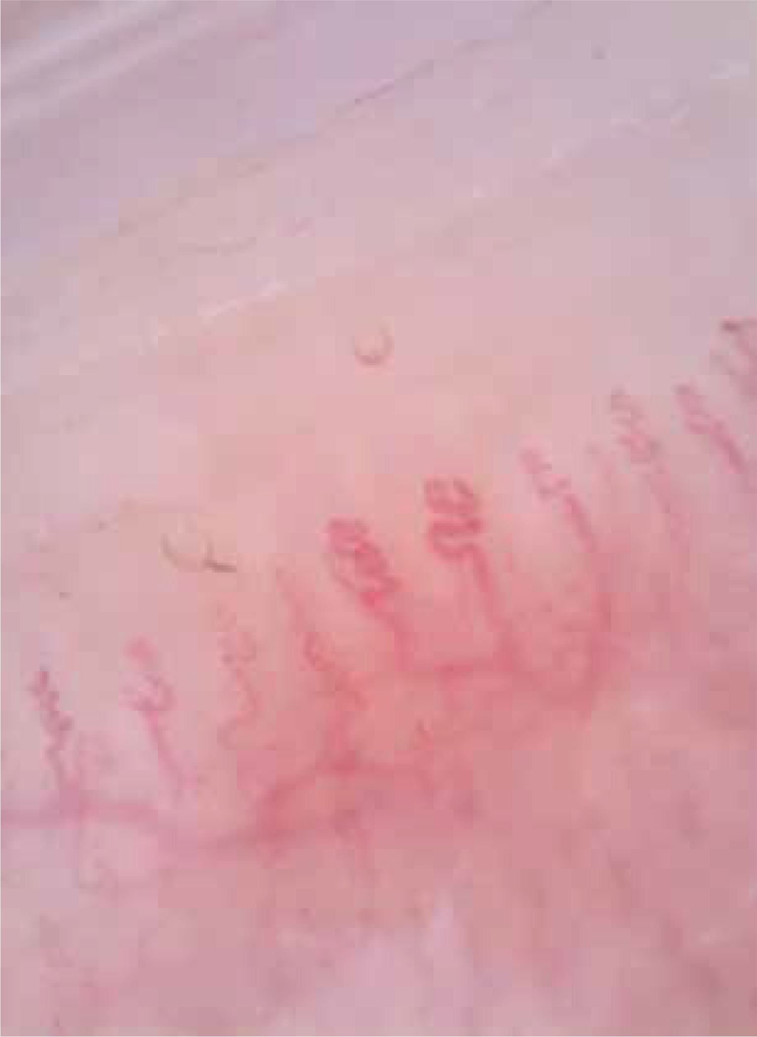
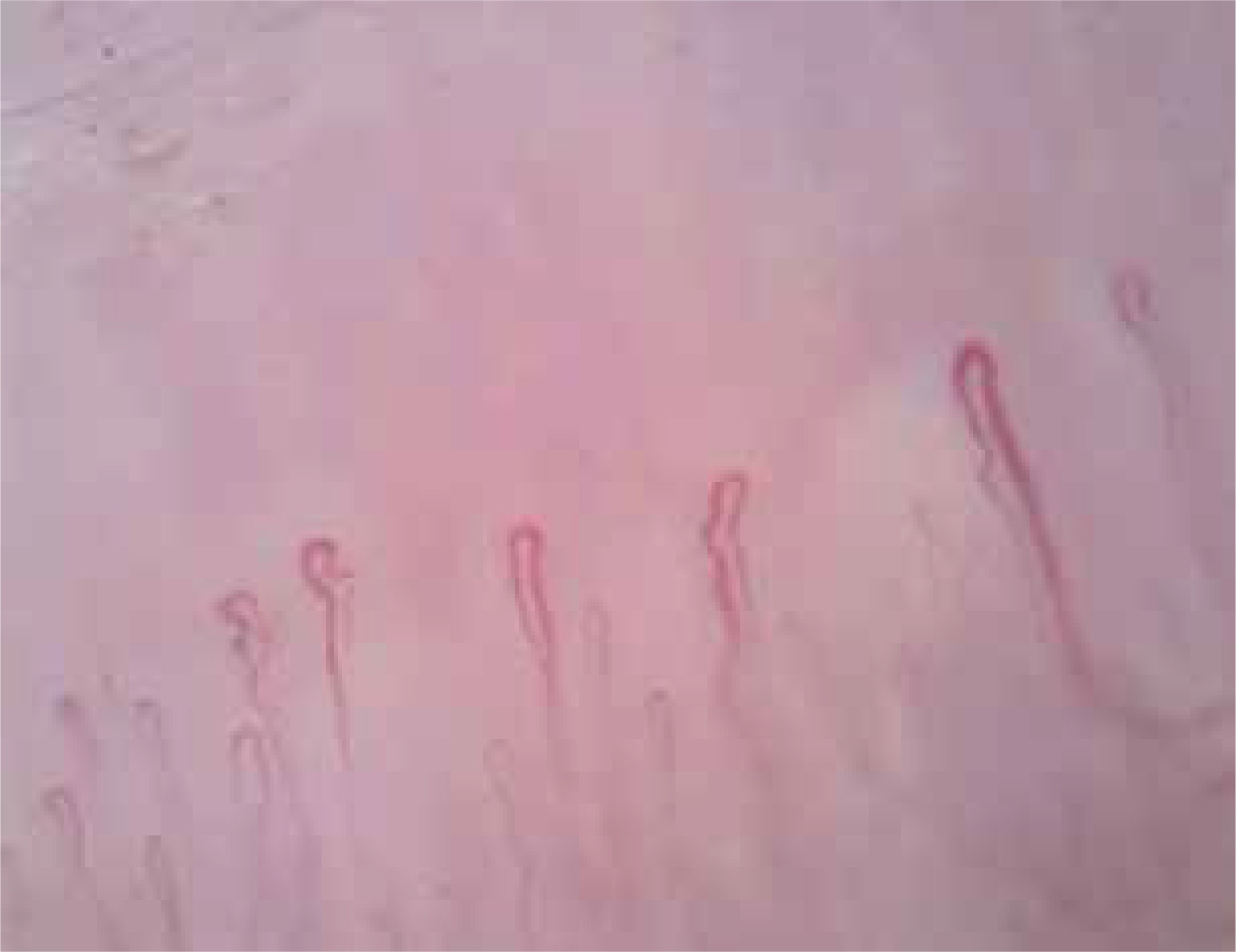
A microhemorrhage was observed in 21% (n = 10) of the patient group and 14% (n = 7) of the control group. Neovascularization was detected in eight patients (17%) in the patient group but not in the control group, and this difference was found to be statistically significant (p = 0.002) (Figure 1). An avascular area was found in three patients (6%) in the patient group but not in the control group (Figure 2).
With regard to the capillaroscopic findings, hairpin-shaped capillaries were observed in 98% of both the patient and control groups. Bizarre capillaries were identified in 12 (26%) patients in the patient group and 11 (22%) individuals in the control group. Notably, no giant capillary was detected in either group. No statistically significant differences were observed between the patient and control groups with respect to other morphologic changes (tortuous, crossed, meandering, elongated, dilated, bushy, or ramified) defined capillaroscopically.
An evaluation of the capillaroscopy findings in terms of pattern analysis revealed that none of the participants in the patient and control groups were included in the scleroderma pattern. In the patient group, 38% (n = 18) exhibited a nonspecific capillaroscopic pattern, while 62% (n = 29) demonstrated a normal capillaroscopic pattern. Conversely, in the control group, 26% (n = 13) exhibited nonspecific capillaroscopic patterns, while 74% (n = 37) demonstrated normal capillaroscopic patterns.
When patients with scalp involvement were classified according to the SALT score, 28 patients were found to be in the S1 group and 7 in the S2 group. Capillaroscopic findings were compared between these two subgroups; however, no statistically significant differences were observed. The results are presented in Table 3.
Table 3
Comparison of capillaroscopic findings according to the SALT score
Discussion
It has been posited that microvascular distribution may also play a role in AA, the pathogenesis of which remains to be elucidated [8, 9]. Although NFC is predominantly utilized in rheumatologic diseases for the evaluation of microcirculation, it has also been employed in diseases such as psoriasis, Behçet’s disease, and rosacea [6, 7, 15]. The number of studies evaluating peripheral microcirculation by nail bed capillaroscopy in AA is limited [8].
Capillary density, capillary density score, and capillary distribution are critical microvascular parameters evaluated using nail bed capillaroscopy [4]. Normal capillary density does not vary significantly according to age or gender [16]. A decrease in capillary density has been observed, especially in connective tissue diseases such as systemic sclerosis [17]. In a study conducted on normal, healthy people in India, capillary density was found to be 7.63 capillary/mm [18]. A capillaroscopic examination of psoriasis patients revealed a significant decrease in capillary density compared to the control group [19].
Gerkowicz et al. reported a decrease in capillary density among AA patients in a videocapillaroscopic study [8]. The study revealed that 22.53% of the AA patient population exhibited altered capillary distribution, while 77.46% demonstrated regular capillary distribution, and the control group exhibited regular capillary distribution [8]. In the study by Cao et al., androgenetic alopecia (AGA) patients and the control group were compared, and the capillary density was found to be similar in both groups [16]. In the same study, 3.8% of capillaries were found to be disorganised in AA patients [16]. Our study revealed a significant decrease in capillary density among AA patients compared to the control group. A statistically significant difference was observed in capillary density score between the patient and control groups. The study also revealed that normal capillary distribution was observed in 78% of the control group and 34% of the AA group. The observation that decreased capillary density and impaired capillary distribution were more pronounced in the patient group underscores a potential link between AA and microcirculation. To the best of our knowledge, our study is the inaugural report from our country that has evaluated capillary density, capillary density score, and capillary distribution in AA. Further studies may offer insights into the relationship between NFC use, microcirculation, and disease in certain dermatologic conditions, such as AA.
The typical structure of nail bed capillaries in healthy individuals is a hairpin or inverted U-shaped [20]. It is thought that minor abnormalities are also observed in the community in addition to normal capillary structure [21]. In a study conducted by Karabay and Demirel on rosacea patients, hairpin-shaped capillaries were observed at a rate of 35.5% in rosacea patients and 78.4% in the control group [7]. In a systematic review, Lazar et al. reported that hairpin-shaped vessels were less prevalent in psoriatic arthritis patients [5]. In our study, hairpin-shaped capillaries were identified in 98% of the patient and control groups, suggesting a potential association with psoriatic arthritis. The observation that hairpin-shaped capillaries, which are considered part of the normal physiological pattern, were also found to be high in the patient group indicates the need for further studies to elucidate the relationship between microvascular changes and AA.
Apart from hairpin-shaped capillaries, some variations within the normal pattern have also been described (tortuous, crossing) [4, 22, 23]. In the study by Gerkowicz et al., the rate of tortuous capillaries was found to be statistically higher in the patient group compared to the control group [8]. In NFC performed on AGA patients, tortuous and crossed capillaries were observed at similar rates in the patient and control groups [16]. A study on rosacea patients reported higher rates of tortuous and crossed capillaries in the patient group compared to the control group [7]. However, our study did not reveal a statistically significant difference in the rates of tortuous and crossed capillaries between AA patients and the control group.
In NFC, nonspecific patterns other than normal and scleroderma patterns may be challenging. Meandering capillaries are considered in the nonspecific pattern and have been demonstrated in normal healthy people [18, 22]. In the NFC study conducted on normal healthy people in India, the meandering capillary rate was reported to be 44.66% [20]. In our study, meandering capillaries were found to be at a rate of 13% in AA patients and 4% in the control group. The higher rate of meandering capillaries in the patient group may be significant but not specific. We think that future studies with a larger number of AA patients will provide detailed information on this subject.
The presence of bizarre, elongated, and bushy capillaries in the nonspecific pattern alone has no predictive value. However, the presence of more than one of these abnormalities in a person may be a sign of connective tissue disease. Bushy capillaries formed as a result of neoangiogenesis are also considered within the spectrum of scleroderma [4, 22, 23]. It has been reported that bizarre and bushy capillaries may be commonly observed in AGA patients [16]. Jakhar et al. found bizarre (2%) and bushy capillaries (4%) in healthy people [18]. Bizarre capillaries, elongated capillaries, and bushy capillaries were found in the patients and control groups in our study. In the anamnesis and dermatologic evaluation of these patients, no findings related to connective tissue disease were found, and ANA values were negative. In addition, these patients were enlightened about possible scleroderma-specific diseases and were taken for follow-up. These results in our study may indicate that microvascular changes may play a role in the pathogenesis of AA.
Giant capillaries, microhaemorrhage, avascular areas, and neoangiogenesis are not expected to be seen in healthy individuals. These are thought to be a part of the scleroderma pattern [22]. Giant capillaries are interpreted as an early indicator of systemic sclerosis [24]. Avascular areas are associated with tissue hypoxia and have been found in diseases such as scleroderma and systemic lupus [4, 25]. Neoangiogenesis is considered an indicator of the effort to compensate for the capillary loss that develops as a result of tissue hypoxia [25]. Microhaemorrhages are not expected in normal healthy people but can sometimes be seen if there is a history of trauma [22]. Despite this information, these capillaroscopic findings have been reported to be seen in normal, healthy people. In the study conducted by Gorasiya et al. in healthy people, no avascular areas and giant capillaries were found, but neoangiogenesis was found in 38.66% of the cases [20]. In NFC studies performed in healthy people, microhaemorrhages were reported in 48% and 14% of the studies [18, 26]. In a study by Cao et al. in patients with androgenetic alopecia, no giant capillary was detected, but 9% avascular areas were found in 9% of patients [16]. In the same study, microhaemorrhages were observed in both AGA and control groups (37.2% vs. 20.5%) [16]. Karabay and Demirel reported the presence of microhaemorrhages and avascular area in the control group, with a higher incidence in patients with rosacea [7]. Santhosh et al. identified avascular areas in psoriasis patients and the control group [19]. In our study, avascular area was detected in 6% (n = 3) of AA patients, and neoangiogenesis was observed in 17% (n = 8) but not in the control group. The presence of giant capillaries was not observed in either the patient or control group. Microhaemorrhages exhibited a higher prevalence in the patient group, though they were also present in the control group (21% vs. 14%). No scleroderma pattern was identified in any patient in the study. These findings may offer valuable insights for future research involving larger patient series, particularly regarding the role of microvascular disorders in the pathogenesis of AA.
Although patients with scalp involvement were classified into S1 and S2 subgroups based on the SALT score, no statistically significant differences were found in capillaroscopic findings between the two groups. This may be attributed to the limited number of patients in the S2 group, which could have reduced the statistical power to detect potential differences.
Studies evaluating the relationship between microvascular changes and AA are scarce in the existing literature. When our data were analysed, statistically significant differences were found in terms of capillary density, capillary density score, neoangiogenesis, and capillary distribution. We think that the data we found as a result of our capillaroscopic study in AA, the pathogenesis of which is still unknown, will set an example for other studies.
The most important limitations of this study are the inability to demonstrate whether the capillaroscopic findings of the patients were influenced by systemic treatment and the lack of comparison with nail findings. Our study includes a limited sample group. In the future, the relationship between nail bed capillaroscopy and microcirculation in AA can be demonstrated with a larger number of patient groups.










