
External ear melanoma (EEM) is a rare type of melanoma (approximately 1% of all cutaneous melanomas) [1] with higher likelihood of an invasive nature compared to other head and neck melanomas [2]. According to previous data, superficially spreading melanoma is the dominant subtype of the EEM, followed by lentigo maligna and nodular subtype, respectively [1, 3, 4]. Patel et al. [5] discovered that EEM prevalence among young adults is currently higher by 111.9% when compared to former data. Herein, we report a nodular melanoma of the external ear in a middle-aged female, which was successfully removed with simple surgical procedure.
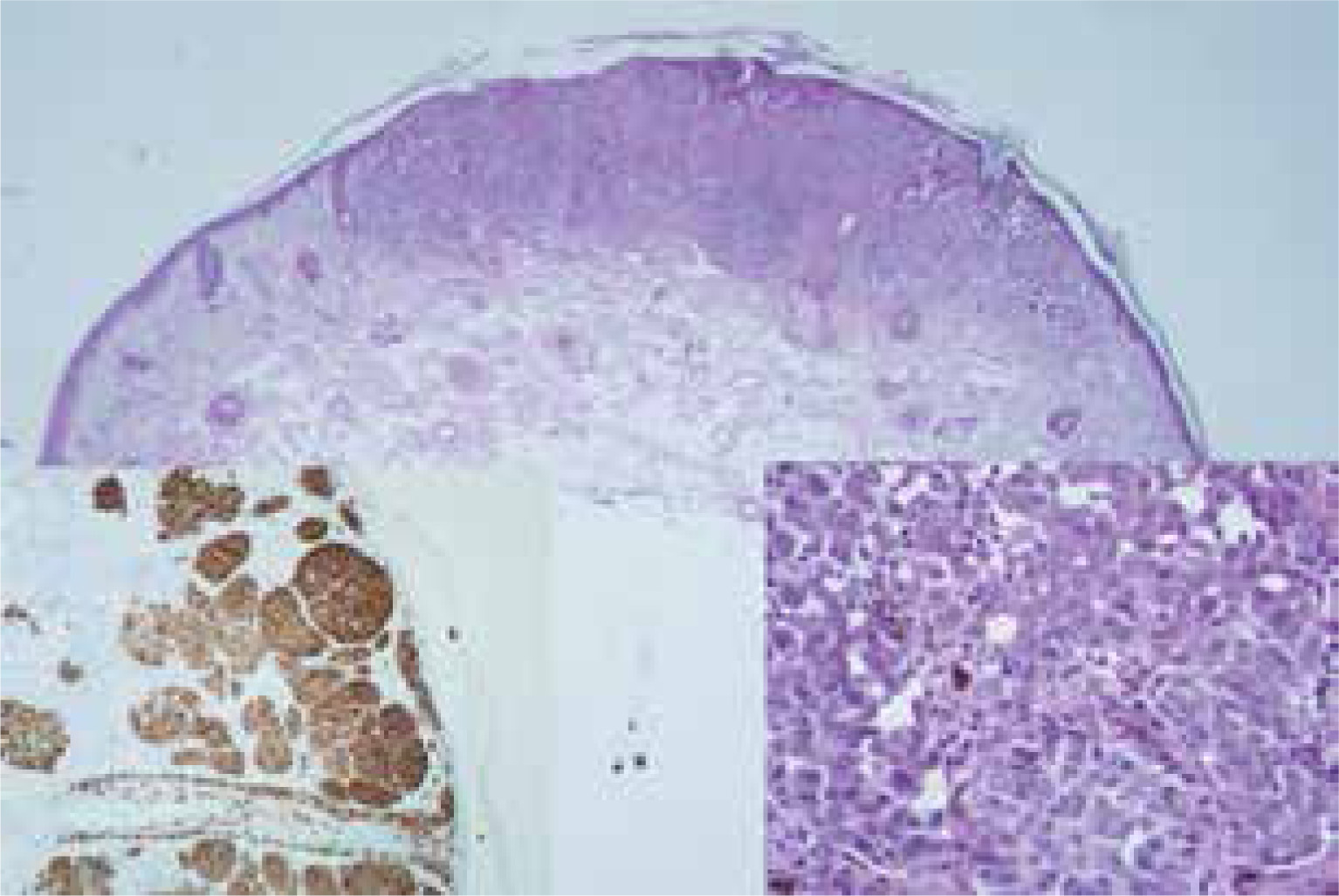
A 43-year-old female patient was admitted to the outpatient clinic with pigmented nevus on the left helical rim (Figure 1). The lesion was noticed by a family member about a month before. The woman’s family medical history was free of melanomas or other skin neoplasms. According to Fitzpatrick classification, the patient had phototype no. 2, and in the past she had experienced numerous sunburns. During consultation a 5 × 6 mm nodular skin lesion placed in the middle of a 10 × 6 mm pigmented mark was revealed in a dermatoscopy, with multiple signs suggesting its malignant character, such as asymmetry, irregular border, multiple range of colours from plain on the outside to the redness on the inside, dark-brownish colour on the lump, and a milky-pink background (Figure 1). The dermoscopic examination revealed a multi-element pattern, blue-whitish blur, small black bodies at the border, and peppering. There was no ulceration or satellite metastasis seen near the main lesion. Regional lymph nodes were not enlarged in physical examination. According to the recommendations of the Polish Society of Oncologic Surgery, the excisional biopsy was performed with 2 mm margins as a first step of treatment [6]. The elliptical wound was closed with 5.0 nonabsorbable sutures. The histological picture (Figure 2) presented with 1 mm depth infiltration of malignant cells in the Breslow scale. Other histological features were Clark level no. 3, no ulceration, brisk type of lymphocytic infiltration, mitotic activity 1 mitosis/mm2. Moreover, an immunohistochemical investigation was done, in which Melan A (+), HMB 45 (+), S100 (+), and Ki67 (15% expression) in hot spots were found. The margins of the resected lesion were free from melanoma cells (from 1.2 to 3 mm). The clinical diagnosis of nodular melanoma of the external ear was confirmed, and the patient was referred to the oncology ward to undergo sentinel node identification process and subsequently biopsy or dissection. Unfortunately, she was lost to follow-up.
Figure 1
Dermoscopic view of nodular melanoma. Nodular melanoma before the excision. After the excision of nodular melanoma
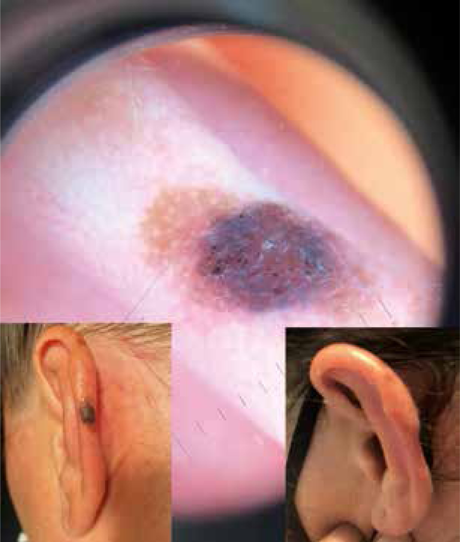
Figure 2
Histological images of nodular melanoma. Haematoxylin and eosin staining 20× – whole lesion. Haematoxylin and eosin staining 400× with cellular nuclear atypia and single mitotic phase figure visible. Melan A immunohistochemical staining with melanocytic cells visible
According to the available data, the most common localization of EEM is the ear helix – particularly the anterior one [3] – as in the described patient. Moreover, some authors pointed out that ear localization had lower survival rate than melanomas located on other parts of the body [1–3].
Surgery (with or without lymph node dissection) is still the most recommended treatment option [1, 5, 6]. Reduction of the excision margins with preservation of the underlying perichondrium and cartilage, if possible, does not affect the overall survival and is considered to have better functional and aesthetic outcomes [1, 7]. A minimal excision margin of 1 cm is usually recommended, while the reconstructive techniques depend on the surgeon’s preferences. In our case the patient was fully satisfied with the cosmetic result, therefore no reconstruction was needed.
Unfortunately, EEM localization is difficult for self-checking, which leads to delayed diagnosis and treatment. As reported by Oliver et al. [2], the mean time from spotting the lesion to the introduction of therapy was approximately 2 weeks for EEM and other head and neck melanomas. That is why it has been suggested that an education campaign about this topic should be addressed to barbers and hairdressers in order to report skin lesions as soon as possible and therefore to refer the patient to undergo a quick diagnostic process and treatment [6]. Surprisingly, the education level did not cause a higher detection rate of EEM [2].
The presented case shows that simple excision could still be a valuable tool for experienced surgeon in terms of therapeutic and cosmetic results among patients with MM.


