Introduction
Brachydactyly is a very rare congenital defect involving shortened fingers and/or toes [1]. It can be a component of congenital or genetic syndromes (e.g., Rubinstein-Taybi syndrome, Down syndrome, Albright hereditary osteodystrophy); however, it can also occur as an isolated inherited defect or as a result of a de novo mutation. Over the years, many monogenic brachydactyly disorders have been described, most of which are inherited in an autosomal dominant pattern. According to the literature, short stature may be associated with some types of the disease [2].
Historically, the first descriptions of the types of brachydactyly were developed by Bell in 1951, and a comprehensive classification was published in 1979 [3]. It distinguished five main types (A–E) based on the characteristic constellations of the affected fingers and bones (Figure 1) [1, 2].
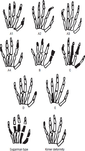
In the European population, the most common type of the disease is brachydactyly type A1 (BDA1), which involves shortening or absence of the middle phalanges [4]. BDA1 is caused by a mutation in the Indian hedgehog (IHH) gene, which encodes a protein belonging to the hedgehog signaling pathway responsible for regulating organogenesis. The IHH gene encodes a paracrine regulator of endochondral ossification, influencing not only the normal development of the bones of the hands and feet but also the overall course of growth. In other ethnic groups, brachydactyly type A3 (BDA3), a hand anomaly characterized by shortening only of the middle phalanx of the fifth finger, may be more common. BDA3 results from a mutation in the HOXD13 gene, a member of the homeobox gene family, encoding transcription factors that also play an important role in morphogenesis [5]. According to the literature, children with BDA1 and BDA3 and short stature respond well to recombinant human growth hormone (rhGH) therapy [6–8].
Short stature can also occur in brachydactyly type C (BDC) [9, 10]; however, data regarding rhGH treatment in children with this condition are difficult to find in the literature. BDC is characterized by brachymesophalangy of the second, third, and fifth fingers, with possible hypertrophy of the index and middle fingers [1, 2, 9, 10]. However, the fourth finger is typically normal and remains the longest finger in patients with this defect. The condition results from a mutation in the growth differentiation factor-5 (GDF5) gene, encoding a protein responsible for cartilage and bone formation (transforming growth factor β, TGF-β). In addition to short stature, BDC can also be associated with broad toes, abnormal tooth eruption, delayed wrist development, and shortened forearms and lower legs (mesomelia) [1, 2, 9, 10].
This paper presents a case report of a child with brachydactyly and short stature, as well as a review of the literature on the treatment with growth hormone (GH) of such patients.
Bioethical standards
The patient’s parents provided written consent for the publication of the case report and use of the photographs in the manuscript. This consent has been documented and is available upon request. The patient’s identity has been fully anonymized to ensure confidentiality. Approval was also obtained from the Ethics Committee at the Medical University of Warsaw (number AKBE/283/2025).
Case report
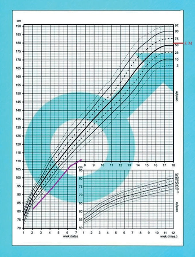
A 7-year-old boy was initially admitted to the pediatric endocrinology department for the diagnosis of short stature. The boy had no significant medical history, no known chronic diseases, and a normal nutritional status (BMI 17.1 kg/m2); also, he did not meet the criteria for hypotrophy at birth (e.g., small for gestational age, intrauterine growth retardation, SGA/IUGR). Available documentation indicated that from the age of two, the child’s growth curve had consistently been below the third percentile (according to the percentile charts for the population of Polish children [11]), with a clear tendency to deviate and deepen the height deficit (Figure 2). At the time of diagnosis, the standard deviation score (SDS) was –3.1 (more than 6 cm below the third percentile).
Figure 2
Growth chart showing the boy's growth before treatment (based on the charts for the population of Polish children by Palczewska and Niedźwiedzka [11])
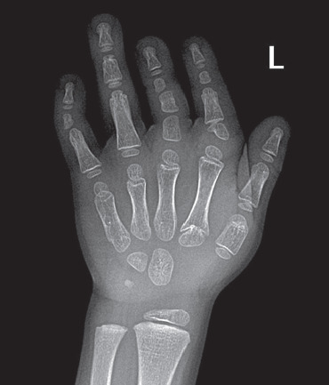
On physical examination, the boy was found to have brachydactyly in both hands, in this case involving shortened fingers I, II, III, and V (Figure 3). X-rays of the wrist bones revealed shortened metacarpal bones and hypoplastic phalanges (Figure 4), and a bone age approximately 3 years younger than chronological age. The child’s mother had the same hand presentation.
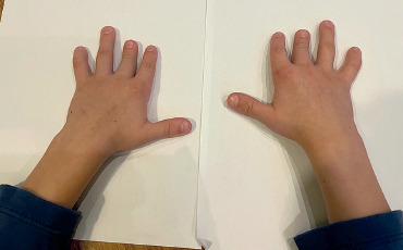
Figure 4
X-ray of the patient's hand. The image shows: shortening of the first metacarpal bone, hypoplastic phalanges of fingers II, III, and V, an extra phalanx of finger III, and deformation of the base of the proximal phalanx of finger II
A complete diagnostic workup for short stature was performed. Laboratory tests ruled out anemia, confirmed normal liver, kidney, and thyroid function, and showed no ion imbalances. Glucose and insulin levels remained within the normal range. Celiac disease and parasitic infestations were ruled out. Echocardiography revealed no evidence of heart defects. Insulin-like growth factor 1 (IGF-1) concentrations, monitored several times, remained at the lower limit of normal (48.6–74.6 ng/ml), while a glucagon stimulation test (at a dose of 0.03 mg/kg body weight) resulted in normal GH release, with a maximum concentration of 10.9 ng/ml at the 90th minute (Table I).
Table I
Result of the glucagon stimulation test at a dose of 0.03 mg/kg body weight. The table shows normal (> 10 ng/ml) growth hormone secretion at the 90th and 120th minute of the test
| minute 0 | minute 60 | minute 90 | minute 120 | minute 150 | minute 180 | |
|---|---|---|---|---|---|---|
| Growth hormone concentration [ng/ml] | 5.2 | 7.2 | 10.9 | 10.1 | 9.8 | 7.5 |
| Glycemia [mg/dl] | 83 | 132 | 72 | 49 | 67 | 79 |
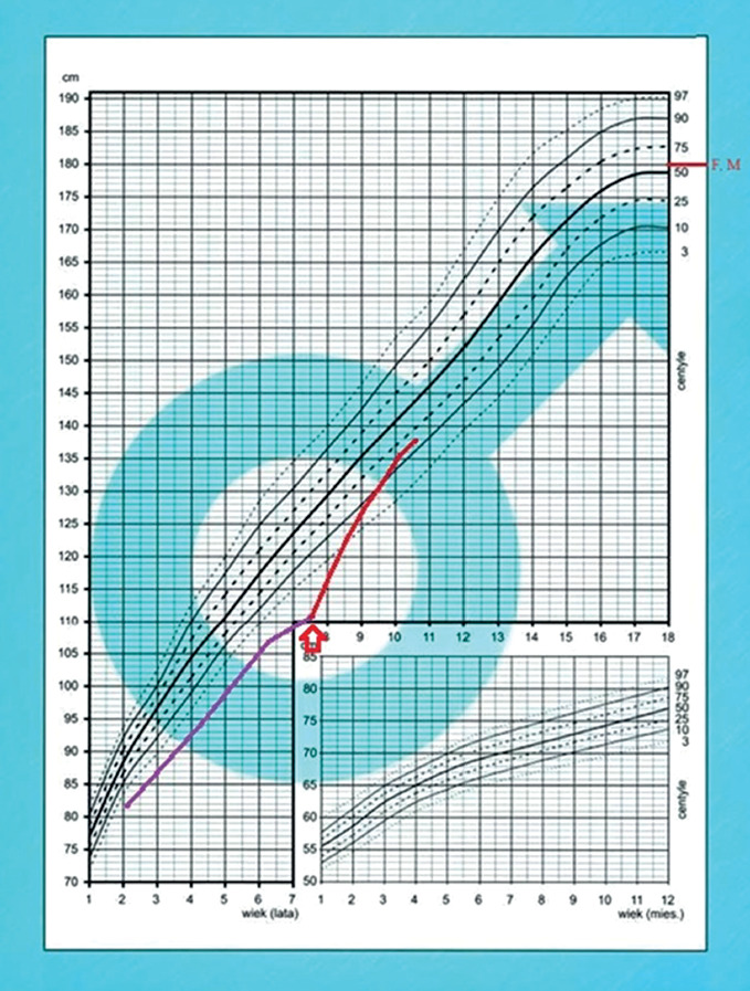
After conducting the literature review and an expert consultation, it was decided to begin rhGH treatment of the child in gradually increasing doses from 0.180 to 0.210 mg/kg/week, with excellent results. The boy tolerates the therapy well, and after three years of treatment, his height has exceeded the 10th percentile (–1.0 SDS, change of the SDS +2.1), with an average annual growth velocity of approximately 9 cm/year (the results are shown in Table II and in Figure 5). During the treatment, no IGF-1 concentrations above the normal range (Table II), significant progression in the child’s bone age, or any disturbances in carbohydrate or lipid metabolism were observed.
Table II
Results of 3 years of recombinant human growth hormone therapy of the patient with brachydactyly type C
Figure 5
The boy’s growth chart after starting recombinant human growth hormone; the red arrow marks the beginning of therapy (based on the charts for the population of Polish children by Palczewska and Niedźwiedzka [11])
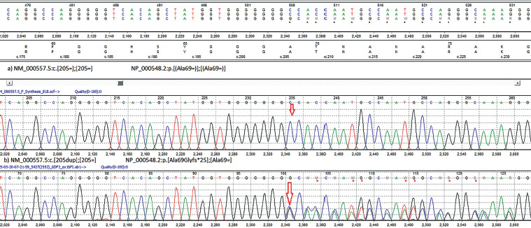
The child also underwent genetic testing. Using the Sanger next-generation sequencing method, a mutation in the IHH gene (BDA1) was excluded, then a pathogenic variant in the GDF5 gene was found – the c.205dup variant (p.Ala69Glyfs*25) was identified in a heterozygous pattern – typical for BDC, which allowed the correct diagnosis to be made in the child (Figure 6).
Genetic testing (methods)
The genomic DNA of the proband was extracted from whole blood using NucleoSpin Dx Blood (Macherey-Nagel, Düren, Germany). The purity of the DNA was checked using a spectrophotometric method with a Nanodrop instrument (Thermo Fisher Scientific, Waltham, United States of America).
Sequencing of the GDF5 gene was performed using the Sanger method. The analysis was performed using the Mutation Surveyor software Version 5.1.0 (SoftGenetics, State College, United States of America).
Using the Sanger method, the heterozygous c.205dup variant in GDF5 was identified. This variant causes a frameshift and introduces a premature STOP codon. The variant is registered in the Single Nucleotide Polymorphism Database (rs753691079), classified as pathogenic in the Clinical Variation Database, and reported in the Human Gene Mutation Database as associated with BDC. The c.205dup variant has also been described in Online Mendelian Inheritance in Man (OMIM) (601146.0009) and in the literature in patients with a clinical diagnosis (PMID: 9288091, PMID: 33333243). According to the American College of Medical Genetics and Genomics classification, the identified variant is pathogenic.
The GDF5 gene (OMIM *601146) is located on chromosome 20q11.22 and encodes a secreted ligand of the TGF-β superfamily of proteins. Mutations in this gene are associated with acromesomelic dysplasia, brachydactyly, chondrodysplasia, multiple synostoses syndrome, proximal symphalangism, and susceptibility to osteoarthritis. BDC (OMIM #113100) is inherited in an autosomal dominant manner.
Discussion
Treatment with rhGH of patients without GH deficiency remains a widely debated issue in the scientific community. On the one hand, concerns about the risks and side effects of such therapy are raised, including persistently elevated serum IGF-1 concentrations and possible excessive bone age progression with shortened effective growth time [12]. On the other hand, certain genetic syndromes or diseases with normal GH secretion (e.g., Turner syndrome, Silver-Russell syndrome, short children with SGA/IUGR) have been widely recognized worldwide as indications for rhGH therapy, where good results and safety of such treatment have been proven [13, 14].
Establishing a cutoff point for normal GH secretion in stimulation tests (including insulin, glucagon, clonidine, and arginine tests) is another widely discussed issue. In many countries, a peak value for GH concentration above 7 ng/ml is considered normal [12, 15]. However, in some countries (including Poland), a concentration above 10 ng/ml is considered normal [16, 17].
In the discussed case, the patient’s glucagon stimulation test revealed a GH concentration above 10 ng/ml, which raises no doubts about the child’s normal secretion of this hormone. However, a separate question remains whether decreased GH sensitivity occurs in brachydactyly, where bone and cartilage development is disturbed (this includes abnormal growth plate organization), and whether such children require higher serum hormone concentrations than their healthy peers (similar to, for example, Turner syndrome, where brachydactyly also occurs).
On the other hand, diagnosing short stature is a complex process that involves not only measuring GH concentrations in tests but also considering IGF-1 concentrations, as only this approach allows for a comprehensive assessment of the pituitary-GH-IGF-1 axis and its impact on the child’s development. In the discussed case, IGF-1 levels remained low (at the lower end of the normal range), with normal GH secretion and a healthy nutritional status. All of this suggests a good response to rhGH treatment of this boy.
Taking all of this into account and the fact that the described patient had a profound height deficiency (over –3 SDS), it was decided to implement rhGH therapy, carefully and frequently monitoring possible side effects, IGF-1 concentration (every 3 months; the values remained normal, Table II) and carbohydrate metabolism parameters (including an oral glucose tolerance test once a year), lipid profile (every 6 months), and bone age (once a year).
The decision was made following a literature review, which was subsequently regularly updated. Data were collected from the literature on rhGH treatment for patients with brachydactyly and separately on BDC. Articles from the PubMed/Medline, Embase, and Scopus databases were searched for the years 1983–2025 to identify studies describing the effects of rhGH therapy in patients with isolated brachydactyly and short stature. Exclusion criteria included articles on genetic syndromes associated with brachydactyly (e.g., Turner syndrome, Albright hereditary osteodystrophy). A total of 168 articles were analyzed, of which only three concerned rhGH therapy for isolated brachydactyly: two papers on the effects in BDA1 [6, 7] and one paper on BDA3 [5]. All three studies reported a good response to treatment and a low risk of adverse events.
No papers were found regarding rhGH therapy in BDC, so the review was expanded to include all articles regarding BDC available in the above-mentioned databases, analyzing a total of 51 papers, among which no articles regarding the effects of GH therapy in BDC patients were identified.
Conclusions
To our best knowledge, this article is the first documented example of the effects of rhGH therapy in a short child with BDC. It is therefore important to emphasize that we have demonstrated both an excellent response and the safety of such treatment in the course of BDC.
Furthermore, the discussed clinical case and the literature review conducted suggest that rhGH therapy should be carefully considered in patients with height deficiency and brachydactyly, even despite normal GH secretion values in stimulation tests. This may suggest the presence of decreased GH sensitivity in these patients, which, however, requires further research and in-depth analysis. However, this does not change the view that such treatment is effective and, equally importantly, safe and well tolerated by patients.