Introduction
Laparoscopic surgery was introduced for colorectal cancer treatment in the 1990s [1–4] and today is performed worldwide. Minimally invasive surgery (MIS), including laparoscopic surgery, was adopted for colorectal cancer treatment because it provides rapid postoperative recovery due to less surgical trauma and guarantees oncological safety. Thus, the scope of MIS is widening, and several different modalities have been developed over the past decades, such as laparoscopic surgery, robotic surgery, natural orifice transluminal endoscopic surgery, and single-incision laparoscopic surgery (SILS).
SILS for colon cancer was first reported in 2008 [5] and for rectal cancer in 2010 [6], and since then clinical studies have been conducted to establish its safety and feasibility for colorectal surgery. Although some meta-analyses have been performed on the topic, the studies were heterogeneous because both colon and rectal cancers were included. Rectal cancer surgery is more technically challenging than colon cancer surgery because space is limited in the narrow pelvic cavity. Before adopting SILS for rectal cancer surgery, we need to determine whether this technical issue could be overcome to guarantee surgical and oncological safety.
Aim
We performed this meta-analysis to compare SILS and conventional laparoscopic surgery (CLS) regarding operative and pathologic outcomes and to verify the feasibility and safety of SILS for rectal cancer treatment.
Material and methods
Search strategy
This meta-analysis followed the Preferred Reporting Items for Systematic Reviews and Meta-Analyses (PRISMA) statement [7]. The PubMed, Embase, CENTRAL, and Web of Science databases were searched systematically until November 2021. Search terms included rectal cancer, rectal carcinoma, rectal neoplasm, single incision, single port, single access, single site, laparoscopic, and laparoscopy. Additional articles from references provided in previous systematic reviews were added. After database searching, duplicates were removed, and the identified articles were screened by reviewing titles and abstracts. Then, full texts of screened articles were reviewed, and ineligible articles were excluded. Two authors (G Kim and KY Lee) independently conducted the screening and review and decided on the articles included in the meta-analysis. Disagreements were resolved by discussion.
Eligibility criteria and outcomes of interest
Eligibility criteria included randomized controlled trials (RCTs) or controlled clinical trials comparing outcomes of single-incision versus conventional laparoscopic surgery for rectal cancer. Exclusion criteria were studies on reduced port laparoscopic surgery or single-incision plus one-port laparoscopic surgery. The primary outcome was an overall perioperative complication rate. The secondary outcomes included operative outcomes (operative time, blood loss, conversion rate, incision length, and ileostomy rate), postoperative outcomes (mortality, complications, hospital stay, reoperation, readmission, postoperative pain, postoperative analgesics requirement, and bowel motility recovery), and pathologic outcomes (number of harvested lymph nodes, specimen length, resection margins, positive circumferential margin, and mesorectal grade).
Risk of bias assessment
Risk of bias of the selected studies was assessed using the Cochrane Collaboration tool for assessing risk of bias [8] for randomized controlled trials and the Risk of Bias Assessment Tool for Nonrandomized Studies (RoBANS) [9] for controlled clinical trials. The Cochrane Collaboration’s risk of bias tool evaluated seven independent sources of bias: i) random sequence generation, ii) allocation concealment, iii) blinding of participants and personnel, iv) blinding of outcome assessment, v) incomplete outcome data, vi) selective reporting, and vii) other bias [8]. The RoBANS tool evaluated 6 independent sources of bias: i) selection of participants, ii) confounding variables, iii) measurement of exposure, iv) blinding of outcome assessments, v) incomplete outcome data, and vi) selective outcome reporting [9]. For the studies included in the meta-analysis, the sources of bias were assessed as being of high, low, or unclear risk.
Data extraction
Two authors independently extracted relevant data from the eligible full-text articles. Extracted data included identification information (name of first author, year of publication, country where the study was conducted, study design, type of surgery, sample size, and follow-up period), patients demographics (age, gender, body mass index (BMI), the American Society of Anesthesiologists (ASA) score, and history of previous abdominal surgery), operative outcomes (operative time, blood loss, conversion, incision length, additional trocar insertions, and ileostomy), postoperative outcomes (perioperative mortality, complications, hospital stay, reoperation, readmission, postoperative pain score, analgesic requirements, recovery of gastrointestinal motility, and diet build-up), pathologic outcomes (number of harvested lymph nodes, tumour size, specimen length, length of resection margins, number of positive margins, mesorectal grade, and R0 resection rate), and oncologic outcomes (overall survival (OS) and disease-free survival (DFS)). The extracted data were cross-checked for discrepancies.
Statistical analysis
Pooled effects are presented as mean differences and 95% confidence intervals for continuous variables and as odds ratios and 95% confidence intervals for dichotomous variables. Heterogeneity among studies was measured using I2 statistic: I2 = 100% × (Q – df)/Q, where Q is Cochran’s heterogeneity statistic, and df is the degree of freedom [10]. When the included studies showed high heterogeneity, a random-effects model was applied for meta-analysis, and when they presented low heterogeneity, a fixed-effects model was applied. The statistical analysis was performed using Review Manager 5.4.1.
Results
Article search results
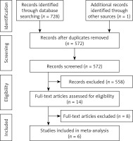
Database searching identified 728 articles (161 from PubMed, 220 from Embase, 118 from CENTRAL, and 329 from Web of Science), and additional searching, as described in the search strategy, added one article. After removing duplicates from the 729 articles using citation manager, 572 articles remained. Title and abstract reviews resulted in the exclusion of 556 irrelevant articles. A full-text review revealed that one of the remaining 16 articles was written in Chinese, and this article was excluded because full text was not available in English. In addition, one duplicate not detected by the citation manager was also excluded. Thus, we reviewed the full text of 14 articles (Table I) [11–24]. Two studies by Sirikurnpiboon [11, 12] had overlapping study populations, and only the most recent study was included for analysis. Also, 2 studies by Tei et al. [14, 15] had overlapping populations, and the most recent study was included. Three studies conducted in Denmark [17–19] had overlapping populations, and only the most recent study (a randomized controlled study) was included. The other 2 articles were a non-randomized study and a poster abstract. A study by Bracale et al. [20] included benign diseases other than rectal cancer and thus was also excluded. Finally, 3 Korean studies [21–23] shared the same population and included sigmoid and rectosigmoid colon cancer, and thus all 3 were excluded. After exclusions, 6 eligible studies remained for meta-analysis [11, 13, 14, 16, 17, 24]. A flow diagram of the article selection is provided in Figure 1.
Table I
A list of the 14 full-text reviewed studies
| Publication year | Author | Country | No. of patients | Study period | Disease besides rectal cancer | Final selection | |
|---|---|---|---|---|---|---|---|
| SILS | CLS | ||||||
| 2021 | Sirikurnpiboon [11] | Thailand | 41 | 43 | 2011–2014 | – | Included |
| 2016 | Sirikurnpiboon [12] | Thailand | 35 | 36 | 2011–2014 | – | Excluded |
| 2020 | Jiang et al. [13] | China | 51 | 51 | 2013–2017 | – | Included |
| 2018 | Tei et al. [14] | Japan | 44 | 49 | 2011–2015 | – | Included |
| 2015 | Tei et al. [15] | Japan | 50 | 50 | 2010–2014 | – | Excluded |
| 2018 | Nerup et al. [16] | Denmark | 12 | 41 | 2009–2012 | – | Included |
| 2015 | Bulut et al. [17] | Denmark | 20 | 20 | 2011–2012 | – | Included |
| 2014 | Levic and Bulut [18] | Denmark | 36 | 194 | 2009–2012 | – | Excluded |
| 2013 | Levic et al. [19] | Denmark | 40, in total | not presented | – | Excluded | |
| 2015 | Bracale et al. [20] | Italy | 21 | 21 | 2010–2012 | Benign diseases | Excluded |
| 2014 | Kim et al. [21] | Korea | 67 | 49 | 2006–2013 | Sigmoid and rectosigmoid cancer | Excluded |
| 2013 | Choi and Lee [22] | Korea | 31 | 49 | 2006–2013 | Excluded | |
| 2013 | Choi [23] | Korea | 31 | 49 | 2006–2013 | Excluded | |
| 2013 | Sourrouille et al. [24] | France | 13 | 32 | 2008–2012 | – | Included |
Characteristics of included studies
Table II shows the characteristics of the 6 studies included in the meta-analysis [11, 13, 14, 16, 17, 24]. These studies included a total of 417 patients who were enrolled in 6 clinical trials. All articles were published between 2013 and 2021. Reported outcomes included; i) operative outcomes (operative times, blood losses, conversions to laparotomy, incision lengths, additional trocar insertions, and ileostomy rates), ii) postoperative outcomes (perioperative mortality, perioperative complications, hospital stays, reoperation and readmission rates, pain scores, analgesic requirements, recovery of gastrointestinal motility, and diet build-up), iii) pathologic outcomes (numbers of harvested lymph nodes, specimen length, length of resection margin, circumferential resection margin (CRM) involvement, mesorectal grade, and R0 resection), and iv) oncologic outcomes (OSs, DFSs, and recurrence rates) (Table III).
Table II
Characteristics of the included studies
| Study ID | Country | Type of procedure | Study type | No. of patients, n | Age [years] | Male, n (%) | BMI [kg/m²] | ASA ≥ III, n (%) | Previous abdominal surgery, n (%) | Neoadjuvant CRTx, n (%) | Tumour size [mm] | Follow up period [months] | |||||||||||||||||
|---|---|---|---|---|---|---|---|---|---|---|---|---|---|---|---|---|---|---|---|---|---|---|---|---|---|---|---|---|---|
| SILS | CLS | SILS | CLS | P-value | SILS | CLS | P-value | SILS | CLS | P-value | SILS | CLS | P-value | SILS | CLS | P-value | SILS | CLS | P-value | SILS | CLS | P-value | SILS | CLS | P-value | ||||
| 2021 Sirikurnpiboon [11]* | Thiland | LAR, AR, ISR, APR | NCT | 41 | 43 | 63.97 ±13.05 | 61.74 ±12.03 | NS | 19 (46.3%) | 20 (46.5%) | NS | 22.20 ±4.00 | 23.14 ±2.89 | NS | 6 (14.6%) | 3 (7.0%) | NS | – | – | – | – | – | – | – | – | – | – | – | – |
| 2020 Jiang [13]** | China | TME | NCT | 51 | 51 | 62 (15) | 62 (14) | NS | 30 (58.8%) | 27 (52.9%) | NS | 23.56 (3.52) | 23.18 (3.39) | NS | 5 (9.8%) | 8 (15.7%) | NS | 12 (23.5) | 14 (27.5) | NS | – | – | – | 30 (20) | 30 (20) | NS | 32.6 (18.3) | 36.8 (23.6) | NS |
| 2018 Tei [14]* | Japan | LAR | NCT | 44 | 49 | 67.3 ±9.3 | 65.0 ±10.1 | NS | 29 (65.9%) | 29 (59.2%) | NS | 23.6 ±3.5 | 22.0 ±3.4 | 0.019 | 7 (15.9) | 3 (6.1) | NS | 13 (29.5) | 13 (32.7) | NS | – | – | – | 39 ±18 | 41 ±19 | NS | 40 (5–63) | 51 (12–70) | 0.017 |
| 2018 Nerup [16]*** | Denmark | APR | NCT | 12 | 41 | 76 (59.5–82.5) | 69 (59–76) | NS | 5 (41.7%) | 28 (68.3%) | NS | 23.5 (19.3–24.8) | 25 (22.5–28.0) | NS | 1 (8.3) | 12 (29.3) | 0.04 | 2 (16.7) | 14 (34.1) | NS | 6 (50%) | 30 (73.2%) | NS | 20 (19.5–27.5) | 25 (20–32.5) | NS | 9 (5.5–16.5) | 14 (8.5–24) | NS |
| 2015 Bulut [17]*** | Denmark | LAR, APR, Hartmann | RCT | 20 | 20 | 69 (50–86) | 73 (50–84) | NS | 8 (40%) | 8 (40%) | NS | 24 (16–32) | 24 (19–29) | NS | 3 (15) | 3 (15) | NS | 3 (15) | 7 (35) | NS | 7 (35%) | 4 (20%) | NS | 25 (10–70) | 40 (20–75) | 0.026 | 12 (6–18) | 15 (6–20) | NS |
| 2013 Sourrouille [24]** | France | TME | NCT | 13 | 32 | 60 (56–65) | 61 (53–64) | NS | 8 (61.5%) | 19 (59.4%) | NS | 23.0 (21.8–23.8) | 24.9 (22.7–26.5) | NS | 2 (15.4) | 4 (12.5) | NS | – | – | – | 9 (69.2%) | 22 (68.8%) | NS | – | – | – | – | – | – |
*** . BMI – body mass index, ASA – the American Society of Anesthesiologists, CRTx. – chemoradiation therapy, SILS – single-incision laparoscopic surgery, CLS – conventional laparoscopic surgery, LAR – low anterior resection, AR – anterior resection, ISR – intersphincteric resection, APR – abdominoperineal resection, TME – total mesorectal excision, NCT – non-randomized clinical study, RCT – randomized controlled study, NS – non-specific.
Table III
Reported outcomes from the included studies
| Study ID | Operative outcomes | Postoperative outcomes | Pathologic outcomes | Oncologic outcomes | |||||||||||||||||||||||
|---|---|---|---|---|---|---|---|---|---|---|---|---|---|---|---|---|---|---|---|---|---|---|---|---|---|---|---|
| Operative time | Blood loss | Conversion to laparotomy | Incision length | Additional trocar(s) | Ileostomy | Perioperative mortality | Perioperative complications | Hospital stay | Reoperation | Readmission | Pain score | Analgesics requirements | Bowel movement recovery | Diet build up | Harvested lymph nodes | Specimen length | PRM | DRM | CRM | CRM involvement | Mesorectal grade | R0 resection | Follow up period | OS | DFS | Recurrence | |
| 2021 Sirikurnpiboon [11] | + | + | – | – | – | – | – | + | + | – | – | + | – | + | – | + | – | – | + | + | + | – | – | + | + | - | |
| 2020 Jiang [13] | + | + | + | + | + | – | + | + | + | – | + | + | – | – | + | + | – | + | + | – | + | – | – | + | + | + | + |
| 2018 Tei [14] | + | + | + | – | + | – | + | + | + | + | – | – | – | – | – | + | – | + | + | – | + | – | – | + | + | + | + |
| 2018 Nerup [16] | + | + | + | – | – | – | + | + | + | – | + | – | – | – | – | + | + | – | + | + | – | + | – | + | – | – | - |
| 2015 Bulut [17] | + | + | + | + | – | + | – | + | + | + | + | + | + | – | – | + | + | – | + | + | – | + | + | + | – | – | - |
| 2013 Sourrouille [24] | + | + | + | – | + | + | + | + | + | + | – | + | + | – | + | + | – | – | – | – | – | + | – | – | – | – | - |
Risk of bias assessment
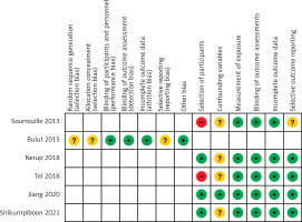
For non-randomized studies, the risk of bias was assessed in 6 independent sources using Ro-BANS [9]. For the selection of participants, 3 studies showed a low risk of bias and 2 showed a high risk of bias; for confounding variables, one study showed a low risk of bias and 4 showed an unclear risk of bias; for measurement of exposure, all 5 studies showed a low risk of bias; for blinding of outcome assessments, all showed a low risk of bias; for incomplete outcome data, all showed a low risk of bias; and for selective outcome reporting, 4 showed a low risk of bias and one showed an unclear risk of bias. For the randomized study, the risk of bias was assessed in 7 independent sources using the Cochrane risk of bias tool [8]. This assessment showed a low risk of bias for blinding of participants and personnel, blinding of outcome assessments, and incomplete outcome data; unclear risk for random sequence generation, allocation concealment, and selective reporting, and no risk of other bias. A summary of the risks of bias for selected studies is provided in Figure 2.
Pooled analysis of measured outcomes
Operative outcomes
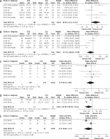
Figure 3 presents pooled analyses of operative outcomes.
Figure 3
Forest plots comparing operative outcomes of single-incision laparoscopic surgery (SILS) vs. conventional laparoscopic surgery (CLS) for rectal cancer. A – Operative time (min). B – Blood loss (ml). C – Conversion to laparotomy. D – Incision length (mm). E – Incidence of ileostomy

Operative time (min): Pooled analysis of the 6 studies showed no significant difference between the SILS and CLS groups. The weighted mean difference was 18.49 min (95% CI: –4.53 to 41.52; p = 0.12), and the included studies showed high heterogeneity (I2 = 94 %). The analysis was conducted using a random-effects model.
Blood loss (ml): Pooled analysis of the 6 studies showed no significant difference between the 2 groups. The weighted mean difference was –41.22 ml (95% CI: –96.82 to 14.38; p = 0.15), and the included studies showed high heterogeneity (I2 = 94%). The analysis was conducted using a random-effects model.
Conversion to laparotomy: Pooled analysis of 5 studies showed no significant difference between the 2 groups. The odds ratio was 0.79 (95% CI: 0.23 to 2.80; p = 0.72), and the included studies showed no heterogeneity (I2 = 0 %). The analysis was conducted using a fixed-effects model.
Incision length (mm): Pooled analysis of 2 studies showed that the incision length was significantly shorter in the SILS group. The weighted mean difference was –49.36 mm (95% CI: –98.58 to –0.14; p < 0.00001), and the included studies showed high heterogeneity (I2 = 97%). The analysis was conducted using a random-effects model.
Ileostomy rate: Pooled analysis of 2 studies showed no significant difference between the 2 groups. The odds ratio was 0.31 (95% CI: 0.06 to 1.72; p = 0.18), and the included studies showed moderate heterogeneity (I2 = 61 %). The analysis was conducted using a random-effects model.
Postoperative outcomes
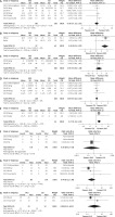
Figure 4 presents pooled analyses of postoperative outcomes.
Figure 4
Forest plots comparing postoperative outcomes of single-incision laparoscopic surgery (SILS) vs. conventional laparoscopic surgery (CLS) for rectal cancer. A – Perioperative mortality. B – Overall complications. C – Hospital stay (days). D – Reoperations. E – Readmissions. F – Pain score (VAS, visual analog scale), postoperative day 1. G – Pain score (VAS, visual analog scale), postoperative day 2. H – Morphine requirement (mg), postoperative day 1. I – Morphine requirement (mg), postoperative day 2. J – Morphine requirement (mg), postoperative day 3. K – Time to first bowel movement (days)

Perioperative mortality: Three studies reported zero perioperative mortality. One study reported 2 perioperative mortalities for SILS and 2 for CLS. The odds ratio was 3.90, and the difference was not statistically significant (95% CI: 0.49 to 31.20).
Overall complications: Pooled analysis of the 6 studies showed no significant difference between the 2 groups. The odds ratio was 0.69 (95% CI: 0.42 to 1.13; p = 0.14), and the included studies showed no heterogeneity (I2 = 0 %). The analysis was conducted using a fixed-effects model.
Hospital stay (days): Pooled analysis of the 6 studies showed that the hospital stays were significantly shorter for SILS than for CLS. The weighted mean difference was –1.20 days (95% CI: –2.02 to –0.39; p = 0.004), and the included studies showed moderate heterogeneity (I2 = 69 %). The analysis was conducted using a random-effects model.
Reoperation rate: Pooled analysis of 3 studies showed no significant difference between the 2 groups. The odds ratio was 1.10 (95% CI: 0.32 to 3.84; p = 0.88), and the included studies showed no heterogeneity (I2 = 0%). The analysis was conducted using a fixed-effects model.
Readmission rate: Pooled analysis of 3 studies showed no significant difference between the 2 groups. The odds ratio was 2.43 (95% CI: 0.64 to 9.14; p = 0.19), and the included studies showed no heterogeneity (I2 = 0%). The analysis was conducted using a fixed-effects model.
Pain score (VAS, visual analogue scale): Pooled analysis of 3 studies showed a significantly lower pain score on postoperative day (POD) 1 after SILS than after CLS. The weighted mean difference was –0.96 (95% CI: –1.18 to –0.74; p < 0.00001). The included studies showed no heterogeneity (I2 = 0%), and the analysis was conducted using the fixed-effects model. Also, pooled analysis of 3 studies showed a significantly lower pain score at POD 2 after SILS than after CLS. The weighted mean difference was –1.43 (95% CI: –2.29 to –0.57; p = 0.001), and the included studies showed high heterogeneity (I2 = 92 %). The analysis was conducted using a random-effects model.
Morphine requirements (mg): Two studies reported postoperative morphine requirements. On POD 1, no significant difference was found between the 2 groups. The weighted mean difference was –0.47 mg (95% CI: –6.34 to 5.39; p = 0.87), and the included studies showed high heterogeneity (I2 = 90%). The analysis was conducted using the random-effects model. On POD 2, postoperative morphine requirements were similar in the 2 groups. The weighted mean difference was –1.46 mg (95% CI: –6.21 to 3.28; p = 0.55), and the included studies showed high heterogeneity (I2 = 87%). The analysis was conducted using a random-effects model. On POD 3, the morphine requirement was significantly lower after SILS than CLS. The weighted mean difference was –1.39 mg (95% CI: –2.30 to –0.48; p = 0.003), and the included studies showed no heterogeneity (I2 = 0%). The analysis was conducted using the fixed-effects model.
Time to first bowel movement (days): Only one study reported a significantly shorter time to first bowel movement after SILS; the mean difference was –0.40 days (95% CI: –0.72 to –0.08).
Complications
Figure 5 presents detailed results for pooled analyses of complications.
Figure 5
Forest plots comparing complications of single-incision laparoscopic surgery (SILS) vs. conventional laparoscopic surgery (CLS) for rectal cancer. A – Intraoperative complications. B – Anastomotic leakage. C – Surgical site infections. D – Gastrointestinal motility dysfunctions. E – Pulmonary complications. F – Cardiovascular complications. G – Urologic complications

Intraoperative complications: Pooled analysis of the 6 studies showed no significant difference between the 2 groups. The odds ratio was 0.46 (95% CI: 0.07 to 3.01; p = 0.42), and the included studies showed no heterogeneity (I2 = 0%). The analysis was conducted using the fixed-effects model.
Anastomotic leakage: Pooled analysis of the 6 studies showed no significant difference between the 2 groups. The odds ratio was 0.71 (95% CI: 0.31 to 1.64; p = 0.42), and the included studies showed no heterogeneity (I2 = 0%). The analysis was conducted using the fixed-effects model.
Surgical site infections: Pooled analysis of the 6 studies showed no significant difference between the 2 groups. The odds ratio was 0.85 (95% CI: 0.39 to 1.86; p = 0.68), and the included studies showed no heterogeneity (I2 = 0%). The analysis was conducted using a fixed-effects model.
Gastrointestinal motility dysfunctions: Pooled analysis of the 6 studies showed no significant difference between the 2 groups. The odds ratio was 0.97 (95% CI: 0.27 to 3.47; p = 0.96), and the included studies showed no heterogeneity (I2 = 0%). The analysis was conducted using a fixed-effects model.
Pulmonary complications: Pooled analysis of the 6 studies revealed no significant difference between the 2 groups. The odds ratio was 1.07 (95% CI: 0.31 to 3.72; p = 0.92), and the included studies showed no heterogeneity (I2 = 0%). The analysis was conducted using a fixed-effects model.
Cardiovascular complications: Pooled analysis of the 6 studies showed no significant difference between the 2 groups. The odds ratio was 2.08 (95% CI: 0.49 to 8.90; p = 0.32), and the included studies showed no heterogeneity (I2 = 0%). The analysis was conducted using a fixed-effects model.
Urologic complications: Pooled analysis of the 6 studies showed no significant difference between the 2 groups. The odds ratio was 0.77 (95% CI: 0.27 to 2.24; p = 0.63), and the included studies showed low heterogeneity (I2 = 5%). The analysis was conducted using a fixed-effects model.
Pathologic outcomes
Figure 6 presents pooled analyses of pathologic outcomes.
Figure 6
Forest plots comparing pathologic outcomes of single-incision laparoscopic surgery (SILS) vs. conventional laparoscopic surgery (CLS) for rectal cancer. A – Number of harvested lymph nodes. B – Specimen length (cm). C – Length of proximal resection margin (cm). D – Length of distal resection margin (cm). E – Length of circumferential resection margin (mm). F – Circumferential resection margin involvement. G – Incomplete mesorectal grade. H – RO resection

Number of harvested lymph nodes: Pooled analysis of the 6 studies showed no significant difference between the 2 groups. The weighted mean difference was –0.26 (95% CI: –1.53 to 1.01; p = 0.68), and the included studies showed moderate heterogeneity (I2 = 51%). The analysis was conducted using a random-effects model.
Specimen lengths (cm): A pooled analysis of 2 studies showed that specimen lengths were significantly longer in the SILS group than in the CLS group. The weighted mean difference was 3.26 cm (95% CI: 2.01 to 4.52; p < 0.00001), and the included studies showed no heterogeneity (I2 = 0%). The analysis was conducted using a fixed-effects model.
Lengths of PRMs (cm): Pooled analysis of 2 studies showed no significant difference between the 2 groups. The weighted mean difference was 0.45 cm (95% CI: –0.29 to 1.18; p = 0.23), and the included studies showed no heterogeneity (I2 = 0%). The analysis was conducted using a fixed-effects model.
Lengths of DRMs (cm): Pooled analysis of 5 studies showed no significant difference between the two groups. The weighted mean difference was 0.04 cm (95% CI: –0.16 to 0.24; p = 0.69), and the included studies showed no heterogeneity (I2 = 0%). The analysis was conducted using a fixed-effects model.
Lengths of CRMs (mm): A pooled analysis of 2 studies showed lengths of CRM was significantly longer in the SILS group. The weighted mean difference was 2.72 mm (95% CI: 1.32 to 4.12; p = 0.0001), and the included studies showed low heterogeneity (I2 = 23%). The analysis was conducted using a fixed-effects model.
CRM involvements: Two studies reported no CRM involvement after SILS or CLS. One study reported CRM involvement in 1 patient in the CLS group and no CRM involvement in the SILS group. The odds ratio was 0.36, and the difference was not statistically significant (95% CI: 0.01 to 9.15).
Incomplete mesorectal grade: Pooled analysis of 4 studies showed no significant difference between the 2 groups. The odds ratio was 0.44 (95% CI: 0.12 to 1.60; p = 0.21), and the included studies showed relatively low heterogeneity (I2 = 49%). The analysis was conducted using a fixed-effects model.
R0 resection rate: Pooled analysis of 2 studies showed no significant difference between the 2 groups. The odds ratio was 0.90 (95% CI: 0.13 to 6.32; p = 0.92), and the included studies showed no heterogeneity (I2 = 0%). The analysis was conducted using a random-effects model.
Oncologic outcomes
Three studies reported oncologic outcomes [11, 13, 14]. Sirikurnpiboon reported that the 3-year and 5-year survival rate, local recurrence rate, distant metastasis rate, and average times to recurrence and metastasis were not significantly different between the 2 groups [11]. Jiang et al. reported that the recurrence rate, 3-year disease-free survival rate, and overall survival rate were not significantly different between the 2 groups [13]. Tei et al. found that distant metastasis and local recurrence rates and 3-year overall survival rates were not significantly different in the 2 groups. However, the 3-year relapse-free survival rate was significantly higher for SILS (94.7% in SILS vs. 78.6% in CLS; p = 0.032). The authors attributed this result to different follow-up periods (40 months after SILS vs. 51 months after CLS; p = 0.008) [14]. Pooled analysis was not performed because the included studies did not provide hazard ratios with standard errors or any variable to estimate their values for performing a meta-analysis of time-to-event data.
Discussion
After laparoscopic colectomy was introduced in the 1990s [1–4], in the early 2000s, the safety and feasibility of laparoscopic surgery for colon cancer were demonstrated by several RCTs [25–28]. These studies showed that laparoscopic colectomy had outcomes equivalent or superior to open colectomy, and subsequently, laparoscopic colectomy gained in popularity for colon cancer surgery. As regards rectal cancer, the feasibility of laparoscopic surgery was shown by some comparative studies conducted in the early 2000s [29–31], and later, the CLASICC, the COREAN, and the COLOR II trials confirmed its oncological safety [32–35].
Laparoscopic surgery may produce superior outcomes because it causes less surgical trauma and thus fewer complications and faster recovery than open surgery. In the case of rectal cancer surgery, its superior outcomes are probably due to the magnified view it provides of limited surgical areas in the pelvis, which enable precise dissection along proper surgical planes. Because of these advantages, MIS is being continuously developed to maximize its potential benefits for colorectal surgery.
About 2 decades after the introduction of laparoscopic colectomy, SILS for colon cancer and SILS for rectal cancer were reported by Remzi et al. [5] and Bulut and Nielsen [6]. Following clinical studies that compared the outcomes of SILS and CLS for colorectal surgery, some meta-analyses reported promising results, although there were some inconsistencies [36–39]. However, no meta-analysis has addressed the outcomes of SILS specifically for rectal surgery, which is technically more difficult than colon cancer surgery. Because rectal resection is performed in the confined space of the pelvic cavity, the range of motion of the working instruments is limited and traction to maintain a proper surgical plane is difficult. It is a matter of concern whether SILS meets this challenge without compromising surgical outcomes. Questions regarding this topic should be answered by studies on rectal surgery alone.
To the best of our knowledge, this study is the first meta-analysis to compare the outcomes of SILS and CLS in rectal cancer. The key findings of this study are as follows: i) SILS showed superior outcomes in terms of incision length, postoperative pain, and hospital stays; ii) SILS and CLS were similar as regards surgical difficulty-related outcomes; and iii) importantly, the perioperative mortalities, complications, and pathologic qualities of SILS and CLS were comparable.
Incision lengths were significantly shorter for SILS, with a mean difference of 49.58 mm. For rectal surgery, SILS uses only one incision for working port insertion and specimen extraction, whereas CLS usually requires 4 or 5 incisions for port insertion. Thus, the incision length is shorter for SILS, and the present study shows this is a clear advantage of SILS over CLS.
Postoperative pain is related to wound length and site, and because SILS involves one wound only, postoperative pain can be reduced compared with CLS. Our analysis showed that pain scores were significantly lower in the SILS group and that the mean intergroup differences increased from POD 1 (MD = 0.96) to POD 2 (MD = 1.43). When the intergroup difference of postoperative pain score is relatively small, it is probably clinically insignificant, and the morphine requirements are probably the same. It may explain why the group morphine requirements were similar on POD 1 and 2, but on POD 3 they were lower in the SILS group than in the CLS group. Specifically, our study showed that postoperative pain reduced faster in the SILS group. Furthermore, hospital stay was significantly shorter in the SILS group (mean difference of 1.17 days). The reduced postoperative pain and lower incidence of overall complications can explain the earlier discharges observed in the SILS group.
Operative time, blood loss, and conversion rate to laparotomy are related to surgical difficulty, and these outcomes were similar in the SILS and CLS groups. The technical difficulties of SILS are due to the parallel entry of the laparoscope and working instruments, which makes triangulation restrictive, traction for proper dissection less effective, and limits the surgical view. Also, collisions between the laparoscope and working instruments can occur outside the trocar. Several adaptive methods can be used to minimize these difficulties. First, curved or articulating instruments can overcome the limitations associated with parallel insertion and allow more effective triangulation. Second, a flexible laparoscope can overcome the limited surgical view caused by parallel insertion of the scope and instruments. Third, a 3-dimensional laparoscope can improve depth perception limitations caused by parallel laparoscope and instrument insertion. Fourth, a laparoscope with a right-angled light cord or a bariatric length laparoscope or instruments can prevent external collisions between the operator and assistant. And finally, operators can use the instruments in a cross-hand manner to overcome range of motion limitations [40]. Most of the included studies did not specify the methods to overcome the technical difficulties of SILS, and only 2 studies identified the use of flexible laparoscopes.
In addition to these technical tips, experience also enables surgeons to cope with the surgical difficulties posed by SILS. Some researchers analysed the learning curve for single-incision colon cancer surgery. In a study by Kim et al., the learning period for single-incision laparoscopic anterior resection of sigmoid colon cancer was between 61 and 65 cases with an operation time of 173 min for the 65th case [41], and Kirk et al. reported a learning period for single-incision laparoscopic right colectomy of 40 cases with an operation time of 97 min [42]. These studies showed that the operation time was optimized after the learning curve in surgeons with experience. In all studies included in our meta-analysis, expert laparoscopic colorectal surgeons performed the operations. We suppose this contributed to the comparable operative outcomes of SILS to CLS.
Perioperative mortalities, reoperation and readmission rates, and times to first bowel movement were similar in the SILS and CLS groups. Regarding complications, no significant intergroup difference was observed for intraoperative complications, anastomotic leakage, surgical site infection, gastrointestinal motility dysfunction, or pulmonary, cardiovascular, or urologic complications. We suppose that the similar postoperative outcomes of SILS to CLS were in part due to the surgeons’ expertise. In a study by Jiang et al. the operators had experience of over 100 laparoscopic colorectal surgeries [13]. In a study by Bulut et al. the surgeons had long-standing experience of laparoscopic colorectal surgery [17], and in a study by Sourrouille et al. a specialized, skilled laparoscopic surgeon performed the operations [24]. Tei et al. stated that they conduct SILS as a reasonable alternative to CLS for upper rectal cancer in their department [14]. Sirikurnpiboon stated that more than 500 colorectal cancer surgeries were performed during the study period (2011 to 2014) [11]. Nerup et al. concluded that SILS for rectal cancer appears safe and feasible when performed by highly experienced laparoscopic colorectal surgeons [16]. Thus, we believe that surgeons’ experience contributes to overcoming the technical issues of SILS and obtaining feasible outcomes for rectal cancer surgery.
In addition, pooled analysis showed that the overall complication incidence was significantly lower for SILS than for CLS (odds ratio = 0.64). We attribute the advantages of SILS over CLS to less surgical trauma, and thus to less systemic inflammatory response, fewer complications, and faster recovery. However, we found little evidence to support the lesser effect of SILS on inflammatory response. Only Bulut et al. assessed postoperative levels of immunologic markers, i.e. of C-reactive protein (CRP), interleukin-6 (IL-6), and tissue inhibitor of metalloproteinases-1 (TIMP-1), and reported that postoperative CRP levels at 6 and 24 h after skin incision were significantly lower after SILS than after CLS [17]. In previous studies that compared laparoscopic and open surgery, inflammatory immune response was consistently shown to be attenuated after laparoscopic surgery [43–49]. However, conflicting results were reported for SILS versus CLS [17, 50, 51]. Additional studies are needed to clarify this issue.
One of the advantages of SILS over CLS is that it better preserves abdominal wall integrity, and thus SILS would be expected to reduce the risk of incisional hernia. This topic was only investigated by Jiang et al. [13], who reported one incisional hernia after SILS and none after CLS, which was not a significant difference.
Pathological outcomes of SILS as assessed by numbers of harvested lymph nodes, PRM and DRM length, CRM involvement, the incidence of incomplete mesorectal grade, and R0 resection rate were similar to those of CLS. Oncologically proper resection is a fundamental principle of rectal cancer surgery, and it includes total mesorectal excision (TME) with proper lymph node dissection. TME quality could be assessed by CRM involvement and the integrity of the TME specimen, which is a key determinant for evaluating the safety of laparoscopic rectal cancer surgery [52, 53]. The National Comprehensive Cancer Network (NCCN) guideline recommendations are as follows: to examine a minimum of 12 lymph nodes; to acquire an adequate CRM and an adequate distal margin (1 to 2 cm for distal rectal cancers); and to evaluate the quality of mesorectum for low rectal cancers [54]. All pathologic results presented from the included studies followed the NCCN guidelines, and there was no difference between SILS and CLS.
We summarize the results of this meta-analysis as follows. First, SILS for rectal cancer has clear benefits over CLS in terms of incision length, postoperative pain, and hospital stay. Second, SILS for rectal cancer produced postoperative clinical outcomes comparable to CLS. Third, SILS for rectal cancer produced acceptable pathological outcomes and maintained oncological principles.
Only a decade has passed since SILS was first introduced to treat rectal cancer in 2010. Thus, relatively few clinical studies have been performed, and no long-term follow-up data are available to assess the oncological safety of SILS versus CLS for rectal cancer. It is the first limitation of this study. However, our meta-analysis revealed promising results of SILS for rectal cancer with respect to perioperative and pathologic outcomes. Also, 3 of the studies included in our meta-analysis showed similar short-term oncologic outcomes for SILS and CLS in terms of recurrence, disease-free survival, and overall survival rates. Thus, we consider the results of this study promising and hope they encourage more surgeons to undertake future clinical studies.
The various modalities used to overcome the technical difficulties associated with SILS might affect surgical outcomes. A flexible laparoscope was used for SILS in 2 studies [13, 14], a 5 mm 30° scope in 2 studies [16, 17], and a 0° scope in one study [24], and 3 different types of ports were used for SILS, namely: the SILS™ Port (Covidien, USA) in 3 studies [13, 16, 17], the E-Z access Port (Hakko, Japan) in one [14], and the GelPOINT Port (Applied Medical, Canada) in one [24]. Hence, the heterogeneity of the surgical instruments used is also a limitation. However, no other instrumental or methodological differences were reported in the included studies, and expert laparoscopic colorectal surgeons performed all operations. Thus, we consider that the use of different surgical instruments is unlikely to impact our findings.
Conclusions
This study is the first meta-analysis to compare the outcomes of SILS and CLS for rectal cancer and was conducted using data from the most recent clinical studies. SILS was found to have the following merits over CLS: smaller wounds, less pain, and shorter hospital stay. Furthermore, the study showed that SILS is a safe treatment for rectal cancer that does not compromise clinical or pathological outcomes when performed by experienced laparoscopic colorectal surgeons. Future studies are required to determine the long-term oncologic outcomes of SILS for rectal cancer.


