
Introduction
Abnormal skin wound healing can lead to pathological scars, namely hypertrophic scars and keloids, which are fibroproliferative disorders caused by excessive collagen deposition from activated fibroblasts [1]. These are not merely cosmetic issues; they often cause debilitating physical symptoms like pain and itching, functional limitations, and significant psychosocial distress [2]. Current treatments, such as surgical excision and corticosteroid injections, are fraught with limitations, including high recurrence rates, adverse effects, and inconsistent results [3, 4], underscoring an urgent need for more effective therapies.
Botulinum neurotoxin type A (BTX-A) has emerged as a transformative agent for scar management [5]. Extensive clinical evidence supports its prophylactic and therapeutic efficacy, showing that it can produce finer scars and significantly improve the volume, erythema, and symptoms of established lesions [6]. The primary accepted mechanism is that BTX-A induces muscle paralysis, which reduces mechanical tension on the wound – a potent stimulus for fibrosis [7]. By disrupting this mechanical signalling, BTX-A is thought to down-regulate fibroblast activation and abnormal matrix deposition [8].
However, accumulating data suggest a more complex mechanism. Pathological scarring also involves persistent low-grade inflammation, where macrophage biology plays a central role [9]. A dysregulated balance favouring pro-fibrotic M2 macrophages perpetuates the fibrotic process through mediators like TGF-β1 [10]. This review will explore the central role of macrophages in scar pathophysiology and critically analyse how BTX-A’s mechanical effects may indirectly modulate this immune response. We propose that by altering the mechanochemical landscape, BTX-A shifts macrophage polarization away from a pro-fibrotic phenotype, and we will discuss the profound translational implications of viewing BTX-A as a macrophage-targeting agent to optimize future scar therapies.
The pathophysiology of pathological scarring
Pathological scar formation represents a fundamental dysregulation of the wound healing process, where normal tissue repair mechanisms become uncontrolled. Central to this intricate biological program is the macrophage, a myeloid immune cell with remarkable plasticity [11]. Macrophages act as the master coordinators of the entire healing cascade, directing the behaviour of other key cells like fibroblasts and endothelial cells through the precise secretion of cytokines and growth factors. Therefore, understanding the pathophysiology of hypertrophic scars and keloids requires a macrophage-centric view, recognizing that a failure in their orchestration of the healing process is a primary cause of skin fibrosis [12].
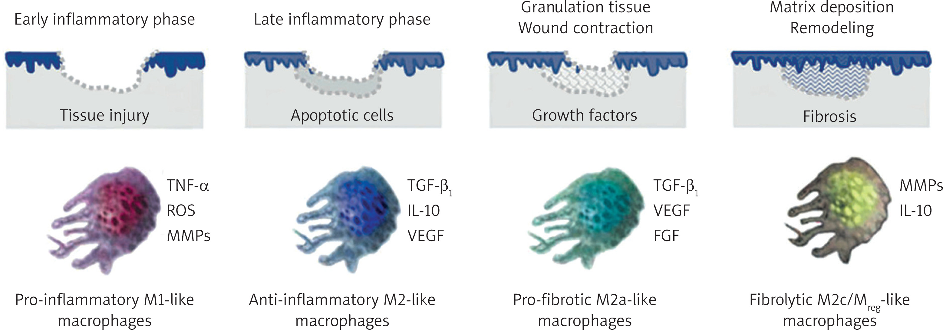
The physiological process of skin wound healing unfolds in four overlapping stages: haemostasis, inflammation, proliferation, and remodelling. Immediately after injury, haemostasis is initiated as the coagulation cascade forms a platelet plug that releases signalling molecules like PDGF and TGF-β, which recruit immune cells [13]. Neutrophils arrive first, followed by circulating monocytes that differentiate into macrophages. In this early inflammatory environment, driven by signals from pathogens and damaged cells, macrophages adopt a classically activated (M1) phenotype [14–17]. These M1 macrophages are potent pro-inflammatory effectors essential for clearing debris and pathogens, characterized by high phagocytic activity and the secretion of pro-inflammatory cytokines such as TNF-α, IL-1β, and IL-6 [18]. The successful transition from inflammation to proliferation depends on a critical phenotypic switch of these macrophages from the pro-inflammatory M1 state to an alternatively activated (M2) state. This shift is triggered by signals like the phagocytosis of apoptotic neutrophils and the presence of cytokines like IL-4 and IL-13.
M2 macrophages are fundamentally pro-resolving and pro-repair [19]. They downregulate inflammation by secreting anti-inflammatory cytokines like IL-10 and TGF-β1, while are concurrently orchestrating tissue construction by releasing growth factors that stimulate fibroblast proliferation and deposition of new extracellular matrix [15]. M2 macrophages also promote neovascularization by secreting VEGF and are marked by high expression of arginase-1, which shunts metabolism towards producing precursors for collagen synthesis. In the final remodelling phase, which lasts for months, the granulation tissue matures as type III collagen is replaced by stronger type I collagen. Throughout this stage, macrophages regulate the balance between matrix synthesis and degradation to ensure the scar gains tensile strength while remaining stable [16]. This timely and balanced M1-to-M2 transition is the cornerstone of physiological wound repair, ensuring inflammation is resolved and replaced by coordinated tissue remodelling [20] (Figure 1).
Figure 1
Functional macrophage phenotypes proposed to be induced at different stages of normal tissue repair [21]
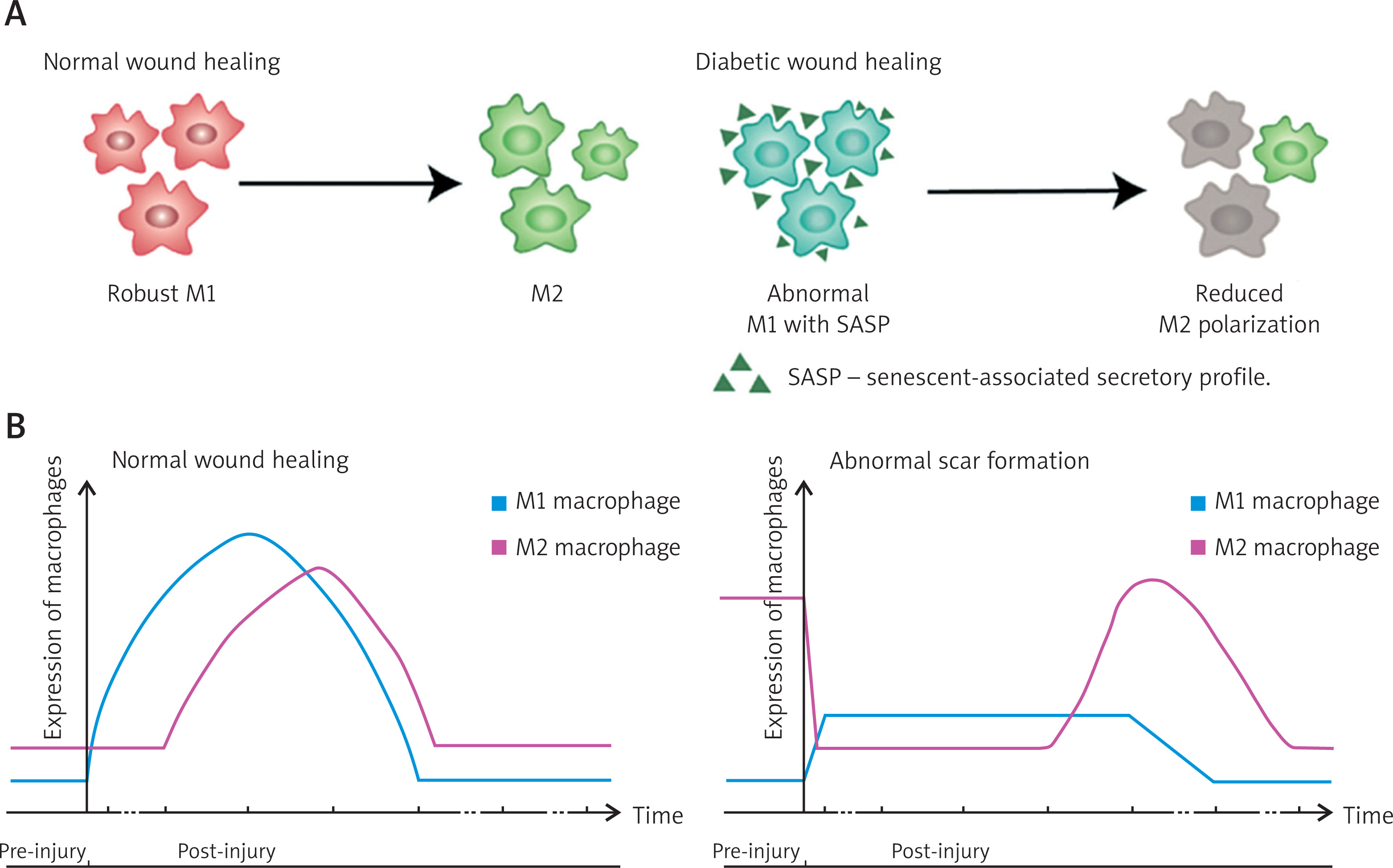
In contrast to this tightly regulated process, the formation of hypertrophic scars and keloids is intrinsically linked to a profound dysregulation of these macrophage polarization dynamics [21]. The core pathology is a failure of the wound environment to transition from a proliferative state to a stable, remodelled state, driven by the persistence of a dysfunctional macrophage population [22]. Histological analyses of hypertrophic scars and keloid tissues consistently reveal an increased density of macrophages that are heavily skewed towards the M2 phenotype, as shown by high expression of markers like CD163 and CD206 [23]. This persistent, M2-dominated infiltration creates a perpetually pro-fibrotic environment. The sustained, high-level secretion of TGF-β1 from these M2 macrophages is arguably the single most important driver of fibrosis, relentlessly stimulating fibroblast proliferation and matrix synthesis. This pro-fibrotic state becomes a self-perpetuating vicious cycle, where the pathologically stiff matrix can itself act as a mechanical signal to promote and stabilize the M2 phenotype, which in turn amplifies TGF-β1 production. Furthermore, the healing process is actively subverted; the initial M1 inflammatory phase may be prolonged or exaggerated, and the M1-to-M2 switch appears defective [24]. Instead of transitioning to a resolving phenotype, macrophages adopt a persistent, non-resolving pro-fibrotic state, resulting in chronic, low-grade inflammation coexisting with severe fibrosis – a hallmark of keloids. This suggests that genetic predispositions may relate to an inherently hyper-responsive macrophage system [25]. Ultimately, the pathological scar is defined by a macrophage population “stuck” in a pro-fibrotic state, firmly positioning dysregulated macrophages as primary cellular culprits and a highly attractive therapeutic target for anti-fibrotic strategies [26, 27] (Figure 2).
Figure 2
Phenotypic dysregulation of macrophages. A – Compared with the spontaneous M1/M2 transition that occurs during normal wound healing, M1 macrophages are abnormal, with a more senescent phenotype and reduced plasticity, which hinders the M1/M2 transition [26]. B – Comparison of macrophage polarization during the wound healing and abnormal scar formation [27]
Botulinum toxin in scar management: clinical applications and mechanistic postulates
Botulinum neurotoxin type A (BTX-A) induces localized chemical denervation by cleaving the SNARE protein SNAP-25 to inhibit acetylcholine release, a mechanism used to treat muscle overactivity [28]. Its application was logically extended to scar management based on the observation that mechanical tension worsens scarring, and a large body of clinical literature now supports its efficacy [29]. This review will synthesise this clinical evidence before exploring both established and emerging mechanistic theories, including its immunomodulatory functions [30]. The clinical utility of BTX-A is supported by over two decades of evidence, including randomized controlled trials [31]. Its efficacy is demonstrated in three primary strategies: preventing excessive scarring in fresh wounds, treating established pathological scars, and as an adjuvant therapy to prevent the recurrence of refractory lesions like keloids [32].
Prophylactic use is based on the principle that mechanical tension drives fibrosis [33]. By injecting BTX-A into underlying muscles at the time of wound closure, a “fixation zone” is created. This has been rigorously studied in high-tension regions [34], with RCTs demonstrating significantly improved aesthetic outcomes for thyroidectomy and median sternotomy scars. The technique is also key in facial procedures like cleft lip repair [35]. These benefits are quantified by validated scales like the VSS and POSAS, as well as objective measures including 3D imaging, confirming a more favourable healing trajectory [36]. Therapeutically, BTX-A is injected directly into established hypertrophic scars and keloids, where it is thought to modulate intrinsic tension and the scar’s hypercellular state [37]. Clinically, this results in scar softening, reduced volume and erythema, and improved range of motion in cases of contracture [38]. A striking benefit is the rapid relief of sensory symptoms like pain and pruritus, suggesting a neuromodulatory mechanism involving the inhibition of neuropeptides like substance P (SP) and calcitonin gene-related peptide (CGRP) [39]. As an adjuvant, BTX-A is crucial for managing keloids, which have a high recurrence rate after excision alone, by breaking the pro-fibrotic cycle [40]. Post-surgical injection creates a low-tension environment, and studies show this significantly reduces recurrence compared to other methods [41]. The safety profile of BTX-A is excellent, with adverse effects being mild and localized, cementing its role as a pivotal therapy [42–52] (Table 1).
Table 1
Strategic framework for botulinum toxin A (BTX-A) in scar management
| Application strategy | Core objective | Clinical implementation and efficacy | Proposed primary mechanism(s) | Key references |
|---|---|---|---|---|
| Prophylactic intervention | To prevent de novo pathological scarring in fresh wounds | Protocol: Intramuscular injection into perilesional muscles at the time of surgical closure, targeting high-tension areas (e.g., neck, chest). Efficacy: Results in significantly narrower, flatter scars with superior aesthetic scores, as validated by VSS, POSAS, and 3D imaging. | Mechanical tension reduction: Minimises tensile forces across the wound during critical healing phases. | [26, 43–46] |
| Therapeutic treatment | To remodel established scars and alleviate symptoms | Protocol: Direct intralesional injection into hypertrophic scars or keloids, often as a series of treatments. Efficacy (Morphological): Leads to progressive scar softening, flattening, reduced volume, and decreased erythema. Improves range of motion in contractures. Efficacy (Symptomatic): Provides rapid and sustained relief from intractable pruritus and neuropathic pain. | Intrinsic tension modulation and direct anti-fibrotic effects (for morphological changes). Neuromodulation (for symptom relief via inhibition of neuropeptides). | [31, 47–49] |
| Adjuvant therapy | To prevent recurrence following keloid excision | Protocol: Perilesional intramuscular injection immediately after surgical removal of the keloid. Efficacy: Significantly reduces high recurrence rates, acting as a cornerstone of multimodal therapy alongside surgery, steroids, or radiation. | Mechanical tension reduction: Creates a low-tension environment for the new wound, mitigating a primary trigger for keloid regrowth. | [50–52] |
The traditional mechanistic explanation for BTX-A’s efficacy is its ability to reduce mechanical tension, a fundamental concept in dermatology [53]. This effect is mediated through mechanotransduction, the process by which cells like dermal fibroblasts convert physical forces into biochemical signals [54]. Mechanical forces, both extrinsic from muscle activity and intrinsic from cellular contraction, are potent activators of fibroblasts [55]. When stretched, fibroblasts proliferate, increase ECM synthesis, and differentiate into contractile myofibroblasts expressing α-smooth muscle actin (α-SMA) [56]. This process begins with integrins activating focal adhesion kinase (FAK) [57]. Downstream signalling includes the RhoA/ROCK pathway, which promotes contractility [58], and the Hippo pathway, where tension leads to the nuclear translocation of the pro-fibrotic transcriptional co-activators YAP and TAZ [59, 60]. BTX-A intervenes by causing muscle paralysis, creating a “splinting” effect that eliminates dynamic extrinsic tension [61]. This keeps pro-fibrotic mechanotransduction pathways quiescent. In established scars, it may also reduce intrinsic tension [62]. This mechanical unloading model provides a powerful explanation for BTX-A’s clinical benefits [63], though it is likely incomplete.
Growing evidence suggests that BTX-A’s effects extend beyond simple mechanical unloading. The rapid relief of sensory symptoms and erythema is not fully explained by the classical model [64], pointing instead to the regulation of neuro-inflammatory and immune axes [65]. A key non-canonical effect is the inhibition of neurogenic inflammation, which is prominent in pathological scars due to excessive sensory innervation [66]. BTX-A is believed to inhibit the release of pro-inflammatory neuropeptides like SP and CGRP from these nerves, which directly reduces pain and pruritus while also removing a potent chemical stimulus for fibrosis [67]. This neuroimmune interaction extends to indirectly inhibiting mast cell degranulation [68]. The observed reduction in scar erythema may result from both reduced neurogenic vasodilation and a potential direct inhibitory effect on angiogenesis [69], as BTX-A can downregulate VEGF, normalizing the scar’s hypervascularity [70]. More direct immunomodulatory evidence comes from in vitro studies. Though its receptor’s presence on immune cells is debated [71], reports suggest BTX-A can regulate the production of pro-inflammatory cytokines like TNF-α [72]. Furthermore, studies show that BTX-A can directly inhibit fibroblast proliferation and TGF-β1 expression, independent of mechanical unloading [73]. Together, these observations portray BTX-A as a pleiotropic modulator that acts as a complex neuroimmunopharmacological agent, not just a mechanical unloader [74]. This expanded mechanistic view provides the crucial bridge to understanding its effects on macrophages, as the altered microenvironment created by BTX-A is expected to have profound downstream effects on macrophage recruitment and polarization, integral to how the toxin promotes regenerative healing [75].
The central mechanism: BTX-A’s modulation of macrophage function in scarring
The therapeutic success of botulinum toxin type A (BTX-A) in scar management is increasingly understood to be mediated through its profound effects on the wound microenvironment, which in turn modulates the function of macrophages, the master regulators of healing. By creating an environment of low mechanical tension and reduced pro-inflammatory signalling, BTX-A removes critical stimuli that sustain the pathologic, pro-fibrotic M2 macrophage phenotype [76]. This BTX-A-macrophage axis is central to its anti-scarring action, interrupting the core fibrotic feedback loop.
The most powerful mechanism by which BTX-A influences macrophage behaviour is through its indirect alteration of the tissue’s mechanical properties. Macrophages, like fibroblasts, are highly mechanosensitive cells whose phenotype is dictated by the physical characteristics of their surroundings [77, 78]. Whereas healthy dermis is soft (1–5 kPa), fibrotic scars are pathologically stiff (often > 30 kPa) [79]. This increased matrix stiffness is an active driver of fibrosis, as numerous studies have shown that macrophages cultured on stiff substrates preferentially polarize toward a pro-fibrotic M2-like phenotype, characterized by upregulated TGF-β1 production, a response often mediated by the YAP/TAZ signalling pathway [80, 81]. BTX-A intervenes by reducing mechanical tension, which inhibits fibroblast activation and excessive collagen deposition. This prevents the pathological stiffening of the extracellular matrix (ECM), ensuring that infiltrating macrophages are not exposed to this potent M2 polarization cue [82, 83]. Furthermore, BTX-A eliminates the pro-fibrotic stimulus of dynamic mechanical stretch from muscle movement, which has also been shown to promote M2 polarization [84–86]. This mechanical unloading also likely reduces the activation of mechanosensitive ion channels like Piezo1, further calming pro-inflammatory signalling [87, 88]. By quieting fibroblasts, BTX-A also disrupts a pathological feedback loop, reducing the secretion of macrophage chemoattractants and polarizing factors [89–91]. Combined with reduced tissue hypoxia and neurogenic inflammation, this comprehensive reprogramming of the microenvironment guides macrophages away from a pro-fibrotic trajectory [45, 92–94] (Table 2).
Table 2
Specific pathways of BTX-A’s indirect modulation of macrophage function via mechanotransduction
| Influencing factor | Pathological state (in high-tension environment) | BTX-A’s intervention | Favourable outcome for macrophages | References |
|---|---|---|---|---|
| Static matrix stiffness | Hyperactivation of fibroblasts leads to excessive collagen deposition, creating a pathologically stiff matrix (> 30 kPa). This stiff substrate drives macrophage polarization towards a profibrotic M2 phenotype via the YAP/TAZ pathway. | By reducing mechanical tension, BTX-A inhibits fibroblast activity, thereby preventing pathological ECM stiffening and maintaining a soft, compliant wound environment (closer to normal dermis at 1–5 kPa). | Macrophages in this “soft” environment do not experience activation of the YAP/TAZ pathway. This fundamentally removes a potent M2 polarization cue, reducing the intrinsic production of TGF-β1. | [79–82] |
| Dynamic mechanical stretch | Periodic stretching from muscle movement acts as a persistent physical stimulus, directly promoting M2-like polarization and secretion of fibrotic growth factors. | By paralyzing local musculature, BTX-A transforms the wound from a “dynamically tensed” to a “mechanically quiescent” state. | Eliminates the driving force for M2 polarization from stretch. Macrophages are shielded from this persistent physical perturbation, allowing for a normal phenotypic transition instead of being locked into a pathological M2 state. | [84–86] |
| Mechanosensitive ion channels | High tension on the cell membrane leads to persistent opening of channels like Piezo1, causing sustained Ca2+ influx that triggers downstream pro-inflammatory and pro-fibrotic signalling pathways. | Mechanical unloading reduces tension on the cell membrane. | Reduces the basal activity of Piezo1 channels and subsequent Ca2+ signalling. This inhibits associated pro-inflammatory/pro-fibrotic pathways, promoting a more quiescent macrophage phenotype. | [87, 88] |
| Fibroblast-macrophage crosstalk | Mechanically stressed fibroblasts secrete high levels of chemoattractants (e.g., CCL2) and M2-polarizing factors, creating a vicious feedback loop that recruits and activates more macrophages. | Maintains fibroblasts in a quiescent state, thereby significantly reducing their secretion of macrophage-activating signals. | Breaks the pathological feedback loop. This leads to reduced recruitment of macrophage precursors (lower cell density) and diminished external M2-polarizing stimuli for infiltrated macrophages. | [89–91] |
| Tissue microenvironment | [92, 93] |
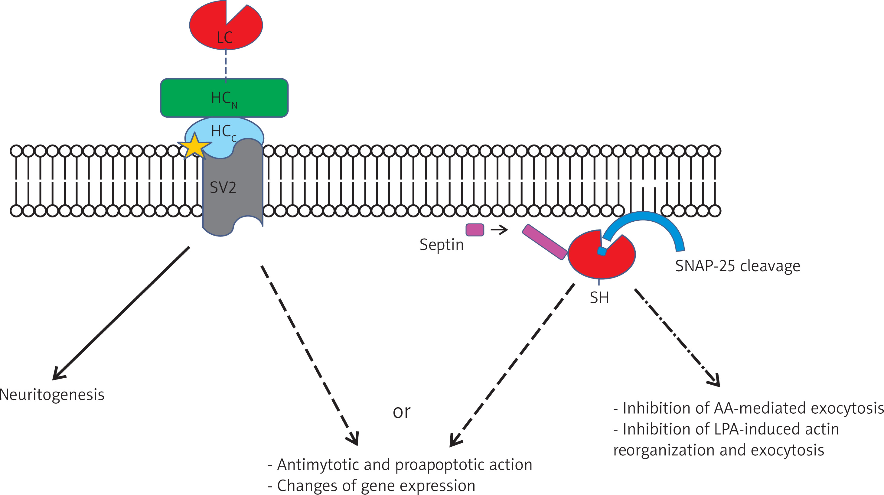
While this indirect modulation is well-established, a transformative hypothesis posits that BTX-A may also interact directly with macrophages [95, 96]. The biological plausibility for this rests on a series of potential steps. For binding and internalization, macrophages express low-affinity ganglioside receptors [97], and their high phagocytic activity may compensate for the uncertain presence of the high-affinity SV2 receptor, allowing toxin uptake, especially at high local concentrations [98–100]. After endosomal escape [101], the toxin’s light chain would find its specific molecular substrate within the macrophage cytoplasm. Crucially, modern evidence shows that macrophages express the canonical BTX-A target, SNAP-25, as well as its homologue SNAP-23 [102–105]. These SNARE proteins are essential for the exocytosis of cytokines and chemokines. Cleavage of these proteins would have profound anti-fibrotic consequences, directly impairing the secretion of key mediators like TGF-β1 [106], PDGF [107], and the monocyte chemoattractant CCL2 [108, 109], thereby striking at the heart of the fibrotic engine. This provides a compelling, though still theoretical, parallel mechanism to mechanical unloading [110–113] (Figure 3).
Ultimately, the most comprehensive understanding of BTX-A’s efficacy comes from a unified model where its distinct biological actions are synergistic [114, 115]. The cascade begins with two concurrent primary events: canonical chemodenervation at the neuromuscular junction, which establishes mechanical quiescence [116], and interaction with sensory nerve endings, which inhibits the release of pro-inflammatory neuropeptides like SP and CGRP to establish neurogenic quiescence [117, 118]. This “calm” microenvironment forms the basis for all subsequent effects. It indirectly modulates fibroblasts by removing both mechanical and chemical stimuli, preventing excessive ECM deposition and subsequent matrix stiffening [119, 120]. This, in turn, prevents the M2 polarization of macrophages that is driven by a stiff substrate [121, 122]. The potential direct interaction provides a powerful “second hit” to this process. For instance, while the indirect pathway reduces the environmental signals for TGF-β1 production, the direct pathway would physically impair the macrophage’s ability to secrete it by cleaving the SNARE machinery [123, 124]. This unified model demonstrates how BTX-A systematically attacks multiple nodes of the fibrotic feedback loop [125, 126], explaining its efficacy in both prophylactic use (where indirect mechanisms dominate) and the treatment of established scars (where direct and neurogenic pathways may be more prominent) [127]. BTX-A is therefore a unique, pleiotropic regulator of wound healing, with the macrophage serving as the central convergence point for its multifaceted and synergistic anti-scarring actions [128] (Table 3).
Table 3
A unified model of BTX-A’s synergistic anti-fibrotic actions
| Stage of action | Mechanism and pathway | Key cellular/tissue target | Primary effect | Synergistic reinforcement | References |
|---|---|---|---|---|---|
| Phase 1: Immediate onset | Mechanical action | Neuromuscular junction | Canonical action: Induces flaccid paralysis of adjacent muscles, creating a mechanically quiescent environment. | Works in parallel with neurogenic calming to establish an overall “calm” microenvironment from the outset. | [116] |
| Neurogenic action | Peripheral sensory nerves | Established neurotropic effect: Inhibits the release of pro-inflammatory neuropeptides (SP, CGRP) by cleaving SNAP-25, creating a neurogenically quiescent state. | Rapidly addresses symptoms (pain/pruritus) and removes a key chemical stimulus for fibroblasts and immune cells. | [117, 127] | |
| Phase 2: Environmental reprogramming | Mechanical pacification | Dermal fibroblasts | Evidence-based indirect effect: Prevents activation of pro-fibrotic signalling (e.g., RhoA/ROCK, YAP/TAZ) by removing tensile forces, keeping fibroblasts quiescent. | Powerfully reinforced by the lack of SP (a known fibroblast mitogen), creating a dual blockade on fibroblast activation. | [119, 120] |
| ECM normalization | Extracellular matrix (ECM) | Downstream consequence: Reduced fibroblast activity leads to less collagen deposition, ensuring the ECM remains soft and compliant and preventing pathological stiffening. | Creates a physically non-permissive substrate for the pro-fibrotic M2 polarization of macrophages. | [121, 122] | |
| Macrophage guidance | Macrophages | Mechanotransduction reprogramming: In a soft, chemically calm environment, macrophages are guided towards a physiological, non-fibrotic phenotype, avoiding the M2 skew. | Macrophages receive fewer external activation signals from both quiescent fibroblasts and calmed nerves. | [114, 122] | |
| Phase 3: Direct cellular intervention | Direct immunomodulation | Macrophages | Plausible mechanism (requires validation): Potential cleavage of SNAP-25/23 within macrophages directly impairs the secretory machinery for cytokines (e.g., TGF-β1). | The indirect pathway reduces the signals for TGF-β1 production, while this direct pathway would block the secretion itself – a robust dual-level blockade. | [123, 124] |
| Overall outcome: Disruption of the fibrotic cycle | Multi-point attack | The entire fibrosis feedback loop | Model’s central thesis: BTX-A systematically breaks key links in the cycle: | The combination of mechanical, neurogenic, and immunomodulatory effects provides a comprehensive therapeutic strategy that is more robust than targeting any single pathway alone. | [125, 126, 128] |
Conclusion and future perspectives
This review has synthesised a broad range of clinical and basic science evidence to construct a new, macrophage-centric framework for understanding the anti-fibrotic efficacy of botulinum neurotoxin type A (BTX-A). The conventional model, which attributes its success solely to the mechanical unloading of healing wounds, is insufficient to explain the full spectrum of its therapeutic effects. We have presented a unified model wherein BTX-A functions as a pleiotropic modulator of the wound microenvironment [129]. This model is built upon two synergistic pillars: the well-documented indirect pathway, through which BTX-A-induced mechanical and neurogenic quiescence reshapes the extracellular matrix and chemical milieu to favour a pro-resolving macrophage phenotype; and a highly plausible direct pathway, wherein BTX-A may directly interact with macrophages to inhibit their pro-fibrotic secretory functions. By positioning the macrophage as the central convergence point for these multifaceted actions, this integrated perspective provides a more complete and mechanistically satisfying explanation for the potent anti-scarring capabilities of BTX-A. This refined understanding not only deepens our appreciation for a widely used clinical tool but also illuminates a clear and compelling roadmap for future research [130].










