
How to cite this article Thirumalaiswamy A., Joshi V.D., Rattan A., Bhoite K., Kharkar V.: The Cryptic Invader: Midfacial Destruction in Extranodal NK/T-Cell Lymphoma – Case Report with Narrative Review. Dermatol Rev/Przegl Dermatol 2026, 113, 143–150. DOI: https://doi.org/10.5114/dr.2026.164355.
INTRODUCTION
Extranodal natural killer/T-cell lymphoma, nasal type (ENKTCL), is a rare and highly aggressive subtype of non-Hodgkin lymphoma that most commonly arises in the nasal cavity and midline facial structures [1]. Less frequently, it may involve the skin, gastrointestinal tract, and other extranodal sites. The disease is strongly associated with Epstein–Barr virus (EBV) infection, which plays a key role in its pathogenesis by promoting the survival and proliferation of malignant lymphoid cells [2].
Nasal involvement typically presents with symptoms such as nasal obstruction, facial swelling, purulent discharge, epistaxis, and malaise. Due to overlapping symptoms with more common infections and inflammatory conditions, diagnosis is frequently delayed, allowing substantial local progression and, in some cases, distant dissemination. Histopathological examination, immunohistochemistry, and imaging, particularly positron emission tomography/computed tomography (PET/CT), are essential for accurate diagnosis and staging.
Current treatment approaches are based on non-anthracycline chemotherapy regimens containing L-asparaginase, often combined with radiotherapy in localized disease. We report this case due to its rare presentation and aggressive clinical course in a middle-aged woman, highlighting the importance of early biopsy, comprehensive immunophenotyping, and prompt diagnosis to improve clinical outcomes.
AIM OF THE REVIEW
The aim of this narrative review is to provide an updated, clinically oriented synthesis of ENKTCL, with particular focus on diagnostic pitfalls, disease biology, prognostic assessment, and current therapeutic strategies. Special attention is given to midfacial destructive presentations, Epstein–Barr virus-mediated oncogenesis, and the clinicopathological features that help distinguish ENKTCL from infectious, inflammatory, granulomatous, dermatological, and otorhinolaryngological mimickers.
The review summarizes current evidence on epidemiology, pathogenesis, histopathology, immunophenotype, imaging and staging, validated prognostic models including PINK and PINK-E, and emerging treatment approaches. By integrating these aspects with the presented case, this review aims to support earlier clinical recognition, more accurate diagnosis, and timely treatment initiation.
SEARCH STRATEGY AND METHODOLOGY
A structured literature search was conducted using PubMed/Medline, Scopus, Web of Science, Embase, Google Scholar, and the Cochrane Library. Reference lists of relevant articles and selected dermatopathology textbooks were also manually screened. The search strategy included combinations of the following keywords and Medical Subject Headings terms: “extranodal natural killer/T-cell lymphoma”, “extranodal NK/T-cell lymphoma”, “ENKTCL”, “natural killer-cell lymphoma”, “non-Hodgkin lymphoma”, “midline lethal granuloma”, “angiocentric lymphoma”, “nasal-type lymphoma”, “Epstein–Barr virus”, “EBV”, and “destructive midfacial lesions”.
English-language publications relevant to epidemiology, pathogenesis, clinical presentation, histopathology, immunophenotype, diagnostic approach, staging, prognosis, and management of ENKTCL were considered. These included original research articles, case series, case reports, review articles, clinical guidelines, and World Health Organization (WHO) classification updates. Given the rarity of this entity, no restrictions were applied regarding year of publication, patient age, or study design. The retrieved literature was screened narratively, with priority given to clinically relevant studies, current classification systems, diagnostic criteria, prognostic models, and contemporary therapeutic recommendations.
CASE DESCRIPTION
A 40-year-old woman presented to the outpatient department with a 6-month history of a progressive, occasionally painful, dark-colored mass in the nasal cavity, with rapid progression over the preceding 3 months. The lesion initially appeared as a pea-sized, asymptomatic swelling in the left nasal cavity. During the first 3 months, it slowly increased in size and was accompanied by intermittent nasal obstruction and purulent discharge. The patient consulted local practitioners and received oral antibiotics, with only partial and temporary improvement.
After 3 months, the lesion began to enlarge, extending to the upper lip and hard palate. This progression was associated with severe nasal obstruction, breathing difficulty, intermittent blood-stained and purulent discharge, hypernasal speech, and regurgitation of food into the nasal cavity. Marked destruction of the nasal architecture resulted in significant facial disfigurement. The patient also reported systemic symptoms, including intermittent high fever, malaise, and significant weight loss. There was no history of nasal trauma, hemoptysis, cough, chest pain, purpuric lesions on the skin, hypoesthetic skin lesions, sensory or motor weakness, or contact with tuberculosis. The patient denied substance use and had no known comorbidities.
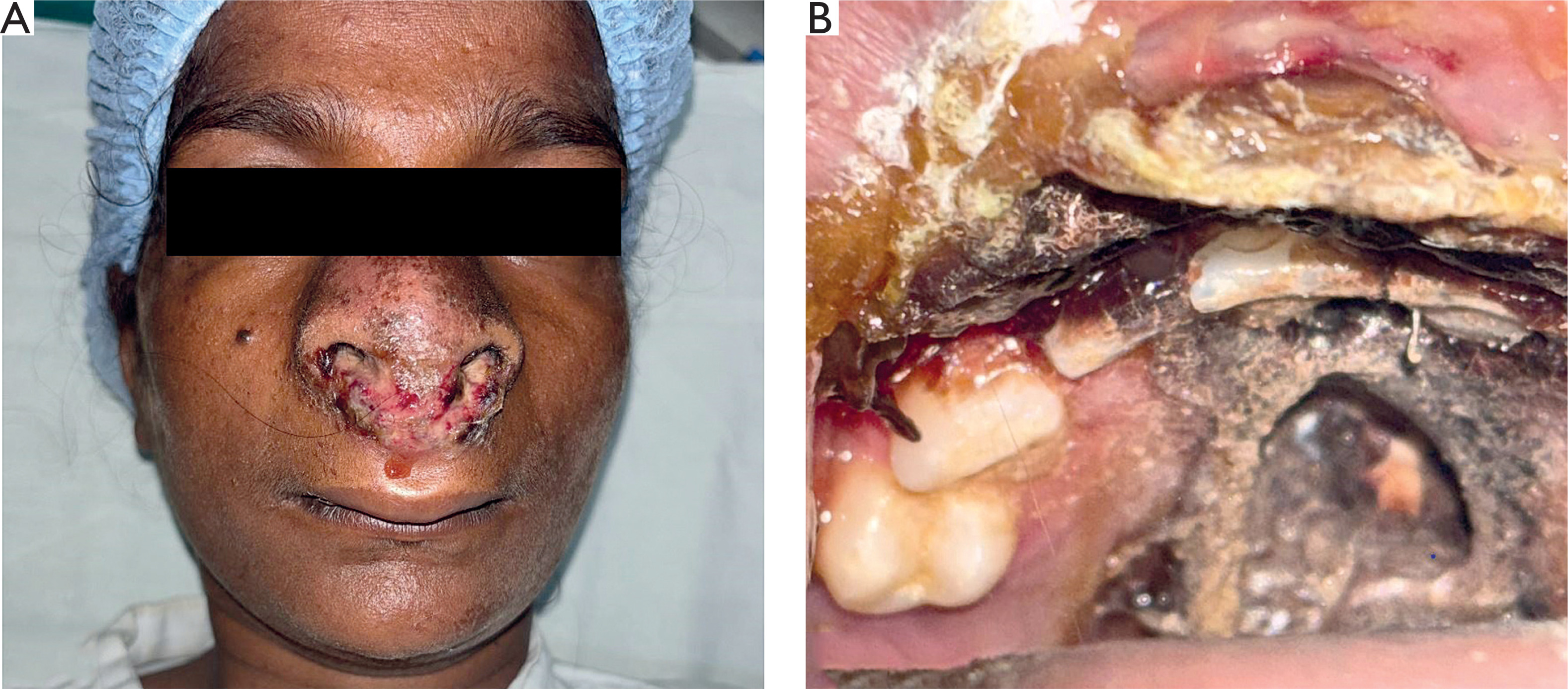
On examination, there was diffuse globular swelling involving the nose, upper lip region, and bilateral infraorbital areas, with marked destruction of the nasal architecture, causing nasal asymmetry. A large, ill-defined, friable, grayish to hyperpigmented mass extended from the nasal cavity to the upper lip and was covered with adherent serosanguineous and purulent crusts (fig. 1 A). Examination of the nasal cavity revealed complete destruction of the nasal septum. Oral examination showed a friable mass involving the hard palate, with associated palatal perforation (fig. 1 B). Systemic examination was otherwise unremarkable, and no cervical lymphadenopathy was detected. The differential diagnoses included lethal midline granuloma, rhinosporidiosis, mucormycosis, rhinoentomophthoromycosis, and granulomatosis with polyangiitis.
Figure 1
Large, friable, grayish to hyperpigmented mass protruding from the nasal cavity and extending to the upper lip, with adherent serosanguineous crusts and associated midfacial distortion and nasal asymmetry (A). Extensive tissue destruction resulting in a large full-thickness palatal perforation with a hyperkeratotic rim (B)
Routine hematological, biochemical, and serological investigations were within normal limits, except for an elevated erythrocyte sedimentation rate of 55 mm/h, C-reactive protein level of 25 mg/dl, and lactate dehydrogenase level of 294 U/l. The rK39 antigen test was negative, arguing against leishmaniasis.
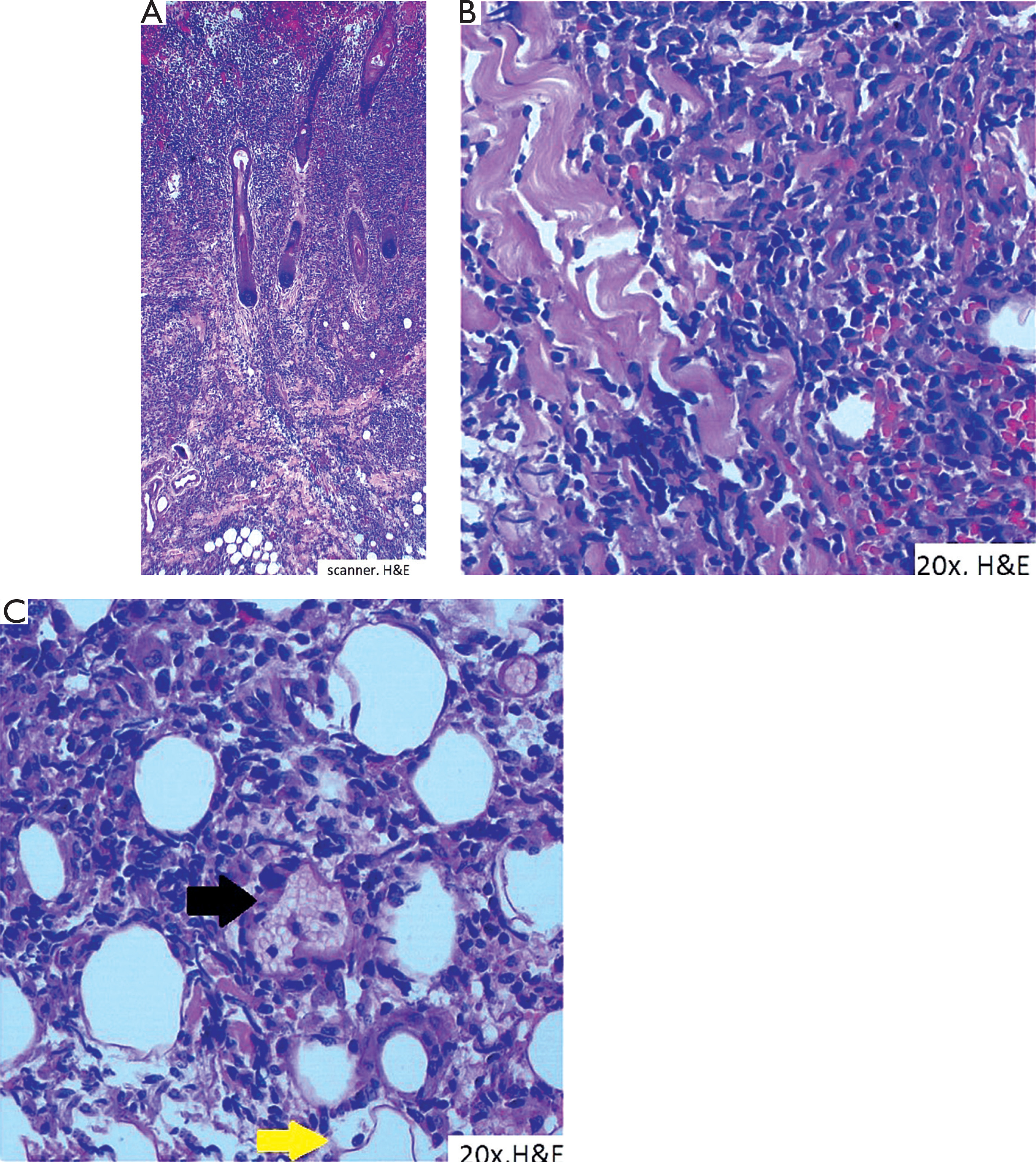
A biopsy of the mass showed a diffuse, dense pandermal lymphoid infiltrate, composed predominantly of lymphocytes of variable size, admixed with plasma cells, eosinophils, and histiocytes (fig. 2 A). The lymphoid cells infiltrated the dermis and included small, medium-sized, and large atypical cells showing pleomorphism, nuclear hyperchromasia, and foci of karyorrhectic debris (fig. 2 B). The infiltrate showed an angiocentric pattern, with focal destruction of panniculus (fig. 2 C). Special stains and cultures for fungi and mycobacteria were negative.
Figure 2
Photomicrograph showing a dense, diffuse pandermal and subcutaneous infiltrate (H&E, ×4) (A). The infiltrate contains numerous atypical medium-sized to large lymphoid cells with nuclear hyperchromasia and pleomorphism, admixed with scattered histiocytes and eosinophils (H&E ×20) (B). Angiocentric infiltrate (black arrow) with destruction of the panniculus (yellow arrow) (H&E, ×20) (C)
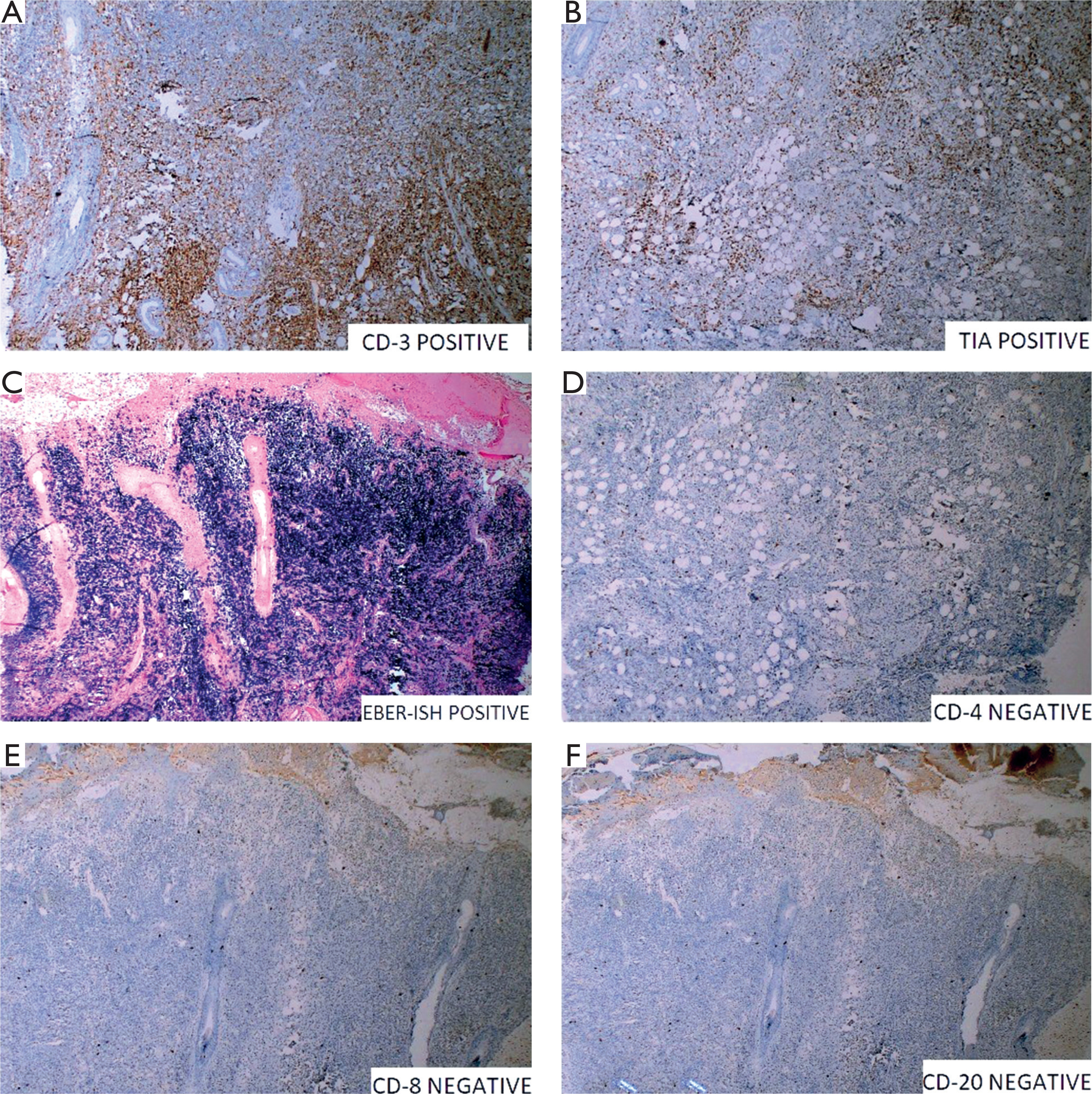
Immunohistochemical studies showed that the atypical lymphoid cells were positive for T-cell markers CD3 and CD2, as well as the cytotoxic marker TIA-1. Epstein–Barr virus-encoded RNA in situ hybridization (EBER-ISH) showed strong positivity. The cells were negative for CD4, CD7, CD8, CD25, and the B-cell marker CD20 (figs. 3 A–F). CD56 staining and T-cell receptor gene rearrangement analysis could not be performed because of limited availability. Based on the clinical presentation, histopathological findings, angiocentric and cytotoxic phenotype, and strong EBER-ISH positivity, a diagnosis consistent with extranodal natural killer/T-cell lymphoma, nasal type, was established. Although CD56 expression and T-cell receptor gene rearrangement status could not be assessed, the clinicopathological profile supported this diagnosis.
Figure 3
Immunohistochemistry showing diffuse CD3 positivity in neoplastic lymphoid cells (A), TIA-1 positivity (B), and strong positivity for Epstein–Barr virus-encoded RNA by in situ hybridization (C). Neoplastic cells are negative for CD4 (D), CD8 (E), and CD20 (F)
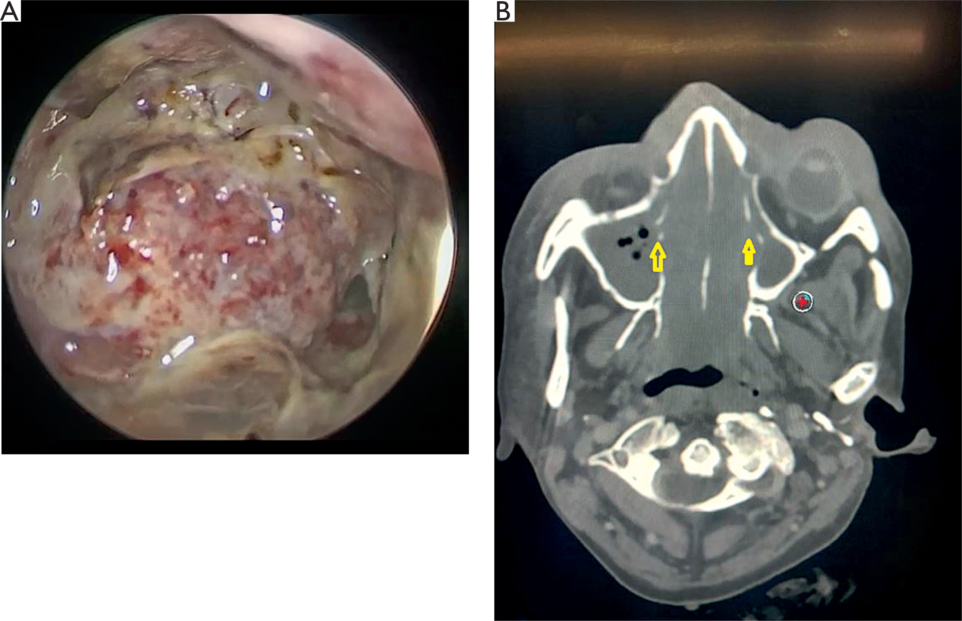
Nasal endoscopy demonstrated a fleshy mass destroying the nasal septum and lateral nasal wall and occupying both nasal cavities, with no reliably identifiable anatomical landmarks (fig. 4 A). A CT of the paranasal sinuses showed a soft-tissue lesion occupying both nasal cavities, with erosion of the turbinates and hard palate and lateral extension into the bilateral maxillary and ethmoid sinuses (fig. 4 B). PET-CT revealed no evidence of extranodal organ involvement outside the nasal region. The staging work-up was consistent with stage I disease, and the prognostic indicator of NK lymphoma (PINK) score was 0.
Figure 4
Nasal endoscopy showing a fleshy mass causing extensive destruction of the nasal septum and lateral nasal wall, with obliteration of normal anatomical landmarks (A). Computed tomography of the paranasal sinuses showing a soft-tissue lesion occupying both nasal cavities, with erosion of the turbinates (yellow arrow) and hard palate, and extension into the maxillary and ethmoid sinuses (B)
The patient was initially treated with an L-asparaginase-based SMILE regimen, consisting of dexamethasone, methotrexate, ifosfamide, L-asparaginase, and etoposide. However, because of poor tolerance, treatment was switched to a planned GELOX regimen (gemcitabine, oxaliplatin, L-asparaginase) followed by local radiotherapy.
DISCUSSION AND REVIEW OF LITERATURE
Mature T and natural killer-cell lymphomas are uncommon, heterogeneous, and often aggressive non-Hodgkin lymphomas (NHLs), accounting for 10–15% of all NHLs. ENKTCL is a rare EBV-associated lymphoma with marked geographic variation. It accounts for less than 1% of NHLs in Western countries, whereas higher frequencies have been reported in East Asia, Mexico, and South America [1, 2].
ENKTCL most commonly involves the nasal cavity and nasopharynx, where it may present as destructive midline facial disease. Historically, it has been described using several terms including “lethal midline granuloma”, “polymorphic reticulosis”, and “angiocentric lymphoma”, reflecting its destructive clinical behavior, polymorphous inflammatory background, and angiocentric growth pattern [3].
Older classifications distinguished between “nasal” disease, arising in the nasal cavity or nasopharynx, and “nasal-type” disease, which showed similar histopathological features but developed at extranasal sites, including the skin, gastrointestinal tract, testis, kidney, and upper aerodigestive tract. In the fifth edition of the World Health Organization classification of haematolymphoid tumours, the qualifier “nasal-type” was removed to acknowledge the occurrence of this lymphoma across a broad range of extranodal sites [4, 5]. After the nasal cavity and nasopharynx, the skin is one of the most frequently involved sites and may be affected primarily or secondarily. Skin involvement has been reported in approximately 10–20% of patients with nasal disease [4].
The most common clinical manifestations include chronic nasal obstruction, purulent rhinorrhea, epistaxis, and facial swelling. Because of its locally destructive growth, ENKTCL may cause perforation of the nasal septum or hard palate, destruction of the paranasal sinuses, and involvement of adjacent bone and cartilage, leading to marked distortion of the midline facial structures. Contiguous spread to the orbit or eyelid may occur, whereas involvement of cranial nerves or the meninges is uncommon. Systemic B symptoms, including fever, night sweats, and weight loss, are more often observed in advanced disease [1, 6].
ENKTCL is characterized by recurrent genetic and epigenetic alterations, including deletions involving chromosome 6q21–q25 and mutations affecting genes and signaling pathways involved in apoptosis, immune evasion, and lymphoid-cell survival. Reported abnormalities include alterations in FAS, TP53, DDX3X, BCOR, and components of the JAK–STAT pathway. Earlier studies also described occasional KRAS and KIT alterations; however, these are less consistent and should not be regarded as defining molecular features of the disease [7].
EBV infection plays a central role in the pathogenesis of ENKTCL. EBV is a ubiquitous gamma-herpesvirus that establishes lifelong latent infection in more than 90% of adults. In B cells, EBV entry is mediated mainly through the CD21/complement receptor 2 pathway, whereas the mechanism by which EBV infects natural killer (NK) and cytotoxic T cells remains incompletely understood. Several mechanisms have been proposed, including infection of precursor or activated lymphoid cells and intercellular transfer from infected B cells or epithelial cells. Once established, latent EBV infection contributes to lymphomagenesis through viral proteins and non-coding RNAs that promote cell survival, inhibit apoptosis, activate oncogenic signaling pathways, and shape an immunosuppressive tumor microenvironment [6, 7].
Latent EBV infection is also implicated in several other malignancies, including Burkitt lymphoma, nasopharyngeal carcinoma, and selected cutaneous lymphoproliferative disorders [8].
ENKTCL most commonly originates from activated NK cells, which lack rearranged T-cell receptor (TCR) genes, and less frequently from cytotoxic T cells with rearranged TCR genes. Because NK cells share part of their developmental and immunophenotypic program with cytotoxic T cells, neoplastic cells in ENKTCL often express T-cell-associated markers, particularly CD2 and cytoplasmic CD3ε, while surface CD3 is typically absent. CD5 is usually negative, whereas CD7 expression may be variable or lost. In cases of NK-cell lineage, TCR genes usually remain in germline configuration.
Most cases express NK-cell-associated marker CD56, although CD56-negative cases have also been reported. CD16 and CD57 are usually negative. In cases of cytotoxic T-cell lineage, surface CD3 may be expressed and TCR gene rearrangement may be detected, most often involving γδ rather than αβ T-cell receptors [7, 9, 10]. Previous studies have suggested that only a minority of ENKTCL cases are of true T-cell origin; however, data on the clinical, pathological, molecular, and therapeutic differences between NK-cell-derived and T-cell-derived cases remain limited [6, 11]. Therefore, in routine practice, diagnosis relies on the integration of clinical presentation, histopathological features, immunophenotype, cytotoxic marker expression, and Epstein–Barr virus status rather than on definitive assignment of cell lineage alone.
Cytotoxic granule-associated proteins, including perforin, TIA-1, and granzyme B, are usually expressed in both NK-cell-derived and T-cell-derived cases [6]. EBV infection is detected by EBER-ISH, which is positive in the great majority of ENKTCL cases and represents a key diagnostic feature [12].
Histologically, ENKTCL typically shows a polymorphous infiltrate composed of atypical lymphoid cells admixed with eosinophils, plasma cells, and histiocytes. Angiocentric and angiodestructive growth is characteristic and may lead to ischemic necrosis. The diagnosis depends on identifying atypical lymphoid cells in the appropriate clinicopathological context and confirming their cytotoxic phenotype and EBV association [13].
Clinical differential diagnoses include granulomatosis with polyangiitis, cutaneous leishmaniasis, fungal infections such as entomophthoromycosis, rhinosporidiosis, tuberculosis, South American blastomycosis, EBV-positive mucocutaneous ulcer, and sinonasal lymphomas [6].
The histopathological differential diagnosis of ENKTCL includes aggressive natural killer-cell leukemia, lymphomatoid granulomatosis, blastic plasmacytoid dendritic cell neoplasm, enteropathy-associated T-cell lymphoma, and diffuse large B-cell lymphoma [2, 3, 9]. Aggressive natural killer-cell leukemia usually presents as a fulminant systemic disease with peripheral blood and bone marrow involvement, hepatosplenomegaly, and multiorgan infiltration; in contrast to localized ENKTCL, it lacks a primary destructive sinonasal mass and is also strongly associated with EBV. Lymphomatoid granulomatosis may show angiocentric and angiodestructive features but represents an Epstein–Barr virus-positive B-cell lymphoproliferative disorder. Blastic plasmacytoid dendritic cell neoplasm may express CD56 but typically shows a distinct immunophenotype, including CD4, CD123, and TCL1 positivity. Enteropathy-associated T-cell lymphoma is usually centered in the gastrointestinal tract, is typically Epstein–Barr virus-negative, and shows clonal T-cell receptor gene rearrangement. Diffuse large B-cell lymphoma is an important sinonasal differential diagnosis and is distinguished by expression of B-cell markers such as CD20 and PAX5.
ENKTCL is staged using fluorodeoxyglucose positron emission tomography/computed tomography according to the Lugano classification, derived from the Ann Arbor system. Although this staging system is routinely used, it does not fully capture the prognostic heterogeneity of ENKTCL. Therefore, prognostic assessment is commonly supported by the Prognostic Index of Natural Killer Lymphoma (PINK), which includes age > 60 years, stage III/IV disease, distant lymph-node involvement, and non-nasal disease. The modified PINK-E model additionally incorporates detectable Epstein–Barr virus DNA [7].
Combined-modality treatment has shown better outcomes than either chemotherapy or radiotherapy alone in localized disease. Because ENKTCL is radiosensitive, involved-site radiotherapy is a cornerstone of treatment in early-stage disease; however, radiotherapy alone carries a substantial risk of relapse outside the radiation field. Anthracycline-based regimens such as CHOP are generally ineffective, partly because ENKTCL cells commonly express multidrug resistance-associated mechanisms. Current treatment strategies therefore favor non-anthracycline, asparaginase-containing regimens, including SMILE, DDGP, P-GEMOX or GELOX, and AspaMetDex [6, 7, 14]. Although SMILE is widely used, it is associated with considerable toxicity, including cytopenia, hepatotoxicity, infections, and pancreatitis. In patients who cannot tolerate intensive therapy, less intensive asparaginase-containing regimens may be considered.
Emerging therapeutic approaches increasingly focus on immune-based strategies, particularly for relapsed or refractory disease. Programmed cell death protein 1/programmed death ligand 1 blockade has shown clinically meaningful activity in ENKTCL, consistent with the frequent immune-evasion phenotype of this Epstein–Barr virus-associated malignancy. More recently, combined programmed cell death protein 1 and CD38 inhibition with cemiplimab and isatuximab has shown encouraging results in relapsed or refractory natural killer/T-cell lymphoid malignancies, with durable responses reported in a subset of patients [15]. These data support further evaluation of immune checkpoint-based combinations, although their optimal place in first-line therapy remains to be defined.
Despite therapeutic advances, ENKTCL remains an aggressive malignancy with a significant risk of relapse and systemic dissemination, particularly in advanced-stage, extranasal, or treatment-refractory disease.
CONCLUSIONS
Extranodal natural killer/T-cell lymphoma is a rare but highly aggressive malignancy that requires early clinical recognition, timely biopsy, and comprehensive immunophenotypic assessment. Its strong association with Epstein–Barr virus and its potential for destructive midfacial involvement underline the importance of distinguishing it from infectious, inflammatory, and granulomatous mimickers. Combined-modality treatment with asparaginase-containing chemotherapy and radiotherapy remains central to management, while emerging immune-based therapies may expand treatment options in relapsed or refractory disease.












