


Introduction
Upper urinary tract calculus is a common disease of the urinary system, mainly manifested as fever, renal pain, and haematuria, and the global incidence rate is 2–20% [1]. Percutaneous nephrolithotomy is usually utilized to treat upper urinary tract calculi in clinical practice, especially for the large ones, and it has become the gold standard for the treatment of large and complex nephrolithiasis [2]. However, percutaneous nephrolithotomy is unsuitable for treating patients with upper urinary tract calculi complicated by renal insufficiency, ectopic migrating kidney, solitary kidney, diabetic nephropathy, hypertensive nephropathy, and spinal deformity leading to difficulty in puncture and risk of bleeding [3]. Characterized by high safety and efficiency, flexible ureteroscopy lithotripsy is preferred for the treatment of nephrolithiasis with a diameter of < 2 cm and upper ureteral calculi with a diameter of < 1.5 cm [4]. Nevertheless, stones cannot be actively removed from the human body after flexible ureteroscopy lithotripsy, which required traditional methods such as drinking large amounts of water and increasing exercise after surgery, accompanied by unsatisfactory outcomes and stone re-formation risk [5]. The external physical vibration machine, which is self-developed in China, uses main and auxiliary vibrators to remove stones actively along the urinary tract. It has been employed to markedly improve the efficiency of removing upper urinary tract calculi in recent years [6]. Combining external physical vibration can notably improve the efficiency and stone clearance rate of flexible ureteroscopy lithotripsy and shorten the removal time [7].
Aim
This study, therefore, aimed to compare the therapeutic effects of flexible ureteroscopy alone and in combination with external physical vibration on upper urinary tract calculi.
Material and methods
Determination of sample size
The sample size was estimated according to the 2 independent sample method 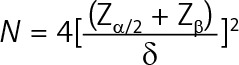 , where N is the number of required cases, Zα/2 is the Z value corresponding to α = 0.05 (Zα/2 = 1.96), Zβ is the Z value corresponding to the probability of type II error β (β = 0.20, Zβ = 0.84), and δ is the allowable error (which is generally 5.4). N was calculated as 67, so the number of samples included in each group should not be lower than 67.
General information
This prospective study was performed following the CONSORT statement. A total of 146 patients with upper urinary tract calculi admitted to our hospital from April 2017 to April 2019 were enrolled. They were divided into control and experimental groups (n = 73) by the random number table method. The control group received flexible ureteroscopy lithotripsy, while the experimental group underwent external physical vibration combined with flexible ureteroscopy lithotripsy. In the control group there were 49 males and 24 females aged 21–78 years, with an average age of 45.42 ±10.64 years. In the experimental group there were 46 males and 27 females aged 20–76 years old, with an average age of 46.08 ±11.16 years.
Inclusion criteria were as follows: a) Patients with upper urinary tract calculi diagnosed by ultrasonography of the urinary system, computed tomography (CT) and CT urography (CTU), b) those with unilateral calculi, c) those who had calculi with a diameter of < 2 cm, d) those who met the indications for surgery after electrocardiography, blood biochemistry, routine blood and urine tests, urine culture, full-size chest radiography, and tests of blood coagulation, liver and kidney, and e) those with complete clinical data.
Exclusion criteria were as follows: a) patients with urinary system obstruction and malformation, b) those with blood coagulation dysfunction, c) those who were in pregnancy, lactation, or menstrual period, d) those with history of severe cardiovascular diseases, e) those with severe pulmonary infection, urinary tract infection, or other serious disease, f) those with severe hydronephrosis, g) those with poor compliance, or h) those with incomplete clinical data.
This study was approved by the institutional ethics committee of our hospital. Written informed consent was obtained from each patient and their families.
Collection of baseline clinical data
The baseline clinical data of patients were collected through electronic medical records. After admission, age and gender were recorded, and height (m) and weight (kg) were measured to calculate the body mass index (BMI = weight/height2). The stone size was detected by B-ultrasonography. The CT values of stone edge, middle area, and core were measured by a CT machine, and the average was recorded.
Treatment methods
The control group received flexible ureteroscopy lithotripsy. After surgery, the patients were required to drink more than 3000 ml of water every day, and to skip rope, jump, and go up and down stairs. The patients were guided to lie on the healthy side when they were resting, and the cases with lower calyx calculi rested upside down.
The experimental group underwent flexible ureteroscopy lithotripsy combined with external physical vibration to remove stone fragments actively.
Flexible ureteroscopy lithotripsy
The patients were sent to the operating room. After successful general anaesthesia, the patients took the oblique supine lithotomy position, followed by using sandbags to raise the shoulders and buttocks, and tilting the affected side up by 30–45°. The lower limbs of the affected side were raised and bent, and the lower limbs of the healthy side were straightened and slightly extended. Sterile towels were routinely draped. A rigid ureteroscope (Olympus, Japan) was used to insert the guide wire retrogradely into the affected ureter to the renal pelvis. An F12 ureteral expansion sheath was inserted along the guide wire, the inner sheath was pulled out to retain the outer one, and an F8 flexible ureteroscope (Olympus, Japan) was inserted along the outer sheath while flushing. After the upper part of the ureter or renal pelvis was observed and the stones were found, the guide wire was pulled out. Subsequently, a 200 µm holmium laser fibre was inserted along the operating sheath of the flexible ureteroscope, followed by adjusting the output power of the holmium laser. Then the stone was crushed to a size of 1–2 mm in a cannibalistic manner. After surgery, the flexible ureteroscope was removed, the guide wire was inserted, the ureteral expansion sheath was withdrawn, the double J tube of the ureter was retained along the guide wire, and the ureter was also retained.
External physical vibration
Thirty minutes before treatment, 20 mg furosemide was intravenously injected and the patients were required to drink a large volume water. Residual stones and hydrops the of upper urinary tract were observed by B-ultrasonography. Then the external physical vibration machine (Zhengzhou Kang Baijia Technology Co., Ltd.) was started. According to the ultrasound results, the patients were asked to lie in the supine position, decubitus position of the healthy side, or prone position on the treatment bed, and the inclination angle of the bed was adjusted. The main and auxiliary vibrators were turned on at the same time. The vibration frequency of the main vibrator was 3000/min, and that of the auxiliary vibrator was 1800/min. The amplitude of both was 5 mm. The pushing force was adjusted according to the patient’s condition. The main and auxiliary vibrators co-ordinately vibrated to push the stones into the renal pelvis, and then the bed was adjusted to a head-high/foot-low position. The vibration continuously drove the stones to run down along the renal pelvis and ureter for about 8–10 min. According to the outcomes, 1 or 2 more cycles were conducted, no more than 3 times, and the stone position was detected in real time by B-ultrasonography during the operation.
Component analysis of urinary tract stones
After treatment, the patients were required to drink a large volume of water and to withold their urine. When they withheld to the limit, the urine was discharged. The stones were collected by a filter, and then infrared spectroscopy of the stone composition was performed.
Measurement of renal function indices
Venous blood was drawn from every patient and placed into an anticoagulation tube 2 h before and 1 week after treatment. After centrifugation at 3000 rpm for 10 min, the serum was separated to detect blood urea nitrogen (BUN) and serum creatinine (Scr) levels before and after treatment using an automatic biochemical analyser (Beckman Coulter, USA).
Follow-up
The patients were asked whether there was stone removal on the day after treatment. On the next day, the stone clearance rate was measured through abdominal CT, and the removal results were followed up for 1 week. If there were residual stones, vibration was performed again. If the stones were removed, the double J tube was withdrawn. After 3 weeks of follow-up, renal function and abdominal CT were re-examined to determine stone removal and recovery, and whether the double J tube should be withdrawn. The postoperative complications of patients were recorded. The patients were followed up until April 2020, at least 4 times for each case. Stone re-formation was examined through ultrasonography, abdominal CT, and CTU within 1 year of the treatment.
Statistical analysis
SPSS 19.0 software was adopted to perform 1-way analysis of variance. GraphPad Prism 5.0 software was used for plotting. The differences between 2 groups were compared by t-test, and those among multiple rates or constituent ratios were compared by χ2 test. The Kaplan-Meier method was utilized to analyse the stone-free rate during 1-year follow-up. P < 0.05 indicated that a difference was statistically significant.
Results
Baseline clinical data
The 2 groups had similar age, gender ratio, BMI, stone size, stone CT value, number of patients with renal insufficiency, and stone type (p > 0.05) (Table I).
Table I
Baseline clinical data
Rate of finding stones in the urine on the day after treatment and stone clearance rate at different time points
The rate of finding stones in the urine on the day after treatment was significantly higher in the experimental group (100%) than that in the control group (29.73%). The stone clearance rates on the day, and 1 week and 2 weeks after treatment in the experimental group were 71.23%, 87.67%, and 95.89%, respectively, which were significantly higher than those of the control group at corresponding time points (p < 0.05). Also, such rates in both groups increased with extended time after treatment (Table II).
Table II
Rate of finding stones on the day of treatment and stone clearance rate at different time points
Components of urinary tract stones
The 2 groups had similar components of urinary tract stones (p > 0.05) (Table III).
Table III
Components of urinary stones
Levels of renal function indices BUN and Scr before and after treatment
The 2 groups had similar levels of renal function indices BUN and Scr before treatment (p > 0.05). Compared with before treatment, the levels of BUN and Scr significantly decreased after treatment in both groups, which were lower in the experimental group (p < 0.05). Moreover, the 2 groups had significantly different improvement of BUN and Scr levels between the pre- and post-operative period (p < 0.05) (Table IV).
Table IV
Levels of renal function indices BUN and Scr before and after treatment
| Variable | BUN [mmol/l] | Scr [µmol/l] | ||||
|---|---|---|---|---|---|---|
| Before treatment | After treatment | Difference between pre- and post-operative period | Before treatment | After treatment | Difference between pre- and post-operative period | |
| Control group (n = 73) | 7.74 ±0.85 | 7.49 ±0.62* | 0.24 ±0.03 | 79.64 ±10.37 | 72.38 ±9.72* | 7.34 ±1.22 |
| Experimental group (n = 73) | 7.82 ±0.73 | 7.25 ±0.68* | 0.68 ±0.11 | 79.85 ±11.41 | 68.43 ±8.69* | 12.45 ±1.34 |
| t | 0.610 | 2.228 | 32.972 | 0.116 | 2.588 | 24.092 |
| P-value | 0.543 | 0.027 | < 0.001 | 0.908 | 0.011 | < 0.001 |
Incidence rates of complications after treatment
The 2 groups had similar incidence rates of complications such as haematuria, dizziness, fever, and low back pain as well as total number of complications (p > 0.05) (Table V).
Table V
Incidence rates of complications after treatment
Discussion
Urinary tract calculus is a common urologic disease induced by multiple factors such as heredity, water quality, climate, nature of work, eating habits, obesity, and diabetes mellitus [8]. Urinary tract calculi easily occur in the upper part, which may cause sepsis and renal damage if not treated promptly [9]. Currently, upper urinary tract calculi are mainly treated by conservative therapy, extracorporeal shock wave lithotripsy, flexible ureteroscopy lithotripsy, percutaneous nephrolithotomy, and open surgery [10]. If stone fragments are not removed in time, they are prone to fusion or increase after 3 months, inducing obstructive or infective symptoms [11]. Although the therapies for urinary tract calculi have developed rapidly, stones still cannot be effectively removed.
In recent years, external physical vibration has been used to remove stones in patients with urinary tract calculi, with good clinical effects [12]. During external physical vibration, the movement space of stones is first expanded by drugs, then the stones are separated from tissues by high-frequency vibrators, and the body position is adjusted in real time according to the movement direction of stones, so the physiological cavity of human body is maintained downward, which is conducive to stone removal. The auxiliary shock wave is conducted to the target location through the conduction of muscle tissue, so stones are suspended in the liquid space. The main shock wave can push stones to move along the physiological space until they are removed [13]. External physical vibration, as a new method for removing stones by simple physical means, is painless and harmless [14]. Wu et al. effectively removed stones by combining flexible ureteroscopy lithotripsy with external physical vibration. Stone fragments were found on the day after treatment, and the stone clearance rate 2 weeks after treatment increased remarkably [15]. In this study, the rate of finding stones in the urine on the day after treatment, as well as stone clearance rates on the day, and 1 week and 2 weeks after treatment were significantly higher in the experimental group than those in the control group. In addition, the components of urinary tract stones of the 2 groups were similar, being consistent with the results of a previous study [16].
If upper urinary tract calculi are not treated in a timely and effective manner, renal insufficiency or even acute obstructive renal failure may occur, which seriously threatens the health, safety, and quality of life of patients [17]. Wang et al. reported that the serum levels of renal function indices BUN and Scr significantly decreased in patients with upper urinary tract calculi undergoing flexible ureteroscopy lithotripsy, and the renal function of cases with renal insufficiency was significantly ameliorated [18]. In this study, compared with before treatment, the levels of BUN and Scr significantly decreased after treatment in the 2 groups, especially in experimental group. It is well-established that haematuria, dizziness, fever, and low back pain usually occur after withdrawal of the double J tube after flexible ureteroscopy lithotripsy. Herein, the 2 groups had similar incidence rates of complications after treatment. Moreover, residual stones are risk factors for the postoperative re-formation of upper urinary tract calculi, suggesting that promoting the removal of stones after lithotripsy for patients with upper urinary tract calculi and increasing the stone clearance rate can reduce the risk of postoperative re-formation and thus relieve pain and economic burden [19]. In this study, the results of Kaplan-Meier method indicated that during 1-year follow-up, the stone-free rate was significantly higher in the experimental group (97.26%) than that in the control group (83.56%).
Conclusions
External physical vibration combined with flexible ureteroscopy lithotripsy can significantly increase the rate of finding stones in the urine on the day after treatment and the clearance rate of upper urinary tract calculi, ameliorate the levels of renal function indices BUN and Scr, and reduce the stone re-formation rate.