


Dear Editor,
With reference to the article entitled “Ultrasound-guided percutaneous tracheotomy” by Plata and Gaszyński [1], I would like to draw the readers’ attention to the fact that eight different techniques of percutaneous tracheotomy are currently available and usable for clinical practice. They are characterised by different indications and different clinical outcomes, including the incidence of complications that are generated. Table 1 of the paper listing indications and contraindications presumably concerns one of these methods of management. For instance, the feasibility of percutaneous tracheotomy in patients with coagulation disorders (BlueDolphin) or in children (TLT-J) has been known for years. Likewise, in other clinical situations described in Table 1, the procedure can be performed depending on the method chosen and operator’s skills [2]. Importantly, ultrasound monitoring should be used in each of these methods, bringing unquestionable benefits and improving the safety of the procedure in a multidirectional manner [2, 3]. The method of tracheotomy by Griggs et al. (1990), described in the context of ultrasound monitoring, gained its popularity mainly due to low costs, use of a reusable dilator, a modification of guidewire dilating forceps (GWDF) and the fact that it is commonly performed without bronchoscopy monitoring, at a hazardously steep learning curve (20 procedures?) (H. Ebbinghaus). However, in this regard, any form of monitoring, including non-invasive US evaluation, undoubtedly improves the safety and conditions of performing tracheotomy [2, 4]. Moreover, it should be emphasised that one of the biggest flaws of the Griggs technique is that the force exerted on the dilating forceps arms is subjectively (sic!) decided during shaping of the stoma (Figures 1 and 2).
FIGURE 1
A two-handed method of shaping the stoma at low tracheal wall compliance during tracheotomy according to Griggs et al.
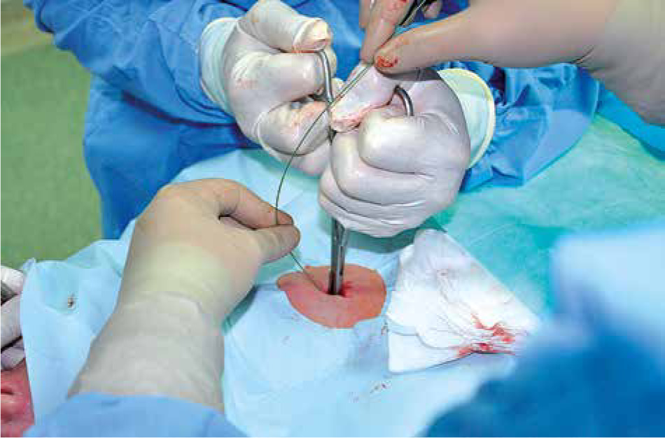
GRIGGS TRACHEOTOMY
In such cases, ischaemia of the tracheal ring ligaments and/or cartilaginous rings can result in the stoma of uncontrollable sizes, disruption of the cartilaginous rings or even separation of the distal and proximal parts of the trachea. This is particularly likely when the procedure described is performed in cases of critical perfusion disorders and prolonged intubation. According to the Authors, tracheomalacia is a relative contraindication for the procedure. To prevent the complications, Tracheo S.E.T (Xmed S.r.l, Mirandola, Italy) was designed characterised by a controllable extent and different mechanics of dilating the forceps. Nevertheless, these changes seem to have slight and still undetermined effects on the manoeuvre’s safety [2]. Unfortunately, the tracheal condition at the site of tracheotomy and possible complications cannot be assessed based on ultrasound monitoring without bronchoscopy; most importantly, however, the change of the method applied cannot be decided based on such a monitoring (e.g. to the centrifugal one). Additionally, we cannot anticipate whether the malacic changes at the procedure site have already occurred (dilemma of early and late tracheotomy). Therefore, I believe that ultrasound can be used as a method for tracheal puncture positioning and visualising the anatomy of the procedure field; nevertheless, it is bronchoscopy that remains the major strategy to visualise percutaneous tracheotomy procedures (including the Griggs method) [2, 3, 5]. The additional use of ultrasound can increase the safety of scheduled procedures and thus, the techniques can be considered complementary in this regard. Furthermore, the Griggs technique can be applied for rescue purposes (as an alternative to conicotomy); in such cases, the use of ultrasound techniques, being less time-consuming, may significantly improve the efficiency of management. Moreover, the Authors’ comments regarding ultrasound monitoring of blood supply to the tracheal puncture site (including the underestimated middle central jugular vein) and to the deeper located vessels, seem essential [6]. Although the interpretation of sonoanatomic images of vessels requires more proficiency, a reduced potentially life-threatening risk of injuring the brachiocephalic trunk is an excellent argument for combining brochoscopic monitoring and ultrasound visualisation.