
INTRODUCTION
Lichen planus (LP) is a chronic, immune-mediated inflammatory dermatosis that may affect the skin, mucous membranes, hair follicles and nails. It is typically characterized by pruritic, violaceous, polygonal, flat-topped papules and plaques, with a predilection for the flexor surfaces of the wrists and ankles and the lower back [1]. Although its etiology remains incompletely understood, the pathogenesis of LP involves a T-cell-mediated cytotoxic immune response directed against basal keratinocytes. Viral infections, medications, and contact allergens have been proposed as potential triggering or modifying factors [2]. LP affects approximately 0.5–1% of the general population, with a slight female predominance and a peak incidence between 30 and 60 years of age [3].
Classical LP is commonly described using the “6 Ps”: planar, purple, polygonal, pruritic papules and plaques. However, numerous clinical and morphological variants have been recognized, including hypertrophic, atrophic, bullous, annular, actinic, follicular (lichen planopilaris), erosive, and pigmented forms [4, 5]. These variants may pose diagnostic challenges because they can mimic other dermatoses, including psoriasis, eczema, and cutaneous lupus erythematosus [6]. Confluent LP is an uncommon clinical presentation in which individual lesions coalesce to form larger plaques. Its distribution may be influenced by friction or trauma through the Koebner phenomenon [7]. The Koebner phenomenon, also referred to as the isomorphic response, is defined as the development of lesions characteristic of an existing dermatosis at sites of cutaneous injury and is observed in conditions such as psoriasis, vitiligo, and LP [8, 9]. In LP, koebnerization may result in linear lesions or accentuation and confluence of lesions in areas exposed to repeated mechanical stress, including friction from clothing [10].
On dermoscopic examination, LP typically demonstrates Wickham striae, which appear as fine white lines or reticular structures and correspond histopathologically to areas of hypergranulosis and hyperkeratosis [3]. The classic histopathological features include compact orthokeratosis, wedge-shaped hypergranulosis, irregular or saw-tooth acanthosis, vacuolar degeneration of the basal layer, a band-like lymphocytic infiltrate at the dermoepidermal junction, and pigment incontinence [11]. Individual variants may demonstrate additional or more pronounced histopathological features, such as marked acanthosis and hyperkeratosis in hypertrophic LP [12]. Differential diagnoses include allergic contact dermatitis (ACD), lichenoid drug eruptions, and graft-versus-host disease. In clinically atypical cases, histological examination may be required to confirm the diagnosis, while patch testing may be useful when ACD is suspected on the basis of the lesion distribution and exposure history [6, 13].
OBJECTIVE
The aim of this case report is to describe an uncommon presentation of confluent LP mimicking ACD, with particular emphasis on the possible contribution of koebnerization in friction-prone areas and the role of histopathological examination in resolving the diagnostic uncertainty. By presenting this case, we aim to expand the available literature on atypical clinical patterns of LP and to facilitate the recognition and appropriate management of similar presentations thereby reducing the risk of misdiagnosis and inappropriate treatment.
CASE REPORT
A 30-year-old female presented to the dermatology outpatient department with a 6-month history of violaceous-to-hyperpigmented, elevated lesions on the trunk. The onset was insidious, and the lesions gradually progressed, with increasing confluence in areas exposed to friction from undergarments. They were intensely pruritic, resulting in excoriations and occasional oozing. There was no history of preceding medication use, systemic symptoms, or similar lesions in family members. The patient denied any recent vaccinations, infections, or exposure to new potential allergens other than routinely worn clothing.
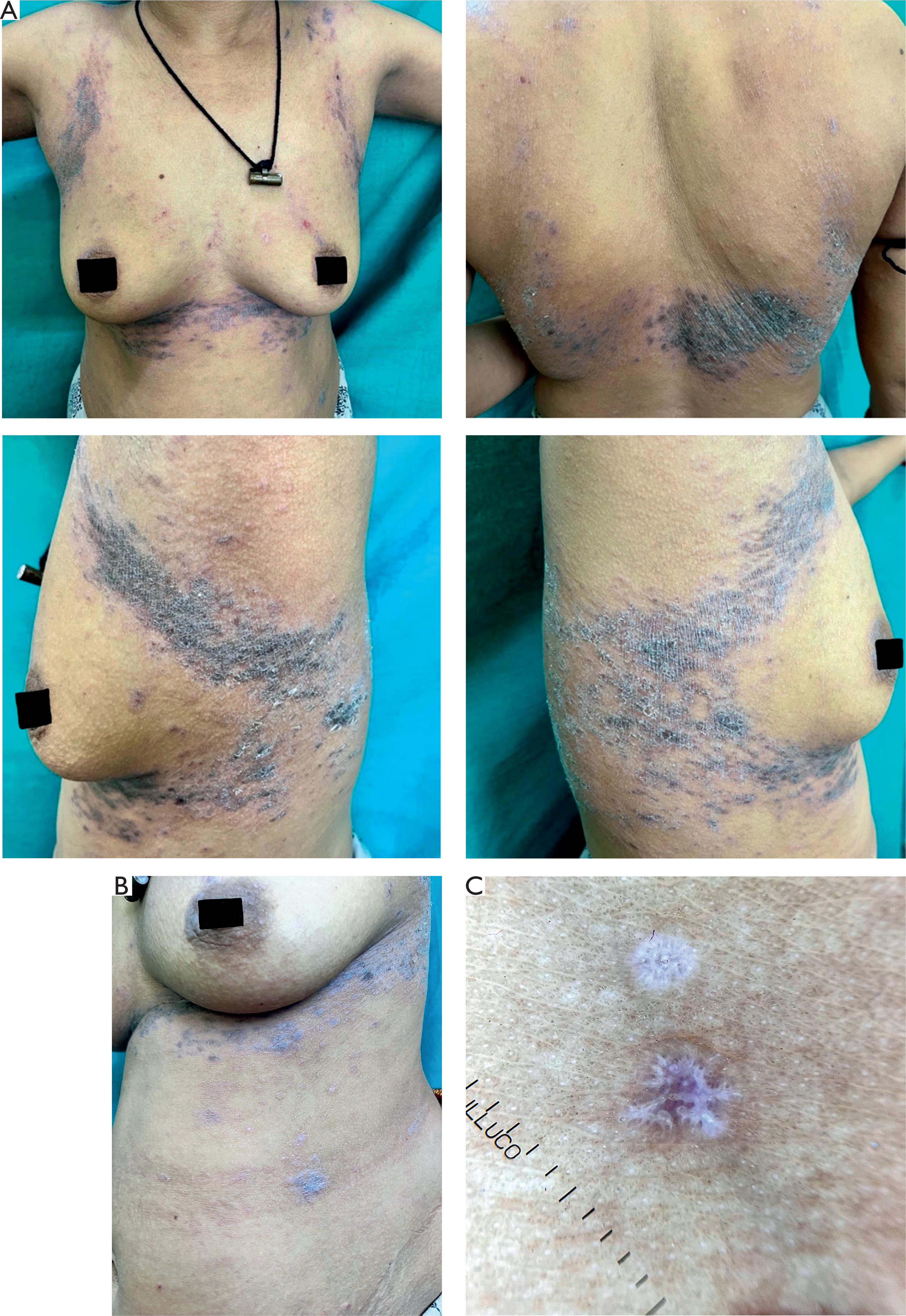
On clinical examination, violaceous, reticulated plaques were noted on the chest and back (fig. 1 A), accompanied by several isolated polygonal papules on the right side of the abdomen (fig. 1 B). The plaques were predominantly confluent in areas corresponding to the bra-straps and waistband, suggesting a koebnerized distribution [14]. No oral, genital, or nail involvement was observed. Dermoscopy revealed structures resembling Wickham striae, supporting the presence of a lichenoid process (fig. 1 C) [3].
Figure 1
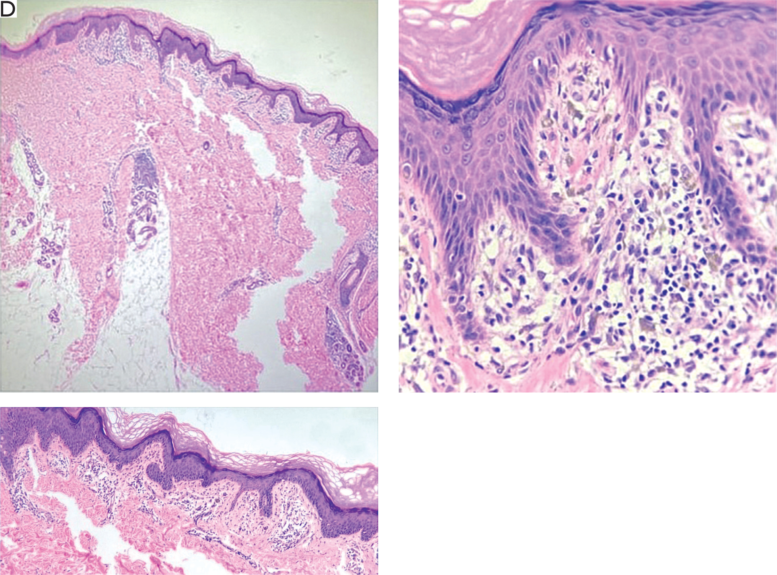
A – Violaceous reticulated plaques on the chest and back; B – isolated polygonal papules on the right side of the abdomen; C – polarized dermoscopy showing structures resembling Wickham striae (DermLite DL3, ×10); D – histological examination showing wedge-shaped hypergranulosis, vacuolar degeneration of the basal layer, a band-like lymphocytic infiltrate, and pigment incontinence (H & E; 4×, 10× and 40× magnification)
Based on the distribution of the lesions, lichenoid ACD secondary to fabrics or textile dyes in the patient’s undergarments was initially considered [13, 15]. Patch testing using a standard series supplemented with textile allergens yielded negative results, making ACD less likely. Owing to the persistent diagnostic uncertainty, a skin biopsy was obtained from a representative plaque on the back.
Histopathological examination demonstrated hyperkeratosis without parakeratosis, wedge-shaped hypergranulosis, vacuolar degeneration of the basal layer, a dense band-like lymphocytic infiltrate closely apposed to the dermoepidermal junction, and prominent pigment incontinence in the superficial dermis (fig. 1 D) [12]. These findings were consistent with LP. The confluent distribution of the lesions in friction-prone areas suggested that chronic mechanical irritation and koebnerization may have contributed to the observed clinical pattern [7, 8].
The patient was treated with a potent topical corticosteroid, clobetasol propionate 0.05% ointment, applied twice daily for 4 weeks, along with an oral antihistamine for symptomatic control of pruritus. She was also advised to avoid tight-fitting clothing to reduce further mechanical irritation. At the 2-month follow-up, marked resolution of the plaques was observed, with residual post-inflammatory hyperpigmentation. No recurrence was reported during the subsequent 6 months of follow-up.
DISCUSSION
Confluent LP, as exemplified in this case, represents a rare and diagnostically challenging variant of LP, where mechanical friction via the Koebner phenomenon drives lesion coalescence in trauma-prone sites [7, 8]. The violaceous-to-blackish hue and pruritic, excoriated plaques initially evoked allergic contact dermatitis (ACD), a common mimic, but negative patch testing and confirmatory histopathology underscored the need for a multimodal diagnostic approach [6, 13]. This presentation aligns with reports of koebnerized LP in clothing-contact areas, emphasizing avoidance of irritants as an adjunct to pharmacotherapy [10, 14].
Treatment of LP is tailored to disease extent, severity, and variant, with the goal of symptom relief, lesion resolution, and prevention of scarring or hyperpigmentation. For localized cutaneous LP, including confluent forms, first-line therapy involves potent topical corticosteroids (TCS), such as clobetasol propionate 0.05% ointment applied twice daily for 4-6 weeks [3, 11]. Adjunctive topical calcineurin inhibitors (e.g., tacrolimus 0.1% ointment twice daily) serve as steroid-sparing agents, particularly for facial or intertriginous involvement [6, 7]. Oral antihistamines effectively control pruritus, as utilized here [11, 15].
Chronic LP, persisting > 6 months, requires maintenance strategies to mitigate relapse, which occurs in up to 20% of cases post-acute therapy [16]. Intermittent TCS or narrowband UVB phototherapy is recommended [11, 12].
Disseminated (generalized) LP, affecting > 10% body surface area, demands systemic intervention due to extensive involvement and heightened pruritus [16]. Oral corticosteroids provide rapid control: prednisone 0.5–1 mg/kg/day tapered over 4–8 weeks achieves resolution in most cases.
This case’s favorable response to topical clobetasol highlights the efficacy of conservative management in koebnerized, localized LP, yet broader variants necessitate escalated therapies. Emerging data on biologics (e.g., dupilumab for pruritus-dominant cases) and apremilast (30 mg twice daily for 12 weeks) show promise in chronic/disseminated disease, pending larger trials [5, 10].
CONCLUSIONS
This case illustrates the diagnostic challenge posed by confluent LP, which may closely mimic eczema or lichenoid ACD, particularly when lesions are distributed over friction-prone areas [6, 13]. The Koebner phenomenon may have contributed to the confluence of the lesions, highlighting how mechanical trauma can influence the distribution and clinical presentation of LP [9]. Clinicians should consider atypical variants of LP in patients presenting with pruritic, violaceous eruptions and use dermoscopy, patch testing, and histological examination, as appropriate, to facilitate differential diagnosis [2]. An accurate diagnosis enables appropriate treatment and may reduce the risk of persistent symptoms, post-inflammatory hyperpigmentation, and other complications. Further studies investigating the role of mechanical friction and other potential triggers in LP may improve our understanding of this condition.












