
Introduction
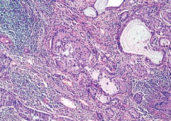
Mucoepidermoid carcinoma (MEC) of the breast is an exceptionally rare and histologically distinct subtype of invasive breast carcinoma, classified among the salivary gland-type tumours of the breast. It accounts for 0.2–0.3% of all invasive breast carcinomas [1–4]. Morphologically, MEC of the breast recapitulates the biphasic architecture of its salivary gland counterpart, comprising a heterogeneous admixture of mucin-secreting, intermediate, and epidermoid cells arranged in cystic, glandular, or solid growth patterns (Figure 1) [5, 6]. The histologic grade, typically defined as low, intermediate, or high according to criteria adapted from salivary gland MEC, has significant prognostic implications, with high-grade tumours showing solid growth, nuclear pleomorphism, necrosis, and increased mitotic activity associated with adverse outcomes [5, 7].
Figure 1
A case of mammary mucoepidermoid carcinoma showing glandular structures with intraluminal mucin and scattered epidermoid-appearing nests; focal lymphocytic infiltration is present in the stroma (HE, 200×)
Courtesy of Dr. Reena Tomar and PathologyOutlines.com.
Immunophenotypically and molecularly, MEC of the breast frequently exhibits a triple-negative profile, lacking expression of oestrogen receptor (ER), progesterone receptor (PR), and HER2, while demonstrating positivity for basal markers (CK5/6 and p63) [4, 6]. These features can lead to diagnostic confusion with other triple-negative breast carcinomas (TNBC) with a basal phenotype, such as adenosquamous or metaplastic carcinoma. However, the presence of mucin-secreting cells and the detection of specific molecular alterations can aid in distinguishing between the two. In particular, MAML2 gene rearrangements, most often the CRTC1-MAML2 fusion, are characteristic of salivary gland MEC and have also been identified in a subset of breast MEC, suggesting a common pathogenesis and association with lower histologic grade and favourable clinical behaviour [4, 8].
Clinically, MEC of the breast most often presents as a palpable, well-circumscribed mass, and while comprehensive demographic data are limited, reported cases involve middle-aged and postmenopausal women [7, 9, 10]. Low- and intermediate-grade MEC of the breast are described in the literature to follow a generally indolent clinical course and may be treated successfully with surgical excision, in some series with breast-conserving surgery and sentinel node evaluation, without evidence of early recurrence [4, 11]. Conversely, high-grade MEC of the breast has been reported to exhibit more aggressive behaviour, including lymph node and distant metastasis, and disease- related deaths [4, 7]. Because of the potentially less aggressive behaviour in the low/intermediate-grade subset of MEC, accurate histologic grading and appropriate molecular/immunohistochemical characterisation are crucial to guide management and follow-up.
A recent population-based analysis by Ch’ng [12] analysed 488 cases of salivary gland-type carcinomas of the breast using the Surveillance, Epidemiology, and End Results (SEER) database and compared them with over 375,000 cases of invasive ductal carcinoma not otherwise specified (IDC-NOS). They identified the tumour grade, American Joint Committee on Cancer (AJCC) stage, HER2 overexpression, and acinic cell differentiation as independent prognostic factors. The study underscored the heterogeneity and variable outcomes across this group, with acinic cell carcinoma showing a worse prognosis compared to adenoid cystic carcinoma. However, only five cases of MEC were included, precluding any meaningful statistical analysis or prognostic inference regarding this specific subtype. Consequently, despite the study’s valuable contribution to the understanding of salivary gland-type breast carcinomas, MEC remains underrepresented and incompletely defined within population-level datasets.
To address this limitation, the present study aims to expand upon prior findings by performing a comprehensive comparative analysis between MEC of the breast and IDC-NOS. By leveraging a larger cohort and extended follow-up data, this work seeks to elucidate the epidemiologic distribution, clinicopathologic spectrum, therapeutic patterns, and survival outcomes of MECB relative to IDC-NOS. This approach not only aims to broaden the current understanding of MEC beyond its traditional comparison with other salivary gland-type tumours but also offers a clinically relevant benchmark against the most prevalent form of invasive breast carcinoma, representing a key step toward clarifying its prognostic behaviour and informing specific management strategies.
Material and methods
Case selection and data acquisition
This retrospective cohort study was conducted using data from the SEER Program of the U.S. National Cancer Institute, which provides population-based information on cancer incidence, demographics, tumour characteristics, treatment, and survival. Data were extracted using SEER*Stat software (version 8.4.2; National Cancer Institute, Bethesda, MD), including all cases diagnosed with primary breast carcinoma between 1 January 1975 and 31 December 2022.
Eligible cases were identified using the International Classification of Diseases for Oncology, Third Edition (ICD-O-3) morphology code 8430/3 for MEC, in accordance with the World Health Organisation classification of salivary gland-type breast tumours. For comparative purposes, cases of IDC-NOS, 8500/3, were also retrieved. Only confirmed cases were included in this study (SEER Diagnostic Confirmation codes indicating histology/cytology); histology for nonsurgical cases was derived from percutaneous/core biopsy or cytology. Cases lacking microscopic confirmation (clinical diagnosis only, death certificate or autopsy only) were excluded.
Collected demographic variables included age at diagnosis, sex, race, marital status, and median household income. Tumour-related characteristics included year of diagnosis, laterality, histologic grade, tumour size, nodal involvement, AJCC 8th edition stage, hormone receptor (ER, PR), HER2 status, presence or absence of distant metastases to liver, lung, brain, and bone, survival duration in months, survival status, and cause of death. Treatment variables included whether they had surgery, radiotherapy, and systemic therapy.
Statistical analysis
Given the rarity of MEC and the limited sample size, establishing a directly matched comparator group was not feasible. To provide context for clinical characteristics and survival outcomes, the MEC cohort was descriptively compared with a larger cohort of IDC-NOS cases diagnosed within the same SEER registry period. This approach was intended to highlight demographic, clinicopathologic, and outcome differences. We acknowledge that IDC-NOS represents a biologically and histologically heterogeneous group and therefore does not accurately reflect the specific molecular or histopathologic features of MEC. Accordingly, no formal statistical matching or subtype-specific stratification was performed, and these comparisons should be interpreted as descriptive rather than inferential.
All statistical analyses were performed using Stata/SE version 18.5 (StataCorp LLC, College Station, TX). Descriptive statistics were used to summarise case and tumour characteristics. Categorical variables were expressed as frequencies and percentages, while continuous variables were assessed for normality of distribution using boxplots and descriptive statistics. Variables demonstrating an approximately normal distribution were summarised as mean ± standard deviation (SD), whereas non-normally distributed variables were presented as median and interquartile range (IQR).
For comparisons between groups, one-way analysis of variance was used for normally distributed continuous variables, and the Kruskal-Wallis test was employed when the normality assumptions were not met. Differences in categorical variables were examined using Pearson’s χ2 test. All statistical tests were two-sided, and a p < 0.05 was considered to indicate statistical significance.
Survival analysis was conducted using the Kaplan-Meier method to estimate overall survival (OS) and disease- specific survival (DSS), defined as the time in months from diagnosis to death from any cause (OS) or death attributable to breast cancer (DSS). The log-rank test was used to compare survival distributions across histologic subtypes.
To evaluate prognostic factors associated with survival, Cox proportional hazards regression models were employed to estimate hazard ratios (HR) and corresponding 95% confidence intervals (CI). Univariate Cox models were initially employed to identify potential predictors of survival across demographic, clinicopathologic, and treatment-related variables. Subsequently, multivariable Cox models were constructed, adjusting for key baseline factors including age group, race, marital status, laterality, median household income, number of primary malignancies, tumour size, nodal and metastatic status (bone, brain, liver, lung), hormone receptor status, HER2 expression, histologic grade, AJCC 8th edition stage, and treatment modalities (surgery, radiotherapy, and chemotherapy). Kaplan-Meier survival curves were generated for both OS and DSS to visualise survival differences between MEC and IDC-NOS, both before and after adjustment for covariates.
Results
Case demographics
This study included 50,923 cases, comprising 42 with MEC and 50,881 with IDC-NOS. Tables 1–3 summarise the cohorts’ demographic, clinicopathologic, treatment, and survival characteristics.
Table 1
Demographic characteristics of mucoepidermoid carcinoma vs. invasive ductal carcinoma not otherwise specified in the Surveillance, Epidemiology, and End Results database
Table 2
Clinicopathologic characteristics of mucoepidermoid carcinoma vs. invasive ductal carcinoma not otherwise specified in the Surveillance, Epidemiology, and End Results databaseI
Table 3
Treatment modalities (surgery, chemotherapy, radiotherapy) and survival outcomes of mucoepidermoid carcinoma vs. invasive ductal carcinoma not otherwise specified in the Surveillance, Epidemiology, and End Results database
Demographic characteristics of the cohorts
The mean age at diagnosis was comparable between the groups (63.7 ±14.9 years for MEC vs. 61.9 ±13.6 years for IDC-NOS, p = 0.648) (Table 1). However, MEC displayed a distinct age distribution pattern, with 91% of cases occurring in cases aged 70 years and older, in contrast to a more even distribution for IDC-NOS (20.3%, 48.6%, and 31.1% in the 18–49, 50–69, and ≥ 70 years age groups, respectively; p < 0.001). Racial composition also differed significantly (p < 0.001). Most MEC cases were White (66.7%), although this proportion was lower than that observed among IDC-NOS cases (74.3%). Marital status did not significantly differ between groups (p = 0.094), although a higher proportion of MEC cases were married (71.4%) compared to IDC-NOS cases (56.2%). The median household income distribution did not differ substantially (p = 0.018), with most cases in both groups falling within the 40,000–120,000 $ income bracket (90.5% for MEC vs. 74.1% for IDC-NOS) (Table 1).
Tumour characteristics of the cohorts
Mucoepidermoid carcinoma tumours predominantly occurred on the left side (78.6%) compared with IDC-NOS (51.0%, p = 0.012) (Table 2). Single primary tumours were observed in 78.6% of cases with MEC and 67.8% of IDC-NOS (p = 0.329). Hormone receptor and HER2 expression patterns differed significantly between the two tumour types. HER2 negativity was observed in 64.3% of MEC cases compared with 31.4% of IDC-NOS (p < 0.001), while none of the MEC cases were HER2 positive. Similarly, ER and PR expression were markedly lower in MEC (ER-positive: 31.0% vs. 60.1%; PR-positive: 0.0% vs. 52.3%; both p < 0.001) (Table 2).
Tumour grade also differed significantly (p < 0.001), although grade information was unavailable for most cases. Low-grade tumours were less common in MEC (7.1%) compared to IDC-NOS (20.3%), with similar patterns observed in intermediate- and high-grade tumours (16.7% and 11.9% in MEC vs. 32.5% and 17.3% in IDC-NOS, respectively). Regarding tumour (T), nodal (N), and metastatic (M) status, MEC tumours were smaller on average, with 35.7% classified as T1, compared to 50.5% in IDC-NOS (p = 0.004). However, MEC demonstrated a significantly lower rate of metastasis. Most MEC cases were N0 (50.0%) and M0 (64.3%), compared with 51.8% and 79.9% in IDC-NOS, respectively. Notably, no M1 or N3 cases were observed in MEC cases. Although the difference in nodal status was not significant (p = 0.298), MEC demonstrated a significantly lower rate of documented metastasis (p = 0.002). American Joint Committee on Cancer stage distribution was similar between the two groups (p = 0.295), although more than half of MEC cases had an unknown stage. Stage I disease was observed in 31.0% of MEC and 44.1% of IDC-NOS cases. No MEC cases were in either stage III or IV. Distant organ metastases (in the bone, brain, liver, and lung) were observed exclusively in IDC-NOS, with none reported in the MEC cohort (all p < 0.001) (Table 2).
Treatment modalities and survival outcomes
Treatment patterns and survival outcomes for MEC and IDC-NOS are summarised in Table 3. Fewer MEC cases underwent surgery compared to IDC-NOS (85.7% vs. 94.0%, p = 0.034). Radiotherapy was less frequently administered in MEC (38.1%) than in IDC-NOS (46.9%, p < 0.001), with all MEC radiotherapy delivered postoperatively; IDC-NOS included preoperative (0.8%) and intraoperative (0.5%) treatments. Chemotherapy use was significantly lower among MEC cases (11.9%) compared to IDC-NOS (38.1%, p < 0.001). Correspondingly, 59.5% of MEC cases had no record of systemic therapy vs. 22.6% of IDC-NOS cases (p < 0.001) (Table 3).
Survival analysis showed no significant difference in cause of death between groups (p = 0.653) (Table 3). The majority were alive at the last follow-up (81.0% MEC; 92.5% IDC-NOS). Breast cancer-specific mortality was 7.1% in MEC vs. 4.3% in IDC-NOS cases. Mucoepidermoid carcinoma demonstrated shorter mean OS (64.9 months [SD 73.6]) vs. IDC-NOS (104.5 months [SD 80.4], p = 0.001). Short-term survival (≤ 25 months) was more frequent in MEC cases (38.1% vs. 16.6%), while long-term survival (≥ 75 months) was higher in IDC-NOS (58.4% vs. 35.7%, p < 0.001). Interpretation of survival outcomes should be cautious due to the limited MEC sample size (Table 3).
Survival analysis
Univariate Cox regression analysis identified several cli- nicopathologic and treatment-related factors significantly associated with OS and DSS (Tables 4, 5).
Table 4
Univariate analysis of demographic risk factors affecting overall survival and disease-specific survival
Table 5
Univariate analysis of clinicopathologic risk factors and treatment modalities affecting overall survival and disease-specific survival
Demographic factors
Age was found to have a strong association with mortality (Table 4). Compared with cases aged 18–49 years, those aged 50–69 years had a modestly higher risk of overall mortality (HR = 1.32, 95% CI: 1.18–1.48, p < 0.001), while those aged ≥ 70 years had a substantially greater risk (HR = 3.57, 95% CI: 3.21–3.98, p < 0.001). A similar but less pronounced trend was observed for DSS, with older cases demonstrating worse outcomes (HR = 1.98, 95% CI: 1.74–2.24, p < 0.001). Race was also a significant factor in determining survival. Using Black cases as the reference group, both White (OS HR = 0.63, 95% CI: 0.57–0.68; DSS HR = 0.50, 95% CI: 0.44–0.56; both p < 0.001) and other races (OS HR = 0.48, 95% CI: 0.42–0.54; DSS HR = 0.45, 95% CI: 0.39–0.53; both p < 0.001) were associated with significantly better OS and DSS, indicating a survival disadvantage for Black cases. Compared with married individuals, those categorised as other or unmarried had more than twice the risk of mortality for both OS (HR = 2.22, 95% CI: 2.07–2.38, p < 0.001) and DSS (HR = 2.00, 95% CI: 1.83–2.18, p < 0.001), underscoring the potential protective effect of marital support in cancer outcomes. Median household income showed a modest but notable effect on survival. While cases in the $40,000–$120,000 income bracket did not differ significantly from those in the lowest income group, individuals residing in areas with a median income above $120,000 demonstrated improved considerably survival (OS HR = 0.43, 95% CI: 0.24–0.75, p = 0.003; DSS HR = 0.44, 95% CI: 0.21–0.93, p = 0.032) (Table 4).
Tumour factors
With respect to laterality, right-sided tumours showed no significant difference in OS compared to left-sided tumours (HR = 0.96, 95% CI: 0.90–1.03, p = 0.272) but were associated with a better DSS (HR = 0.85, 95% CI: 0.78–0.93, p < 0.001) (Table 5). In contrast, bilateral and paired-side tumours demonstrated markedly worse OS (HR = 15.58, 95% CI: 7.42–32.74) and DSS (HR = 11.00, 95% CI: 7.93–15.27). Cases with multiple primary tumours exhibited significantly poorer OS (HR = 1.59, 95% CI: 1.48–1.70, p < 0.001), but the effect on DSS did not reach statistical significance (p = 0.071). Higher tumour grade correlated strongly with reduced OS (OS: HR = 3.34; disease-specific survival: DSS: HR = 15.48; both p < 0.001). Regarding receptor status, ER-positive and PR-positive tumours were associated with markedly improved OS and DSS compared with receptor-negative tumours (ER: HR = 0.47 and 0.31; PR: HR = 0.48 and 0.30; all p < 0.001). HER2 positivity had no impact on OS (p = 0.558) but was linked to a slight worsening in DSS (HR = 1.14, 95% CI: 1.01–1.28, p = 0.028) (Table 5).
Tumour size and local extension demonstrated a clear and progressive association with both OS and DSS (Table 5). Compared with cases presenting with T1 tumours, those with more advanced T stages had significantly higher risks of mortality. Specifically, T2 tumours were associated with more than a twofold increase in overall mortality (HR = 2.35, 95% CI: 2.17–2.55, p < 0.001) and a fourfold higher disease-specific mortality (HR = 4.28, 95% CI: 3.78–4.85, p < 0.001). This risk escalated further for T3 and T4 lesions, with OS hazard ratios rising to 4.82 and 11.08, and DSS hazard ratios reaching 10.31 and 26.73, respectively (all p < 0.001). These findings indicate that increasing tumour size and local invasion are strong independent predictors of adverse outcomes (Table 5).
Similarly, lymph node involvement emerged as a powerful prognostic factor (Table 5). Compared with node-negative (N0) disease, progressive nodal involvement was associated with a stepwise decline in survival. Cases with N1 disease had an OS HR of 2.22 (95% CI: 2.07–2.40) and a DSS HR of 4.03 (95% CI: 3.64–4.46), while those with N2 involvement had HRs of 3.32 and 6.24, respectively. The most pronounced survival disadvantage was observed in N3 disease, where cases experienced a nearly eightfold increase in all-cause mortality (HR = 7.72, 95% CI: 6.78–8.78) and a fourteenfold increase in disease-specific death (HR = 14.15, 95% CI: 12.05–16.61; all p < 0.001) (Table 5).
Metastatic and staging factors
The presence of distant metastasis (M1) was also a major determinant of poor prognosis (Table 5). Compared with cases without metastasis (M0), those with M1 disease had a ninefold higher risk of overall mortality (HR = 9.37, 95% CI: 8.70–10.09, p < 0.001) and a 17-fold higher risk of breast cancer-specific death (HR = 17.40, 95% CI: 15.93–19.01, p < 0.001).
The presence or absence of organ-specific metastases had a significant impact on case outcomes (Table 5). The absence of metastases to the bone, liver, or lungs was associated with substantially improved survival metrics across all endpoints. Compared to cases with bone metastases, those without exhibited an 89% reduction in the risk of all-cause mortality (HR = 0.11, 95% CI: 0.11–0.13, p < 0.001) and a 94% reduction in disease-specific mortality (HR = 0.06, 95% CI: 0.06–0.07, p < 0.001). Similarly, the absence of liver metastasis decreased the risk of death by approximately 91% for OS (HR = 0.09, 95% CI: 0.08–0.10) and by 94% for DSS (HR = 0.06, 95% CI: 0.05–0.07; both p < 0.001). The same pattern was observed concerning lung metastasis, wherein the lack of pulmonary dissemination was associated with an approximately 90% lower risk of overall mortality (HR = 0.10, 95% CI: 0.09–0.12) and a 94% lower risk of disease-specific mortality (HR = 0.06, 95% CI: 0.05–0.07; both p < 0.001) (Table 5).
A clear stepwise decline in survival was observed with advancing AJCC stage. Compared with stage I disease, cases with stage II tumours had more than double the risk of death (OS: HR = 2.29, 95% CI: 2.07–2.53; DSS: HR = 5.21, 95% CI: 4.46–6.08; both p < 0.001). This risk increased sharply in stage III disease, where cases demonstrated a fivefold higher overall mortality (HR = 5.55, 95% CI: 5.01–6.14) and nearly a 16-fold higher disease- specific mortality (HR = 15.78, 95% CI: 13.63–18.27; both p < 0.001). Cases with stage IV disease had the poorest outcomes, exhibiting a 15-fold higher risk of death from any cause (HR = 15.08, 95% CI: 13.85–16.42) and nearly a 50-fold increased risk of breast cancer-specific death (HR = 49.84, 95% CI: 43.81–56.70; both p < 0.001) (Table 5).
Treatment-related survival factors
Concerning treatment, surgical intervention was significantly associated with improved outcomes; cases who did not undergo surgical intervention encountered a substantially higher risk of mortality, with a 13-fold increase in overall mortality (HR = 13.40, 95% CI: 12.54–14.32, p < 0.001) and a 20-fold increase in disease-specific mortality (HR = 19.97, 95% CI: 18.29–21.82, p < 0.001) in comparison to those who underwent surgery (Table 5). Radiotherapy was similarly correlated with survival benefits. Cases who did not receive radiotherapy faced a 3-fold higher risk of death overall (HR = 3.17, 95% CI: 2.77–3.63, p < 0.001) and a 2.6-fold higher risk of disease-specific death (HR = 2.57, 95% CI: 2.13–3.09, p < 0.001) relative to patients who received radiotherapy. Conversely, chemotherapy presented a differential effect on survival. Absence of chemotherapy was not significantly associated with OS (HR = 0.96, 95% CI: 0.90–1.03, p = 0.275) but was linked to improved DSS (HR = 0.57, 95% CI: 0.52–0.62, p < 0.001), indicating that its impact may vary contingent upon other treatment modalities and case characteristics (Table 5).
Kaplan-Meier analysis
Kaplan-Meier survival curves and corresponding Cox proportional hazards models comparing MEC and IDC-NOS are presented in Figures 2 A–D. The small number of events among MEC cases (five deaths for OS and three for DSS) limits direct comparability with IDC-NOS, where mortality events were substantially more frequent. Consequently, survival estimates for MEC should be interpreted with caution.
Figure 2
A–D. Kaplan-Meier survival curves and corresponding Cox proportional hazards models comparing overall and disease-specific survival (DSS) between mucoepidermoid carcinoma and invasive ductal carcinoma not otherwise specified. A) Unadjusted overall survival (HR = 1.65; 95% CI: 0.83–3.30). B) Adjusted overall survival (HR = 3.80; 95% CI: 1.18–12.22). C) Unadjusted DSS (HR = 1.94; 95% CI: 0.62–6.00). D) Adjusted DSS (HR = 2.32; 95% CI: 0.71–7.58)
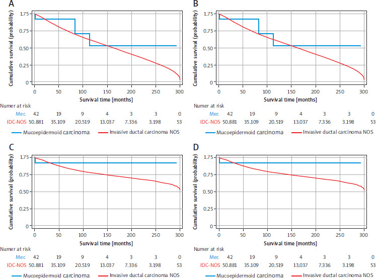
For OS, the unadjusted Kaplan-Meier curves (Figure 2 A) showed that the survival trajectory for IDC-NOS generally paralleled that of MEC during the early follow-up period but continued to decline steadily beyond approximately 120 months, while the MEC curve plateaued, reflecting no further deaths among these cases. Similar patterns were observed in the adjusted model (Figure 2 B), with IDC-NOS demonstrating a persistently steeper decline over time. In both analyses, IDC-NOS was associated with a higher hazard of all-cause mortality compared with MEC (unadjusted HR = 1.65; 95% CI: 0.83–3.30; adjusted HR = 3.80; 95% CI: 1.18–12.22), with statistical significance reached only after adjustment for covariates.
For DSS, the unadjusted curves (Figure 2 C) indicated an early small drop in survival for MEC at the beginning of follow-up, after which the curve plateaued, consistent with the small number of disease-specific deaths. In contrast, IDC-NOS showed a gradual, continuous decline throughout the study period, although the rate of decrease was less marked than that observed for OS. The adjusted DSS curves (Figure 2 D) followed a similar trend: MEC survival stabilised after the initial decline, while IDC-NOS continued to decrease modestly over time. Consistent with the OS findings, IDC-NOS demonstrated a higher, although not statistically significant, risk of cancer-specific mortality compared with MEC (unadjusted HR, 1.94; 95% CI: 0.62–6.00; adjusted HR, 2.32; 95% CI: 0.71–7.58).
Discussion
This population-based analysis, to our knowledge, is one of the most extensive comparative evaluations of breast MEC to date, utilising the SEER registry to characterise the demographic, clinicopathologic, treatment, and survival features of MEC relative to IDC-NOS. Although MEC of the breast is an exceedingly rare subtype of salivary gland-type breast carcinoma, elucidating its clinical behaviour is of practical importance for diagnosis, treatment planning, and prognostication.
Consistent with earlier published case series and small institutional reviews, our results indicate that breast MEC manifests a distinctive clinicopathologic profile and generally a more favourable prognosis than IDC-NOS. Mucoepidermoid carcinoma was more frequently diagnosed in older women, predominantly White, and typically presented as unilateral, low-grade, and early-stage disease with limited nodal involvement and no distant meta- stasis at diagnosis. Hormone receptor and HER2 negativity were strikingly common, confirming MEC’s classification within the triple-negative spectrum. However, in contrast to the aggressive biology typically associated with TNBC, MEC displayed a more indolent course, with lower rates of metastasis and mortality, aligning with prior reports suggesting a better outcome for this histologic subtype despite its triple-negative profile [13].
The survival analysis further supports this observation. Kaplan-Meier and Cox proportional hazards modelling revealed that, although OS and DSS appeared numerically shorter in MEC, the small number of events limits direct comparability. Importantly, adjusted models showed that IDC-NOS was associated with significantly higher overall mortality compared with MEC, while differences in DSS did not reach statistical significance. The plateauing of the MEC survival curves after ~120 months suggests that most MEC-related deaths occur early, with long-term survivors experiencing excellent outcomes thereafter. This temporal pattern contrasts with IDC-NOS, which exhibited a progressive decline in survival over time, reflecting its broader biological diversity and the higher risk of late recurrence characteristic of this subtype.
These findings reinforce the concept that mammary MEC may follow a relatively indolent clinical trajectory, particularly when diagnosed as low-grade and without metastasis. This is further supported by the observation that no MEC cases demonstrated bone, liver, lung, or brain metastasis at presentation, whereas such involvement was common among IDC-NOS cases. Moreover, the absence of stage III or IV disease in the MEC cohort and the lower incidence of high-grade tumours underscore its less aggressive phenotype.
Within the broader category of salivary gland-type carcinomas of the breast, MEC represents one of several rare histologic subtypes that share morphologic and immunophenotypic parallels with their salivary gland counterparts but differ in their molecular alterations and biological behaviour. Among these, adenoid cystic carcinoma (AdCC) typically harbours the MYB-NFIB fusion and demonstrates a biphasic epithelial-myoepithelial pattern, corresponding to its well-recognised indolent clinical course despite triple- negative status [14, 15]. Secretory carcinoma, defined by the ETV6-NTRK3 fusion, is also low-grade and associated with an excellent prognosis, even in node-positive disease [14, 16]. Conversely, acinic cell carcinoma frequently shows high-grade cytology, serous differentiation, TP53 or PIK3CA mutations, and more aggressive behaviour, aligning it more closely with conventional TNBC [14, 17, 18].
In this molecular and biological spectrum, MEC occupies an intermediate position, displaying distinctive CRTC1-MAML2 fusion in a subset of cases and typically low-to-intermediate histologic grade, features that may underlie its generally favourable outcome compared with most TNBC. However, unlike AdCC or secretory carcinoma, MEC shows greater heterogeneity in grade and morphology, which may translate into broader variability in prognosis. These distinctions emphasize the importance of precise histologic classification and, when feasible, molecular confirmation, as management strategies and long-term outcomes differ considerably among these rare salivary gland-type breast tumours.
Treatment patterns observed in this study reflect both the rarity of breast MEC and its favourable clinicopathologic presentation. Surgical resection was the predominant treatment modality, aligning with standard practice for localised breast tumours. Compared with IDC-NOS, a smaller proportion of MEC cases received surgery, radiotherapy, or chemotherapy, likely reflecting smaller tumour size, lower histologic grade, and the perception of a more indolent biological behaviour. Given the absence of distant metastases at diagnosis and the predominance of low-grade lesions, adjuvant systemic therapy may be unnecessary in many cases. In the absence of formal management guidelines, owing to the extreme rarity of this entity, treatment recommendations are generally extrapolated from limited case series. Most reported MEC are diagnosed at early stages, where surgical excision remains the mainstay of therapy. For tumours exhibiting high-risk features such as large size, high-grade, elevated Ki-67 index, or lymphovascular invasion, surgery with sentinel lymph node evaluation is recommended [6]. Conversely, in low-risk MEC lacking these adverse factors, breast-conserving surgery with negative margins is often sufficient to achieve durable disease control while avoiding overtreatment [6]. These observations align with previous reports emphasising the excellent long-term outcomes of low-grade MEC following complete surgical excision alone [1, 7, 10]. In all cases, individualised multidisciplinary decision-making and personalised follow-up are advisable, while the role of adjuvant systemic therapy should be cautiously de-escalated pending further molecular and outcome data from larger multi-institutional series.
The molecular biology of MEC may offer a mechanistic basis for its relatively favourable behaviour. While most conventional TNBC are associated with basal-like or claudin-low phenotypes and carry high mutational burdens, MEC of the breast frequently demonstrate low mutational burden and harbour characteristic gene fusions such as CRTC1-MAML2 (and occasionally CRTC3-MAML2). For example, reported two breast MEC, both harbouring the CRTC1-MAML2 fusion, with rare other somatic alterations [1]. A subsequent work by Venetis et al. [6] found that breast MEC with MAML2 rearrangements were associated with lower histologic grade and better outcomes. In salivary-gland MEC, CRTC1-MAML2 fusion has been shown experimentally as a driver of tumourigenesis, because its knockdown suppresses tumour growth, and co-targeting CDK4/6 and EGFR pathways has shown therapeutic promise [19]. Therefore, it is plausible that MEC of the breast is biologically distinct from most TNBC. It may not rely on ER- or HER2-driven proliferation, may carry fewer “aggressive” genomic events (e.g. TP53 mutations), and may thereby underpin the more favourable clinical phenotype. Reported breast MEC generally lack TP53 mutations, which may contribute to their more favourable clinical behaviour [20]. Although not assessable in SEER, incorporation of molecular profiling in future studies may refine prognostic stratification and therapeutic tailoring.
Despite the generally favourable characteristics of MEC, several important clinical considerations remain. First, a minority of MEC cases are high-grade, and these have been reported to behave more aggressively, with higher risks of nodal and distant metastasis and poorer outcomes [21]. Accurate histologic grading is therefore essential because it should guide both management intensity and follow-up strategies. Second, diagnostic challenges exist. Mucoepidermoid carcinoma shares some morphological features with other TNBC, such as adenosquamous or metaplastic carcinoma, making misclassification possible unless careful morphological, immunohistochemical, and molecular analyses are performed. Third, the rarity of MEC imposes limitations on study interpretation. The relatively small number of cases in our series restricts statistical power, particularly for subgroup analyses by grade or molecular status. In addition, missing data in SEER, such as for tumour grade and AJCC stage, further limit certainty, and as a registry-based study, information on recurrence, specific systemic therapy regimens, and molecular characteristics is lacking. Taken together, while our findings support an overall favourable prognosis for MEC, they should be interpreted with appropriate caution.
From a practical perspective, our study implies that for a case diagnosed with MEC of the breast, particularly if low-grade, node-negative, and with no distant spread, a more conservative management strategy may be reasonable. This might include breast-conserving surgery (if anatomically feasible) and omission of adjuvant systemic therapy in selected cases – a strategy supported by smaller case series [10, 22]. Nevertheless, high-grade MEC or node-positive disease may warrant more aggressive therapy, mirroring management of higher-risk TNBC. Also, given the molecular uniqueness of MEC, there is a rationale for future therapeutic trials exploring fusion-directed or targeted strategies, analogous to fusion-positive salivary gland MEC. The finding of effective EGFR + CDK4/6 blockade in preclinical MEC models offers a promising path for future translational work [19].
The present study has several limitations inherent to the SEER database. For a subset of cases, predictive molecular biomarkers (ER, PR, HER2) were unavailable, and data on endocrine or targeted therapies were also lacking. In addition, TNM staging (particularly pT stage) was missing for a substantial proportion of the MEC patients, which may influence the interpretation of our findings and the strength of our conclusions. Furthermore, the long study period (1975–2022) spans several decades during which diagnostic criteria, molecular features of mammary MEC, staging practices, and treatment paradigms have evolved substantially. Although a temporal subgroup analysis was considered, this was not feasible due to the very small number of cases diagnosed in the earliest decades, which precluded meaningful survival comparisons. This temporal imbalance may introduce historical bias and should be considered when interpreting the findings.
Looking ahead, further multicentre collaborative efforts are needed to accumulate larger MEC cohorts with central pathology review, integrated molecular profiling, and long-term follow-up, including data on recurrence, metastatic progression, and systemic therapy response. Prospective registries focusing on rare breast carcinoma subtypes could provide such a platform. Such studies should aim to validate the prognostic impact of MAML2 status in breast MEC and explore whether MEC truly warrants de-escalated therapy in select cases.
Conclusions
Mucoepidermoid carcinoma of the breast is an exceptionally rare and biologically distinctive subtype of invasive breast carcinoma, typically presenting with favourable clinicopathologic features, including low-grade histology, early- stage disease, limited nodal involvement, and absence of distant metastasis. Despite its frequent triple-negative immunophenotype, MEC generally exhibits a more indolent clinical course compared with IDC-NOS, with long-term survivors demonstrating excellent outcomes. Nevertheless, a minority of high-grade MEC cases may behave aggressively, highlighting the importance of accurate histologic grading, careful morphological and molecular evaluation, and individualised management strategies. Treatment patterns in our cohort reflect both the rarity of MEC and its typically favourable prognosis, with surgical excision remaining the cornerstone of therapy and adjuvant systemic treatment potentially de-escalated in selected low-risk cases. These findings underscore the clinical and biological distinctiveness of breast MEC and emphasise the need for continued multicentre studies, molecular characterisation, and long-term follow-up to refine prognostic assessment and guide evidence-based management.










