
A 65-year-old male patient was admitted to our institute for diagnostic evaluation. The patient had undergone surgical revascularization 23 years earlier with a left internal thoracic artery (LITA) graft to the left anterior descending artery (LAD) and a saphenous vein graft to the left circumflex artery (Cx), after having experienced two myocardial infarctions a year earlier. He complained of shortness of breath, anginal pain, and paroxysmal nocturnal dyspnea. Transthoracic echocardiography showed severe aortic stenosis. Computed tomography (CT) coronarography was performed as part of the routine assessment of patients with aortic valve stenosis, especially those with previous heart surgery, to determine anatomical relationships and to guide treatment decisions. CT coronarography revealed an occluded saphenous vein bypass graft to Cx and a LITA-to-LAD bypass with progression of native coronary artery disease. During this examination, a proximal anastomosis saphenous vein graft aneurysm (SVGA) with a diameter of about 3 × 2.5 cm was incidentally detected (Figure 1 A). There was no thrombus in the aneurysm. Coronary angiography showed significant triple-vessel coronary artery disease (Figure 1 B). Given that the patient was 65 years old and had severe aortic stenosis and three-vessel coronary artery disease, as well as an SVGA, the heart team decided on surgical treatment.
Figure 1
A – Computed tomography coronarography – a proximal anastomosis saphenous vein graft aneurysm (SVGA) (arrow). B – Coronary angiography showing significant stenosis of the right coronary artery (RCA) (B1), the left anterior descending (LAD) (B2), and the left circumflex (Cx) coronary artery (B3). C – Intraoperative image of SVGA, shown with surgical forceps. D – Intraoperative image of resected proximal anastomosis of SVGA, measuring approximately 3 × 2.5 cm. E – The proximal anastomosis of the vein graft was constructed to the Dacron patch (arrow)
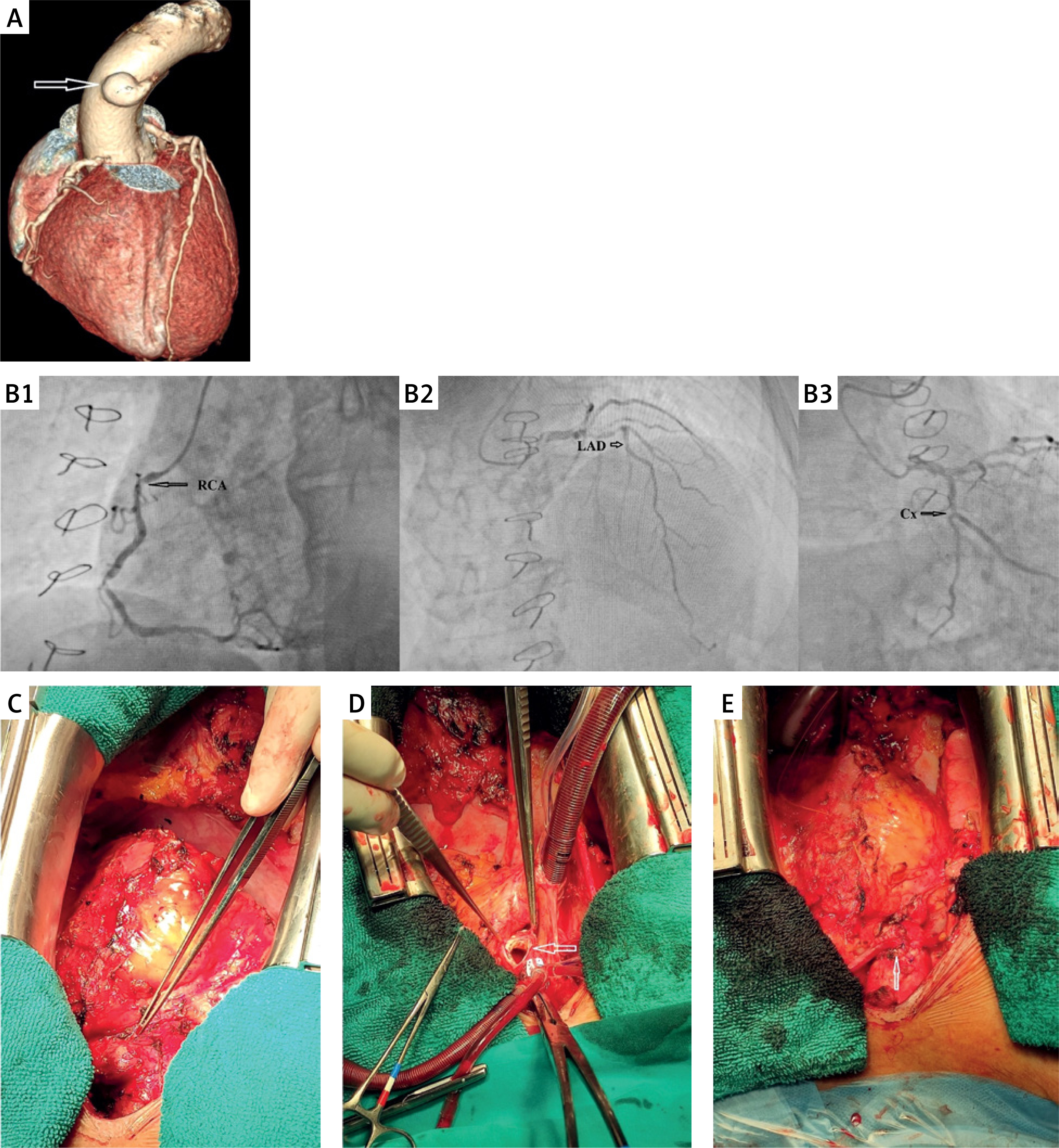
The surgical procedure was performed through a full median re-sternotomy. After pericardial dissection and adhesiolysis, proximal anastomosis of the SVGA was revealed (Figure 1 C). Aortic valve replacement with a mechanical bileaflet valve (St. Jude Regent No. 23) and double coronary artery bypass graft (CABG) with a reversed saphenous vein graft to the right coronary artery (RCA) and LAD was performed. The Cx was diffusely diseased without possibility of revascularization. The proximal anastomosis of the SVGA was resected (Figure 1 D). Reconstruction of the aortic defect was performed with a Dacron patch. The proximal anastomosis of the vein grafts was constructed separately, with the RCA anastomosed to the ascending aorta and the LAD anastomosed to a Dacron patch (Figure 1 E). The subsequent postoperative course was uneventful.
SVGA is an extremely rare and usually late complication of CABG [1]. It is seen in 0.07% of patients undergoing CABG [2]. The pathogenesis of vein graft aneurysms is multifactorial [1]. The clinical manifestation of SVGA is heterogeneous, depending on the size, localization, the part of the myocardium that is supplied with this graft, etc. Symptoms of angina pectoris, myocardial infarction, and atypical chest pain, as well as symptoms of congestive heart failure, may be present. They are often asymptomatic and are detected as an incidental finding during routine chest imaging. Differential diagnostics can be misrepresented as aortic aneurysm, thyroid tumor, lymphoma, bronchogenic cyst, etc. [3]. Venous graft aneurysms, especially if they are larger in diameter, can lead to fatal consequences if rupture or distal embolization occurs. Given the infrequent occurrence of complications and relatively heterogeneous clinical manifestations, there is no consensus regarding management strategies. Most of the cases described so far have been treated surgically. Surgical procedures include ligation or resection of the graft followed by new graft implantation, graft repair, or placement of an interposition graft.










