
Summary
The branched thoracic endovascular aortic repair technique using double- and triple-branched stent grafts in the treatment of patients with aortic arch pathology is a relatively effective method that prevents arch degeneration and offers acceptable medium-term results. This method can only be used by experienced vascular units due to the high risk of neurological complications and postoperative mortality.
Introduction
The treatment of aortic arch pathologies is a continuously evolving field within vascular surgery. The introduction of endovascular treatment methods has made it possible to perform this complex procedure in elderly patients with comorbidities who were previously ineligible for open surgery due to the high rate of complications. Currently, new techniques of thoracic endovascular aortic repair (TEVAR) are being incorporated into surgical standards for the management of aortic arch dissections and aneurysms. Given the limited patient population and the relatively small number of centers specializing in endovascular aortic arch interventions, these techniques warrant further investigation to comprehensively assess their long-term safety and clinical efficacy [1].
Among the techniques employed in the management of aortic arch pathologies, open repair remains a well-established approach. This procedure provides complete access to diseased vessels originating from the aortic arch and allows for effective aortic prosthetic reconstruction while maintaining adequate perfusion of supra-aortic branches. Unfortunately, as an open surgical technique, it necessitates the use of cardiopulmonary bypass and full sternotomy, and it is associated with a considerable risk of perioperative mortality. Consequently, it may be contraindicated in patients with significant comorbidities, in whom the operative risk is substantially elevated [2].
The chimney and periscope techniques were designed to preserve perfusion of supra-aortic arteries. They constitute an important component of endovascular management for aortic arch pathologies. However, they are increasingly being supplanted due to their higher incidence of endoleaks (EL) and mortality when compared with physician-modified stent grafts (PMSGs) and branched stent grafts (bTEVAR). Nonetheless, these approaches remain viable therapeutic options in patients requiring urgent intervention or in clinical settings where PMSG and bTEVAR techniques are not readily available [3, 4].
The PMSG technique is employed in situations where an appropriate patient-specific stent graft is not available at a specific center and the patient requires immediate intervention. These stent grafts are modified by the operator based on computed tomography angiography assessment or on 3D-printed models of the patient’s aorta. This method offers high efficacy and low mortality in patients requiring urgent treatment. However, its primary limitation lies in the level of expertise of the operator responsible for preparing the customized stent graft [5].
The bTEVAR technique enables endovascular repair of aortic arch pathologies in cases where the stent graft landing zone corresponds to Ishimaru zones 0-2. This approach allows for the treatment of aortic disease in patients with significant comorbidities who are not suitable candidates for open arch repair. Reported outcomes regarding the efficacy of branched stent grafts are highly promising: a meta-analysis conducted by Kwan et al. demonstrated a mean technical success rate exceeding 95% [6]. The most common complications following such endovascular procedures are stroke, the occurrence of type I and III endoleaks requiring reintervention for leak sealing, and a risk of mortality of approximately 5% within 30-day follow-up. Additional limitations of this method include the level of institutional experience with the bTEVAR procedure and the waiting time for a custom-made device (CMD), which may range from 6 to 12 weeks.
Despite favorable results reported in the literature regarding the safety of the bTEVAR technique, data on long-term outcomes remain limited, with a mean patient follow-up of approximately 20.7 ±13.5 months.
Aim
In this single-center study, we present the early- to mid-term outcomes of patients treated using the bTEVAR technique.
Material and methods
This single-center study was conducted at the Department of Vascular Surgery, General Surgery and Angiology in Szczecin, Poland. The study included patients who underwent planned endovascular treatment for aortic arch pathology using bTEVAR technique between May 2021 and September 2024. The study group consisted of 15 patients, including 11 (73.33%) men and 4 (26.67%) women. The mean age of the patients was 67.4 years (median: 70, range: 44–77). Patients underwent implantation of double-branched stent grafts (4 patients) and triple-branched stent grafts (11 patients). The inclusion criteria for the study are defined as follows:
Patients with type B aortic dissection with degeneration of the aortic arch or;
Patients with non-A non-B aortic dissection or;
Patients with penetrating atherosclerotic ulcer (PAU) or intramural hematoma (IMH) of the aortic arch or;
Patients with an aortic arch aneurysm meeting the following criteria:
– proximal landing zone exceeding 20 mm;
– stable patients in a subacute or chronic stage of an aortic arch pathology requiring treatment due to aortic arch degeneration;
– patients meeting the anatomic criteria for endovascular treatment (no aortic arch kinking, aortic arch diameter of not more than 40 mm, sufficiently wide iliac or femoral arteries for system insertion);
– patients ineligible for open surgery due to multiple comorbidities;
– patients aged over 18 years.
The exclusion criteria were: a) patients with a ruptured aorta, b) patients requiring urgent surgery, c) pregnant patients, d) patients with allergies to iodine contrast agents.
All the patients participating in the study have been assessed and deemed eligible for bTEVAR by vascular surgeons and cardiac surgeons working together as part of the Aortic Team in Szczecin, Poland. Eleven patients were scheduled for triple-branch bTEVAR treatment, 4 patients for double-branch bTEVAR.
The results of patient treatment were evaluated based on computed tomography angiography results 1 month, 6 months, and 12 months after surgery. The following criteria were established to evaluate patients after bTEVAR:
Primary outcomes – first 30 days after surgery:
Technical success – correct stent graft placement, no detected migration, persistent flow in supra-aortic vessels; effective management of any endoleaks confirmed in angiography during the procedure; no endoleak present during the first month after the procedure;
Early death related to stent graft placement.
Secondary outcomes – after 30 days following surgery:
Incidence of stroke – severity of stroke was evaluated using the NIHSS scale [7];
Incidence of myocardial infarction;
Incidence of stent graft-related death;
Incidence of endoleaks, retrograde type A aortic dissection (RTAD), stent graft-induced new entry (SINE);
Number of stent graft-related reinterventions due to persistent type I or type III endoleak or stent graft migration.
The procedure for implanting triple-branched and double-branched stent grafts in the aortic arch is described below.
Triple-branch stent graft implantation technique
Patients were selected for the procedure based on computed tomography angiography. The procedures were performed under general anesthesia in a hybrid operating room. The sterile surgical field was prepared using an iodine-based solution. The procedure was performed by accessing the common femoral arteries transcutaneously. A Lunderquist guidewire was inserted into the left ventricle through the AL1 catheter sheath. A pacing electrode was inserted into the right ventricle to enable blood pressure reduction using the rapid pacing method. Access to the common carotid arteries was obtained surgically or via the ProGlide system. Guidewires were inserted into the brachiocephalic trunk (BCT) and the left common carotid artery (LCCA). Then, the left brachial artery was cannulated, allowing the guidewire to access the left subclavian artery (LSA).
The delivery system was positioned using prosthesis markers at the openings of the supra-aortic vessels. The correct positioning of the prosthesis was verified in LAO and RAO projections. During rapid pacing, the sheath was removed and the stent graft was released in the aortic arch, followed by angiography to verify the correct flow in the main arteries. The inner branches of the prosthesis were cannulated from the cervical access and bridging stent grafts were implanted, achieving continuity of flow to the left common carotid artery and brachiocephalic trunk. From the femoral side, the branch to the left subclavian artery was cannulated. The guidewire was captured using “through and through wire”, and Viabahn stent grafts were implanted into the left subclavian artery. After securing all arteries, completion angiography was performed, confirming the seal of the prosthesis and flow in the main arteries of the aortic arch. If an endoleak was detected in the completion angiography, the prosthesis was sealed by ballooning the area of the leak.
After obtaining the correct result, the vascular systems were removed, and the vascular accesses were closed with ProGlide systems or surgically. After the procedure, the patients were transferred to the Intensive Care Unit. After discharge from the hospital, patients were advised to undergo follow-up computed tomography angiography at 1, 6, and 12 months.
During bTEVAR procedures, Bolton Relay Arch Devices (CMD, Bolton Medical, Florida, USA), Cook Arch Devices (CMD, Cook Medical, Indiana, USA) and Viabahn stent grafts (Gore, Delaware, USA) were used.
Double-branch stent graft implantation technique
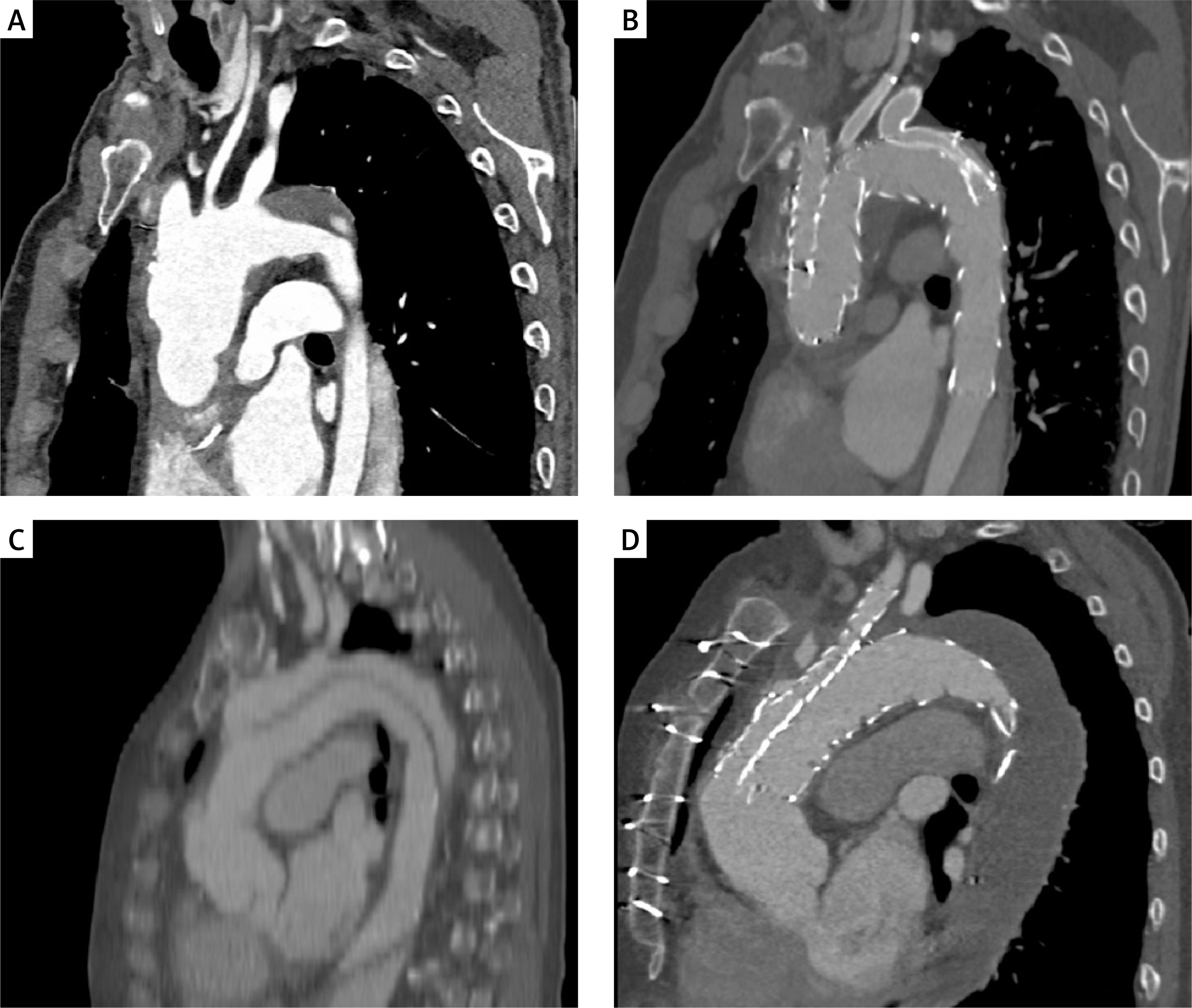
The technique for implanting double-branched stent grafts is very similar to the technique described above. However, the difference in implantation lies in the fact that only the brachiocephalic trunk and the left common carotid artery are accessed for the implantation of the Viabahn stent graft. After correct positioning of the main stent graft in the aortic arch, the device is implanted in the aortic arch, leaving the BCT and LCCA patent. The LSA is covered by the stent graft. Next, a hybrid LSA-LCCA bypass was created after closing the initial segment of the LSA with an Amplatzer vascular occluder. Figure 1 presents the results of implantation of a triple-branched stent graft and a double-branched stent graft.
Results
Study group and concomitant diseases
Fifteen patients from double- and triple-branched bTEVAR treatment were included in the study based on inclusion criteria. Table I presents comorbidities and indications for bTEVAR treatment. The study group included 7 patients with degenerating aortic arch dissection, 7 patients with aortic arch aneurysm, and 1 patient with PAU. In addition, 1 patient was found to have LSA pathology in the form of dissection, and 1 patient developed aortic aneurysm as a result of Marfan syndrome. All patients underwent elective surgery after prior evaluation with computed tomography angiography.
Table I
Patients’ comorbidities and indications for bTEVAR treatment
Early results
In the early postoperative period, one patient death related to the stent graft implantation procedure occurred. The patient experienced complete occlusion of the left anterior descending artery, which led to a massive cardiac infarction 3 days after bTEVAR despite attempts at coronary angioplasty. This patient suffered from Marfan syndrome and had undergone open ascending aorta repair in the past, with bTEVAR treatment intended as the second stage of treatment for aortic arch pathology. In the follow-up CTA, no stent graft migration, RTAD endoleak, or SINE was observed in this patient.
During the 30-day postoperative period, two strokes were observed. One patient developed a severe stroke due to migration of a mural thrombus in the ascending aorta, which led to cerebral artery embolism. This patient developed symptoms of aphasia and right-sided paresis (NIHSS score > 15 points). The second patient suffered a stroke 3 days after stent graft implantation. However, the stroke was non-disabling, and the patient regained his functional capacity during further follow-up (NIHSS score < 15 points). In the follow-up CTA, no stent graft migration, RTAD endoleak, or SINE was observed in either patient.
One patient experienced a complication at the access site: a large introducer system led to dissection of the iliac artery. However, despite the complication at the access site, it was possible to successfully perform stent graft implantation in this patient, with preserved perfusion in all arteries of the aortic arch.
In the perioperative period, technical success was achieved in 80% of cases (12 of 15 patients). The remaining cases were classified as failures due to one patient death related to a myocardial infarction and two strokes – all complications involved patients from the triple-branched stent graft group. No other complications related to bTEVAR treatment were observed. No endoleaks, RTADs, or SINEs were reported.
Follow-up
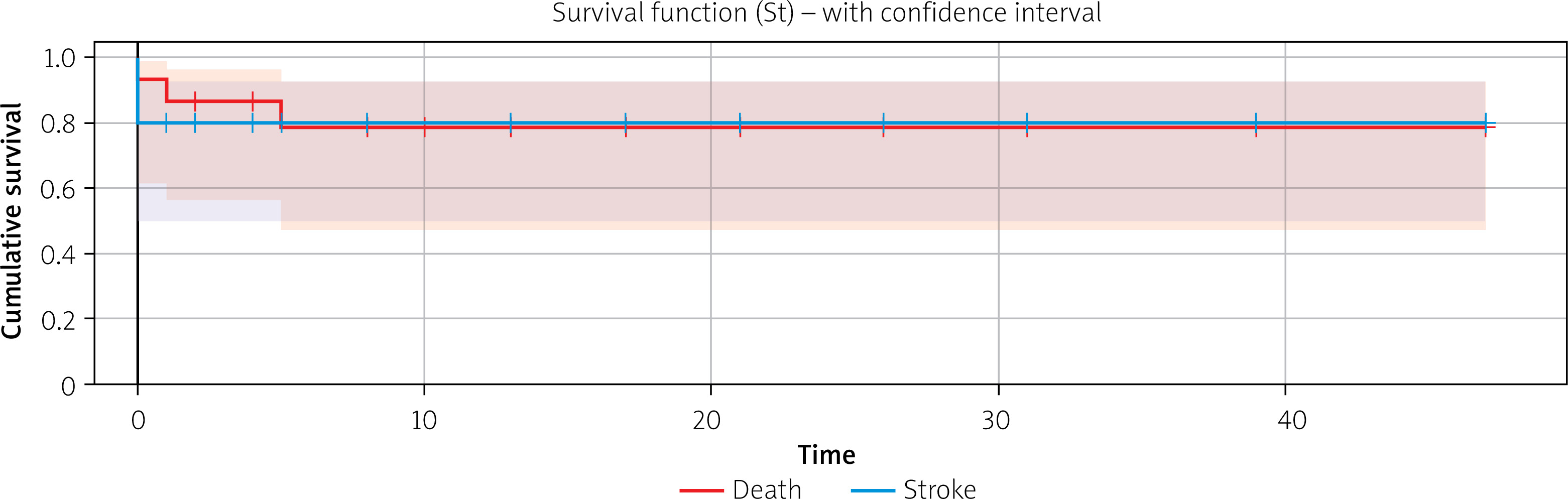
The follow-up period for patients began in May 2021 and ended in November 2025. The follow-up period averaged 15.6 months (median: 10 months, range: 0–47 months). Ten patients exceeded the 6-month follow-up period, and 7 patients exceeded the 12-month follow-up period. Two patient deaths occurred during this period. One patient, after the first stage of aortic aneurysm treatment with bTEVAR, was scheduled for the second stage of TEVAR treatment with landing in zone 4 due to a large degenerating thoracic aortic aneurysm. During the second stage of treatment, the patient suffered a massive stroke and died after the procedure. However, no complications related to the first stage of treatment, which was bTEVAR, were observed in the patient. The second patient died one month and 3 days after the branched stent graft implantation procedure. No stent graft migration was observed in the CTA, all supra-aortic vessels were patent, and only old ischemic lesions were found in the cerebral tomography. The direct cause of death could not be determined. However, this patient had a number of comorbidities (diabetes, hypertension, renal failure, atherosclerosis, previous stroke), and his death is not considered to be related to the vascular intervention. The Kaplan-Meier curve illustrates overall survival and freedom from stroke (Figure 2).
Three patients were diagnosed with endoleaks – one with type III EL, another with type Ia EL, which disappeared spontaneously during further follow-up, and the third with type Ia and Ic EL. Two patients required reintervention to seal the endoleaks – in both cases, the leak was successfully sealed, and no further complications related to vascular treatment were detected in follow-up CTAs.
In addition, 3 patients required further treatment for dissection or aneurysm of the descending aorta due to degeneration. In all cases, the bTEVAR procedure was the first stage of treatment for aortic pathology. In all patients, the aortic pathology was successfully treated in subsequent stages of endovascular treatment.
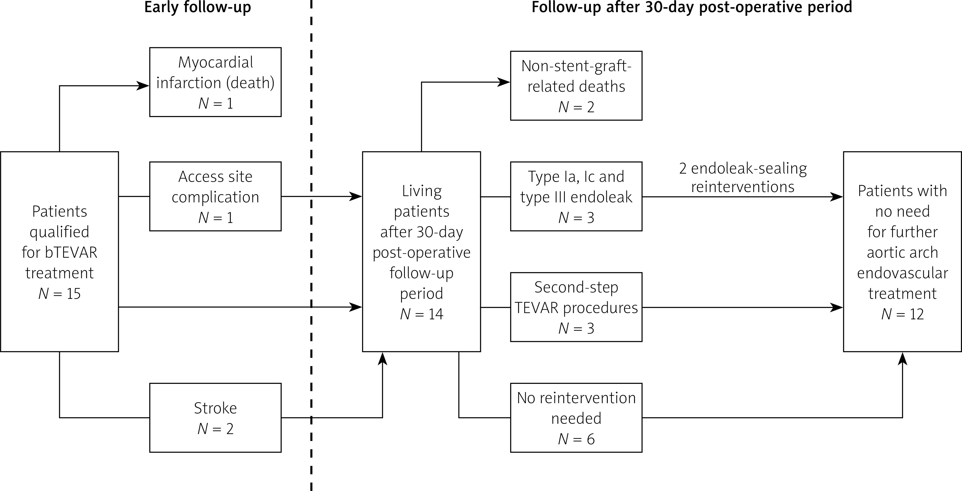
No additional stent graft infections, branch occlusions, RTADs, or SINEs were observed in the study. Stent graft patency was maintained in all patients. The results and complications of bTEVAR treatment during the whole follow-up period are summarized in Figure 3.
Discussion
The single-center study on the outcomes of aortic arch pathology treatment presents the safety, efficacy, and complications associated with bTEVAR therapy using double- and triple-branched stent grafts. The study identified a group of patients with multiple comorbidities who were eligible for bTEVAR treatment as open surgery was considered not feasible. The patients did not require urgent vascular intervention; after evaluation of aortic arch pathology, they were scheduled for elective treatment. Despite adequate preparation for surgery, complications associated with endovascular treatment of the aortic arch remain a major challenge in vascular surgery due to the anatomy and disease burden of patients.
In the study group, technical success was achieved in 80% of cases (12 out of 15 patients). One patient who died a few days after endovascular treatment suffered a massive myocardial infarction associated with LAD occlusion. However, this patient did not suffer a myocardial infarction immediately after the procedure. In the past, this patient required open ascending aorta replacement. Endovascular treatment was the second stage of treatment due to aneurysmal degeneration of the aortic arch associated with Marfan syndrome. The follow-up CTA did not reveal any stent graft migration or RTAD that could have caused coronary artery dissection and myocardial infarction. Due to the patient’s death, the procedure was not classified as a technical success according to SVS Reporting Standards [8]. Two patients suffered a stroke despite receiving standard anticoagulant therapy both during and after surgery. In one case, migration of a mural thrombus from the ascending aorta was observed, which led to a stroke. In the second case, the patient suffered a minor stroke despite patency of all branches. In this case, the patient’s symptoms resolved during follow-up. These strokes were most likely related to manipulation of the delivery system in the aortic lumen, as no abnormalities in stent graft implantation were observed in the follow-up CTAs.
The meta-analysis by Kwan et al. compared data from 618 patients treated with bTEVAR for aortic arch pathologies. In this study, most patients were treated with double-branched stent grafts. In such a large population, an average technical success rate of 97.4% was achieved. It also emphasizes that the 30-day mortality rate was approximately 5.5%, and the incidence of stroke after endovascular treatment remained at 10.5%. It is therefore important to analyze the criteria for technical success defined by the authors of the publication regarding the efficacy of the bTEVAR procedure. With such a high incidence of stroke and death in the postoperative period, technical success below 90% may be considered. In the case of the evaluation carried out by our center, if we were to consider only the stent graft implantation procedure and ignore the clinical aspect of postoperative complications, the technical success rate would reach 100%.
The most common complication associated with the use of branched stent grafts is stroke. The authors of the meta-analysis report a 10–11% risk of neurological complications after bTEVAR surgery in the postoperative period [6, 9, 10]. By comparison, the stroke rate in our study was 13.3%. An important aspect of neurological complications following surgery is the incidence of stroke despite patency of the inner branches of supra-aortic vessels. The pathophysiology of stroke in bTEVAR procedures may therefore result from the formation of mural thrombi during the stent graft implantation process itself. The risk of stroke therefore depends on the experience of the center performing bTEVAR procedures on the aortic arch, including the duration of the procedure, the amount of manipulation within the aortic lumen, and the selection of appropriate anticoagulant therapy for individual patient groups. The risk of stroke can also be reduced by focusing on the further development of neuroprotective techniques, adequate preparation of the patient for the procedure, and the selection of an appropriate endovascular treatment method for a given patient. In the case of endovascular treatment, the physician-modified stent graft (PMSG) technique, which has been used in the treatment of patients requiring urgent intervention, appears to offer promising results [11]. In centers specializing in stent graft modification, the risk of neurological complications in the perioperative period is reported to be below 4%, with a technical success rate of 98% [12].
Despite the full effectiveness of stent graft implantation verified by arteriography, endoleaks were observed in 3 patients during the observation period. One patient did not require reintervention, as the endoleak resolved spontaneously during the observation period. In 2 patients, type III, Ia, and Ic endoleaks required sealing. In both cases, the leaks were successfully sealed, and no lesions requiring reoperation were observed during the follow-up. The literature reports a high incidence of endoleaks, especially in medium-term follow-up [6]. These leaks often require reintervention, although their clinical significance has not been fully understood due to the lack of reports on the effectiveness of long-term treatment. However, the cause of such frequent leaks should be carefully determined in a situation where branched stent grafts are usually CMDs tailored specifically to the individual patient.
The study conducted by our center has certain limitations that may affect the interpretation of the results. Due to the single-center nature of the study, the group of 15 patients scheduled for bTEVAR due to aortic arch pathology is relatively small, which may limit the statistical power and generalizability of the findings. The small diversity of patients may therefore affect the exact number of complications occurring with particular endovascular procedures. Moreover, selection bias must be considered, as only selected patients meeting strict anatomical and clinical criteria were included. Referral bias may also be present, as our center specializes in complex aortic pathologies. It could influence both procedural outcomes and complication rates. In addition, the average follow-up period for patients was 15.6 months, which makes it impossible to draw conclusions about the long-term effectiveness of treatment. However, the follow-up period is comparable to other mid-term follow-up periods published by researchers.
Furthermore, our data reflect the most difficult period of the steep learning curve of this new method. They may serve as an encouragement or a warning to centers considering implementing this technology, especially since this also applies to cardiac surgery centers, which typically do not have extensive experience with endovascular techniques. Nevertheless, with the cooperation of a cardiac surgeon and a vascular surgeon, satisfactory results can be achieved from the outset.
Future research should focus primarily on reducing the incidence of neurological complications among patients. Educating facilities in bTEVAR procedures, defining standardized indications for endovascular treatment based on a comparison of its effectiveness with open and hybrid methods, and extending the observation period for patients will help determine the exact risk and safety of using branched stent grafts in the aortic arch.
Conclusions
The bTEVAR technique using double- and triple-branched stent grafts in the treatment of patients with aortic arch pathology is a relatively effective method that prevents arch degeneration and offers acceptable medium-term results. This method can only be used by experienced vascular units due to the high risk of neurological complications and postoperative mortality.










