
Introduction
Breast cancer is the most common cancer worldwide, with 2.3 million new cases reported in 2020 [1]. Patients diagnosed at advanced stages, particularly with distant metastases, have limited treatment options. Chemotherapy remains the standard treatment for metastatic breast cancer; however, it is associated with significant side effects, including nausea, vomiting, alopecia, and bone marrow suppression [2, 3]. These limitations highlight the need for safer and more effective targeted therapies.
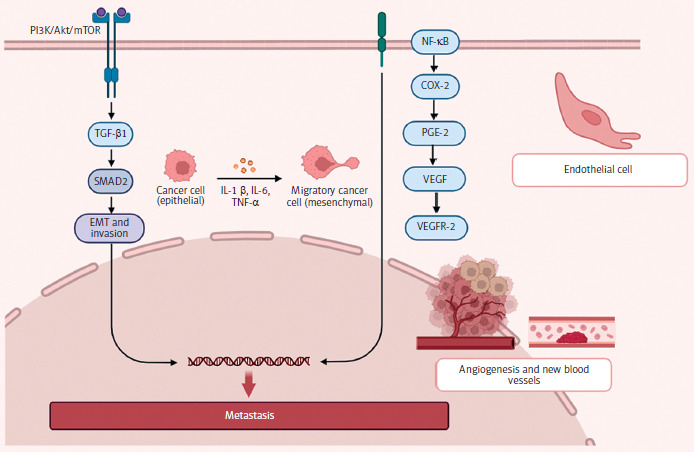
Natural compounds, particularly bisbenzylisoquinoline alkaloids derived from Stephania tetrandra, have shown potential anticancer activity. These compounds are reported to modulate key molecular pathways involved in cancer progression, including matrix metalloproteinases and the PI3K/Akt/mTOR signalling cascade, which are associated with tumour growth, survival, and metastasis [4]. In addition, TGF-β1 signalling plays a critical role in promoting breast cancer progression through epithelial-mesenchymal transition and enhanced cell migration [5, 6]. Targeting these pathways may provide a strategic approach to inhibit tumour progression (Figure 1).
Figure 1
Targeting these pathways may provide a strategic approach to inhibit tumour progression
EMT – epithelial-mesenchymal transition, PGE – prostaglandin E2, SMAD – small mothers against decapentaplegic proteins, VEGF – vascular endothelial growth factor, VEGFR-2 – vascular endothelial growth factor receptor 2
Fangchinoline, a bisbenzylisoquinoline alkaloid isolated from the dried roots of Stephania tetrandra, has demonstrated anticancer activity in various malignancies and is widely used in traditional Chinese medicine [7, 8]. Previous studies have reported its anti-proliferative effects in several human cancer cell lines, including MDA-MB-231 (breast), A549 (lung), PC3 (prostate), and leukemia cells [9–15]. Recent evidence also suggests its potential activity related to HER-2 (Human Epidermal Growth Factor Receptor 2) signalling [16]. Despite these findings, the molecular mechanisms of fangchinoline in triple-negative breast cancer (TNBC), particularly in the murine 4T1 model, remain poorly understood. Specifically, the involvement of key signalling pathways such as TGF-β1 and PI3K/Akt/mTOR has not been systematically investigated in this model. Furthermore, studies integrating in vitro experimental approaches with in silico molecular docking to elucidate the interaction between fangchinoline and these molecular targets are still limited.
Therefore, this study aims to evaluate the anticancer effects of fangchinoline in 4T1 TNBC-cells and to explore its potential molecular mechanisms through a combined in vitro and in silico approach.
Material and methods
Acquisition of biomolecular structures and preprocessing
PubChem provided a fangchinoline (CID: 73481) chemical structure, whereas protein data bank provided the three- dimensional structure of the target protein. Co-crystallized ligands were removed from mTOR, TGF-β1, and COX-2 using AutoDock 1.5.7 under physiological circumstances. Polar hydrogen atoms and protein water removal were performed using AutoDock 1.5.7. Root-mean-square deviation (RMSD) values < 2.0 Å were obtained using PyMOL to validate the protein structures. BIOVIA Discovery Studio 2021 was used to visualize and interact with the structures.
Molecular docking
Molecular docking and binding affinities were calculated using AutoDock Vina 1.2.5, considering ligand flexibility and receptor rigidity [17]. Ligands and protein structures were prepared using AutoDockTools. The binding sites were defined based on the positions of co-crystallized ligands in each target protein. The grid box parameters for each protein, including center coordinates and dimensions, are presented in Table 1, with an exhaustiveness value of 100 applied to improve search accuracy [18].
Table 1
Grid box parameters used for molecular docking
To validate the docking protocol, redocking of the native (co-crystallized) ligands was performed. The accuracy of the docking method was evaluated by calculating the RMSD between the redocked and original ligand conformations, as shown in Table 2.
Molecular dynamics
The selected protein-ligand complexes were subjected to 100 ns molecular dynamics simulations using YASARA version 23.9.29.64 with the AMBER14 force field under physiological conditions (pH 7.4, 0.9% NaCl, 310 K, 1 bar, and 0.997 g/ml water density). The systems were solvated using an explicit water model (TIP3P) with periodic boundary conditions applied in all directions. A timestep of 2 × 1.25 fs (2.5 fs) was used, and non-bonded interactions were calculated with a cutoff distance of 8 Å. Long-range electrostatic interactions were treated using the Particle Mesh Ewald method. Each system was first subjected to energy mini- mization,followed by equilibration under the constant num- ber of particles, volume, and temperature ensemble and subsequently the constant number of particles, pressure, and temperature ensemble to stabilize temperature and pressure before the production run. Structural snapshots were saved every 0.05 ns (1000 frames) throughout the simulation. Structural stability and flexibility were valuated using RMSD and root mean square fluctuation (RMSF) analyses. Protein compactness was assessed by the radius of gyration (Rg), while interaction stability was analysed through hydrogen bond evaluation. In addition, binding free energy was estimated using the Poisson-Boltzmann method.
Cytotoxicity assay
3 × 10³ cells/ml (0.1 ml/well) of 4T1 TNBC-cells were seeded in 96-well plates at a density of 3 x 10³ cells/ml. The cells were treated with sample solution for 48 h. After adding 3-(4,5-dimethylthiazol-2-yl)-2,5-diphenyltetrazolium bromide reagent, the cells were incubated at 37°C for 4 h. The reaction was terminated by adding 10% SDS in 0.01 N HCl. Cell viability was evaluated by measuring absorbance at 595 nm absorbance [19].
Cell cycle analysis
4T1 cells 3 × 10⁵ cells/well were incubated for 48 h, fixed in 70% ethanol, stained with propidium iodide/RNase, and analysed using flow cytometry at 3 × 10⁵ cells per well [20].
Apoptosis analysis
Annexin V-FITC and propidium iodide stained 4T1 cells (3 × 10⁵ cells per well) were incubated for 48 h. Flow cytometry was performed to evaluate apoptosis. After 48 h on titanium disks, 4T1 cells (3 × 10⁵/well) were permeabilized and stained for PI3K, Akt, mTOR, p53, and TNBC. We assessed protein expression by FACS [21].
Protein expression analysis (PI3K, Akt, mTOR, and p53)
Total protein expression was measured using specific antibodies. Phosphorylated protein forms were not evaluated in this study. Untreated (cell-only) samples were used as controls, and data were normalized relative to the control group. Appropriate gating strategies were applied to minimize non-specific signals. 4T1 in 6-well plates, 3 × 10⁵ cells/well were seeded overnight and replenished with a drug-containing medium for 48 h. The adherent and suspended cells were collected using 0.025% trypsin, washed twice with cold water and PBS, and centrifuged for 5 min at 2500 rpm. A FACScan flow cytometer (15 min, 37°C, 10 min) was used to examine the cell pellet resuspended in PermWash buffer with PI3K FITC, Akt APC, mTOR PE, p53 FITC, and HER-2 FITC antibodies [22].
Results
Molecular docking and dynamics
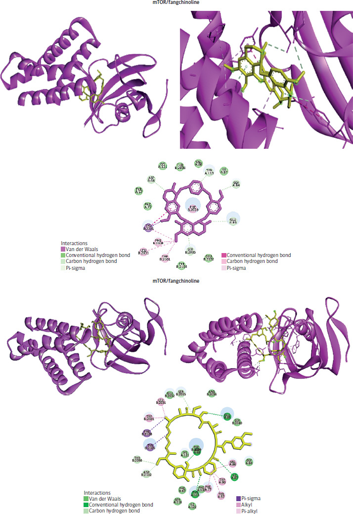
The natural ligand had a higher binding affinity for TGF-β1 than fangchinoline, but mTOR had a lower affinity (Table 3, Figure 2). Docking results suggest that fangchinoline could interact with TGF-β1 and mTOR pathways in TNBC; however, further experimental validation is needed to confirm its biological activity.
Table 3
Binding affinity of fangchinoline and native ligands in complex with the target proteins
| Ligand | Binding affinity [kcal/mol] | |||||
|---|---|---|---|---|---|---|
| mTOR | TGF-β1 | COX-2 | MAPK-14 | RAS | VEGFR-2 | |
| Fangchinoline | –12.1 | –11.3 | –9.4 | –8.9 | –9.1 | –11.7 |
| Native ligands | –17.6 | –10.3 | –8.9 | –11.4 | –11.6 | –8.0 |
| Protein inhibitor | –10.3 | –11.8 | –8.9 | –9.5 | –8.2 | –8.0 |
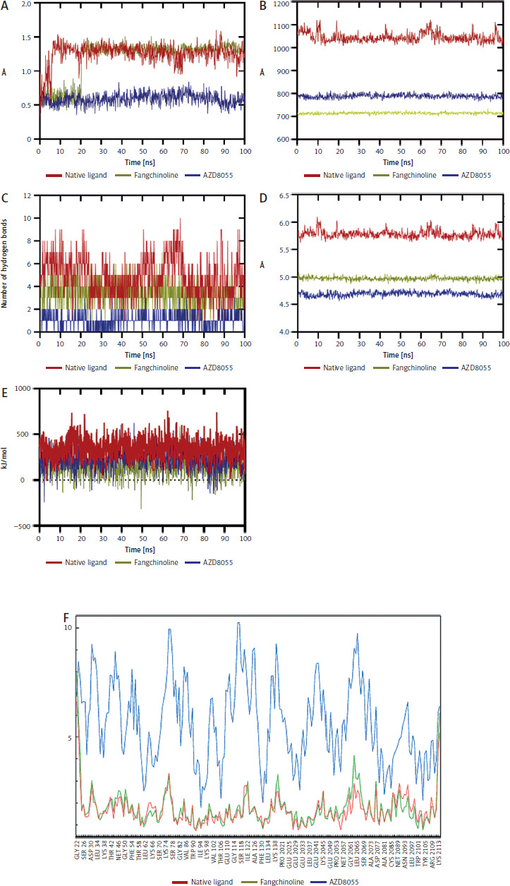
The root-mean-square deviation values and radius of Rg indicated that the mTOR–fangchinoline complex was more stable than the native ligand and protein inhibitor complexes (Figure 3). The fangchinoline complex contained few hydrogen bonds but similar bond energies, indicating robust protein-ligand interaction.
Figure 3
Molecular simulations demonstrated the stability of the mTOR-fangchinoline complex compared with the mTOR-native ligand and mTOR-AZD8055 complexes under physiological conditions fangchinoline/native ligand under physiological conditions. A) Stability of the complexes according to the root-mean-square deviation of backbone atoms. B) Solvent-accessible surface area. C) Number of formed hydrogen bonds. D) Radius of gyration. E) Molecular mechanics Poisson-Boltzmann surface area. Cont. F) Free binding energy estimations of mTOR/fangchinoline compared to mTOR/native ligand
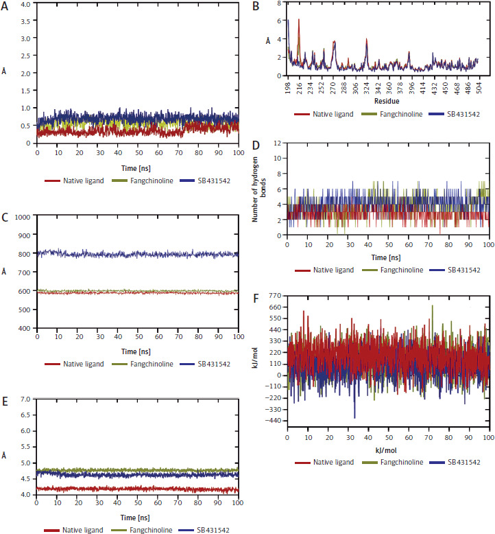
The root-mean-square deviation of backbone atoms, RMSF values, ligand conformation stability, hydrogen bond formation, and free binding energy estimations of TGF-β1/fangchinoline vs. TGF-β1/native ligand were analysed to assess complex and protein stability (Figure 4).
Figure 4
Molecular simulations show the stability of TGF-β1/fangchinoline vs. TGF-β1/native ligand under physiological conditions. A) Stability of the complexes according to the root-mean-square deviation of backbone atoms. B) Root mean square luctuation. C) Solvent-accessible surface area. D) Numberof formed hydrogen bonds. E) Radius of gyration. F) Molecular mechanics Poisson-Boltzmann surface area
Cytotoxicity and cell cycle
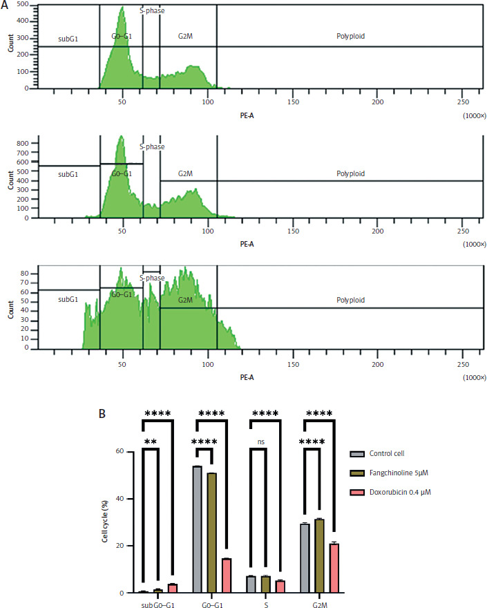
Fangchinoline significantly suppressed 4T1 cell growth, with an IC50 value of 25.95 µM (Figure 5). In comparison, doxorubicin exhibited an IC50 value of 2.29 µM. Fangchinoline is considered a cytotoxic agent against cancer cells, with an effective concentration range of 10–100 µM. Treatment with fangchinoline for 48 h significantly increased the proportion of 4T1 cells in the G2/M phase compared to the control group, without significantly affecting the G0/G1 or S phases (p < 0.001).
Apoptosis and protein expression
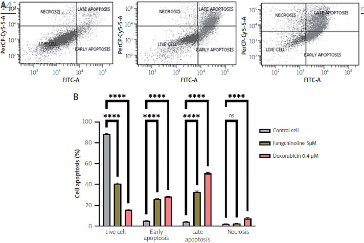
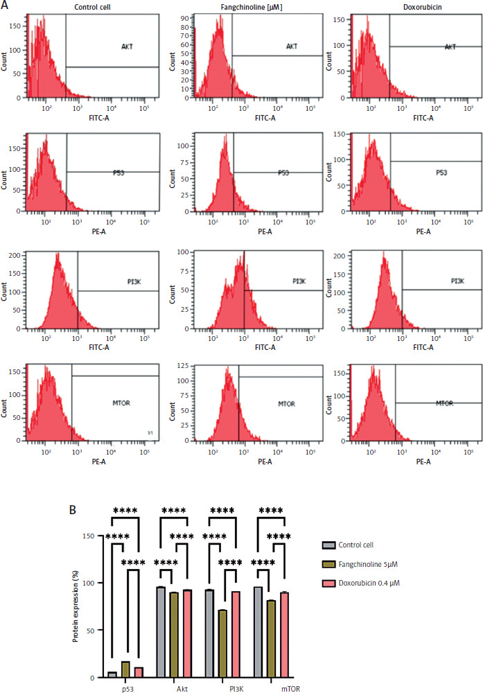
Flow cytometry analysis using Annexin V-propidium iodide staining showed an increase in both early and late apoptotic cell populations, with relatively low levels of necrosis, indicating that apoptosis was the predominant mode of cell death (Figure 6). Furthermore, the expression levels of PI3K, Akt, and mTOR were decreased, whereas p53 expression was increased in 4T1 cells following treatment (Figure 7). These changes indicate altered expression of PI3K, Akt, mTOR, and p53 proteins in treated cells. However, as only total protein levels were measured, these findings do not confirm direct inhibition of the PI3K/Akt/mTOR signalling pathway.
Discussion
Fangchinoline has been reported to exert anticancer activity by affecting cancer cell growth, signalling pathways, and apoptosis. Previous studies have shown that fangchinoline inhibits cell proliferation and induces apoptosis in various cancer cell lines, supporting its potential as an anticancer compound. Several studies have selected mTOR, VEGFR-2, TGF-β1, and p38 MAPK (MAPK14) as molecular targets for docking analysis based on their roles in cancer progression [23–25]. In comparison with native ligands, fangchinoline exhibited relatively lower binding affinity toward VEGFR-2, p38 MAPK, mTOR, and COX-2, but a higher affinity toward TGF-β1 at the same binding site. These proteins are known to be involved in aggressive cancer cell signalling [26, 27].
In the present study, fangchinoline demonstrated favourable binding affinity toward TGF-β1, suggesting a potential interaction with this target. However, molecular docking results alone do not confirm direct biological targeting or functional modulation of the pathway. Previous studies have also suggested that fangchinoline may be associated with the modulation of key signalling pathways, including PI3K/Akt/mTOR [16, 23–25, 28–32]. In line with these reports, our findings indicate a possible association between fangchinoline and TGF-β1-related signalling, which is known to regulate cancer cell invasion and metastasis. Nevertheless, this interaction should be interpreted cautiously and requires further experimental validation.
Molecular dynamics simulations further supported the stability of the fangchinoline-protein complex. In addition, flow cytometry analysis showed that fangchinoline treatment was associated with G2/M phase arrest in 4T1 cells, consistent with the effects of other bisbenzylisoquinoline alkaloids. Apoptosis induction was confirmed by a marked increase in early and late apoptotic cell populations, with relatively low levels of necrosis as assessed by Annexin V-PI staining. The predominance of apoptotic over necrotic cell death suggests a regulated cell death mechanism, potentially triggered under cellular stress conditions.
Furthermore, our results demonstrated that fangchinoline treatment was associated with changes in protein expression related to PI3K/Akt/mTOR and p53, along with increased ROS levels in 4T1 cells. These findings suggest a potential involvement of these pathways in mediating the observed anticancer effects. Previous reports have also indicated that fangchinoline can influence cancer cell migration and enhance the efficacy of chemotherapeutic agents such as doxorubicin [11, 12]. However, it is important to note that the present study assessed only total protein expression without evaluating phosphorylated forms or conducting functional pathway validation.
Taken together, these findings suggest that fangchinoline exerts anticancer activity through multiple cellular mechanisms, including cell cycle arrest, apoptosis induction, and modulation of cancer-related signalling pathways. However, further studies, including phosphorylation analysis and functional assays, are required to confirm these mechanisms and to better understand the molecular basis of its activity in TNBC.
Conclusions
Fangchinoline showed potential for treating triple-negative breast cancer via the induction of G2/M-cell cycle arrest and apoptosis, which had a stable and significant interaction with key signalling proteins, especially TGF-β1. These findings highlight the potential of fangchinoline as a candidate compound for further investigation in TNBC treatment.










