
Introduction
Colorectal cancer (CRC) is a significant global health concern, ranking as one of the leading causes of cancer-related mortality. A substantial proportion of CRC patients develop colorectal liver metastases (CRLM) during their disease course, which considerably worsens their prognosis [1]. Advancements in surgical techniques, systemic chemotherapy, and targeted therapies have improved outcomes for selected patients, but CRLM still represent a major clinical challenge with highly variable prognosis in which identifying robust prognostic biomarkers is essential [2]. In recent years, body composition analysis has emerged as a promising tool for oncologic patients’ evaluation, providing insights into sarcopenia, frailty, nutritional status, metabolic reserves and overall prognosis [3]. The distribution of muscle mass, adipose tissue, and intramuscular fat infiltration has been increasingly recognised as a key determinant of treatment response and survival in cancer patients [3, 4].
Computed tomography (CT) is a widely used imaging modality in oncology, routinely performed for tumour staging and treatment planning [5]. Beyond traditional radiologic assessment, CT-derived body composition analysis allows for a quantitative evaluation of skeletal muscle, visceral adipose tissue (VAT), and subcutaneous adipose tissue (SAT), offering a more detailed understanding of patient physiology [6]. Among the most extensively studied parameters, reduced skeletal muscle mass, and myosteatosis have been associated with worse surgical outcomes, increased chemotherapy toxicity, and shorter overall survival in patients with CRC [7, 8]. Additionally, visceral obesity has been implicated in systemic inflammation and tumour progression, further underscoring the importance of body composition assessment in oncologic care [9, 10]. Historically, body composition analysis has relied on manual or semi-automated segmentation of a single axial CT slice at the level of the third lumbar vertebra (L3), which serves as a surrogate for whole-body muscle and adipose tissue distribution [11]. However, this approach is inherently limited by inter-observer variability, time constraints, and potential inaccuracies in estimating total body composition [12].
The emergence of fully automated artificial intelligence (AI)-driven segmentation has revolutionised this field, enabling rapid, reproducible, and volumetric assessment of muscle and adipose compartments across larger anatomical regions. Recent studies suggest that volumetric body composition analysis, as opposed to single slice approaches, provides a more comprehensive representation of metabolic health and oncologic prognosis [11]. In the context of CRLM, assessing body composition is particularly relevant given the high prevalence of cachexia, treatment-related muscle depletion, and systemic metabolic alterations [13, 14]. Patients undergoing liver metastasectomy often experience significant physiological stress, and their preoperative body composition may influence postoperative recovery, complications, and long-term survival [15]. Furthermore, preoperative chemotherapy – administered in a substantial proportion of CRLM patients – can induce reduction of skeletal muscle mass and myosteatosis, which in turn may affect treatment tolerability and outcomes [16]. Despite increasing recognition of body composition as a prognostic factor in oncology, there remains a gap in the literature regarding its application in fully automated volumetric assessments in CRLM patients.
The primary objective of this study is to evaluate the prognostic significance of CT-based volumetric body composition parameters in CRLM patients undergoing partial hepatectomy. By integrating an AI-driven, fully automated workflow, this study seeks to enhance the predictive accuracy of body composition metrics and facilitate their integration into routine oncologic decision-making.
Material and methods
Material
Clinical and imaging data were retrieved from a publicly available dataset provided by The Cancer Imaging Archive (TCIA) [17]. All data available in TCIA is anonymised with consent obtained and ethical approval ensured by source institution. The dataset consisted of 197 patients with CRLM who underwent CT within six weeks prior to undergoing open partial hepatectomy for metastasis resection. Detailed information about the data acquisition process and inclusion criteria is available as part of the data descriptor provided by the dataset authors [18]. For the purposes of this study, an additional 20 patients were excluded from the analysis due to incomplete data or inadequate imaging quality.
The final cohort consisted of 177 patients, with a male predominance (59.32%) and a mean age of 59.72 ±12.19 years. All patients had pathologically confirmed resected CRLM corresponding to stage IV CRC [19]. Patients’ characteristics are summarised in Table 1. Among the analysed cohort, 95 deaths were observed, of which 88 were attributable to progression of colorectal cancer. The median overall survival was 75.3 months. The total follow-up time ranged 3.8–131.9 months. For patients who died, survival times ranged 3.8–112.4 months, whereas for censored patients (alive at last follow-up), survival times ranged 13.1–131.9 months.
Table 1
Characteristics of the analysed patients
Methods
Each included patient had a conventional portal venous phase contrast-enhanced CT scan within 6 weeks of surgery. A multidetector CT scanner was employed for abdominal imaging. DICOM files were processed using a custom Python script that utilises the pydicom, nibabel, and cv2 libraries to standardise the images and save them in NIfTI format [20–22]. The imaging data were utilised as test datasets for models developed on independent datasets in prior studies [23, 24].
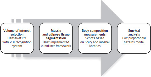
For each study, a volume of interest (VOI) was defined as the abdominal region between the basal margin of the lungs and the upper margin of the iliac bones. An automated VOI classifier based on MONAI library [25] labelled each slice as either within or outside the VOI. It utilised a DenseNet121 architecture with Cross-Entropy as the loss function, the Adam optimiser, and receiver operating characteristic-area under the curve as the evaluation metric. The classification results were then used to create a VOI recognition system, which solved an optimisation problem to refine the VOI selection [23]. The auto- mated and manual annotations were compared using standard evaluation metrics and manually reviewed. Subsequently, all VOIs were segmented using an automated muscle and adipose tissue segmentation model, based on a U-net deep neural network architecture implemented within the nnU-Net framework [24, 26]. Segmentation quality was assessed by calculating the Dice coefficient, and the results were cross verified by an experienced radiologist to ensure reliability. Custom scripts, based on the SciPy and nibabel libraries [21, 27], were developed to obtain body composition measurements from abdominal muscle, paraspinous muscle, and SAT and VAT segments. Initially, the paraspinous and abdominal muscle segments were merged into a single muscle category. A Hounsfield unit (HU) filter was then applied to differentiate intramuscular adipose tissue (IMAT) from skeletal muscle tissue. The predefined HU thresholds were set at 29–150 for skeletal muscle and –190 to –30 for adipose tissue. Finally, four distinct tissue compartments were separated: skeletal muscle, IMAT, VAT, and SAT. For muscle, VAT, and SAT, both volumetric percentage and mean HU values were computed. Intramuscular adipose tissue was quantified as a percentage of the total muscle volume.
Patient data, including body composition measurements, clinical characteristics, and histopathological findings, were analysed to assess their association with overall survival using the Cox proportional hazards model. Continuous variables were standardised using z-score normalization to ensure comparability across different scales. To refine the model and enhance predictive accuracy, stepwise selection based on the Akaike information criterion (AIC) was employed, allowing the identification of the most relevant predictors while balancing model complexity and goodness of fit. Model validation was performed using multiple approaches to ensure the robustness and reliability of the results. The proportional hazards assumption was tested using Schoenfeld residuals to verify that the hazard ratios remained constant over time. Collinearity among predictors was assessed with the variance inflation factor (VIF) to identify potential multicollinearity issues that could distort the model estimates. Model discrimination was evaluated using the concordance index (C-index). Statistical significance was set at p < 0.05. All analyses were performed in R using the survival, survminer, and MASS packages [28]. A summary of described methods can be found in Figure 1.
Results
Volume of interest detection model demonstrated good performance, achieving up to 0.93 accuracy, 0.93 precision, 0.92 recall, and a 0.92 F1 score on the test set. Additionally, its detection capabilities were manually reviewed by an experienced radiologist and deemed satisfactory. The automated segmentation model achieved a median Dice coefficient of 0.99 across all segmented regions, indicating a minimum 99% overlap between automated and manual segmentations. All segmentations were additionally manually inspected and received positive evaluations from an experienced radiologist. The results of body composition measurements based on acquired segmentation, including volumetric percentages and mean HU values, are summarised in Table 2.
Table 2
Automatically acquired body composition parameters
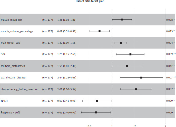
A Cox proportional hazards model was developed to assess factors influencing overall survival. After stepwise selection based on the AIC, the final model included nine covariates: mean HU of muscle segment, volume percentage of muscle segment, maximum tumour size (cm), sex, multiple metastases (≥ 2), extrahepatic disease (considered resectable at the time of surgery), chemotherapy before liver resection, presence of non-alcoholic steatohepatitis (NASH), and histopathological treatment response greater than 50%. The model demonstrated a concordance index of 0.683, indicating moderate predictive ability. Likelihood ratio, Wald, and log-rank tests were all statistically significant (p < 0.05), confirming the model’s overall fit.
Among the statistically significant predictors, higher mean HU of segment muscle was associated with an increased hazard of death (HR = 1.36), whereas a higher volume percentage of segment muscle was protective (HR = 0.69). Larger tumour size also increased hazard (HR = 1.30). It is important to note that these hazard ratios correspond to standardised values of continuous variables, meaning that the effect size reflects a one-standard-deviation increase in each respective variable. Regarding categorical predictors, male sex (HR = 1.75), multiple metastases (HR = 1.58), and the presence of extrahepatic disease (HR = 2.44) were associated with significantly worse prognosis. Additionally, preoperative chemotherapy increased the hazard of death (HR = 2.08). Conversely, patients with NASH (HR = 0.63) and those who demonstrated a treatment response greater than 50% (HR = 0.61) had a significantly lower hazard of death. All hazard ratios were estimated over the entire follow-up period. Detailed results are presented in Figure 2.
Figure 2
Cox proportional-hazards model results
# Events: 95; Global p-value (log-rank): 9.5181e-06 AIC: 868.83; Concordance index: 0.68
extrahepatic_meta – presence of extrahepatic metastases, max_tumor_size – maximum tumour size, multiple_meta – presence of multiple metastases, muscle_mean_HU – mean muscle attenuation in Hounsfield units, muscle_volume – muscle volume percentage, NASH – non-alcoholic steatohepatitis, neoCTx – neoadjuvant chemotherapy, response ≥ 50% – pathologically confirmed tumour response of at least 50%
The proportional hazards assumption was tested using Schoenfeld residuals and showed no significant violations (global test p = 0.88), confirming the model’s validity over time. Additionally, VIF were all below 2.2, indicating no evidence of multicollinearity among the included variables.
Discussion
The findings of this study contribute to the growing body of evidence supporting the prognostic significance of body composition parameters in oncologic outcomes. In the context of colorectal liver metastases, our results underscore the relevance of skeletal muscle volume and composition on overall survival. Our study aligns with previous research showing that a reduced muscle tissue volume percentage is a predictor of poor prognosis in CRC patients [8, 29, 30].
Existing literature links lower muscle volume to increased chemotherapy toxicity, higher rates of postoperative complications, and reduced overall survival [7, 16, 31–36]. Similarly, myosteatosis has been associated with worse oncologic outcomes, including shorter disease-free and overall survival [33]. Our findings confirm the prognostic significance of muscle volume but not fat infiltration; patients with lower muscle volume had reduced survival rates, whereas IMAT had no significant impact. Interestingly, higher muscle density (higher HU) was associated with an increased hazard ratio, which may be explained by an underlying hypercatabolic state leading to muscle depletion, chronic inflammation-induced fibrosis, or treatment-related muscle alterations caused by chemotherapy [37, 38]. These factors could contribute to a decline in muscle function and overall prognosis despite the appearance of higher muscle density on imaging. In contrast, VAT has demonstrated a more complex relationship with oncologic outcomes. While visceral obesity has been linked to systemic inflammation and tumour progression [39], some studies suggest a paradoxical protective effect in certain cancer subtypes [40, 41]. Our data indicate that VAT volume percentage was not a significant prognostic factor.
The observation that patients with NASH exhibit a significantly lower hazard of death (HR = 0.63) is both unexpected and intriguing. Traditionally, hepatic steatosis has been associated with adverse outcomes in liver surgery due to concerns about impaired liver regeneration and increased postoperative complications. However, emerging evidence suggests that the presence of NASH, particularly when it progresses to advanced fibrosis or cirrhosis, may influence tumour biology and the hepatic microenvironment in ways that could impact metastatic progression [42]. For instance, a study demonstrated that patients with advanced fibrosis or cirrhosis had a significantly lower risk of synchronous colorectal liver metastasis compared to those without advanced fibrosis. This counterintuitive finding may be attributed to alterations in hepatic blood flow, immune surveillance, and extracellular matrix composition in fibrotic or cirrhotic livers, potentially creating a less favourable environment for metastatic tumour cells [43]. Consequently, these factors could contribute to improved survival outcomes in CRLM patients with NASH, warranting further investigation into the underlying mechanisms and their potential therapeutic implications. It should also be noted that chemotherapy itself may contribute to the development of steatohepatitis [44]. Previous studies have reported that approximately 20% of patients may develop steatohepatitis following neoadjuvant chemotherapy for colorectal liver metastases. In our study, however, the presence of NASH was assessed using the Kleiner-Brunt scoring system based on histopathological evaluation of liver specimens obtained during partial hepatectomy [45]. As pre-treatment liver biopsies were not available, it was not possible to determine whether NASH was present prior to chemotherapy or developed as a consequence of systemic treatment. Therefore, our study could not differentiate between pre-existing and chemotherapy-associated NASH.
A key strength of our study is the utilisation of an AI-driven volumetric body composition analysis, which overcomes the limitations of traditional single-slice assessments. The standard practice of evaluating body composition at the L3 vertebral level has been widely adopted due to its correlation with whole-body muscle and adipose distribution [32]. However, this approach is inherently limited by inter-observer variability and potential inaccuracies in estimating total body composition. Recent advancements in automated segmentation have enabled volumetric assessments across larger anatomical regions, providing a more comprehensive representation of meta- bolic health [11]. Our automated methodology demonstrated high reliability, with a Dice coefficient of 0.99 across all segmented regions. This level of precision supports the clinical applicability of AI-driven body composition analysis, offering a standardised and reproducible approach. Despite the growing recognition of body composition parameters as critical prognostic factors, their routine incorporation into clinical workflows remains limited. The automation of volumetric segmentation presents an opportunity to streamline body composition analysis, allowing for potential development of decision support systems based on acquired measurements. It is also worth mentioning that the prognostic implications of body composition analysis extend beyond survival predictions. In the setting of CRC, patients often experience significant physiological stress, and preoperative body composition may impact postoperative recovery and complication rates [33]. Given the association between sarcopaenia and increased chemotherapy toxicity, personalised treatment strategies incorporating nutritional and physical rehabilitation interventions could mitigate adverse outcomes [16, 36, 46, 47]. Unfortunately, it was not possible to link out automated measurements to those outcomes due to lack of appropriate data.
Additionally, it needs to be noted that aside from CT several different methodologies can be employed to measure body composition, each with unique advantages and limitations [48, 49]. However, in the context of CRC management, CT imaging plays a pivotal role. It is routinely performed for diagnosis, staging, and treatment planning, offering detailed insights into tumour location, size, and potential metastasis [50]. This routine use of CT scans allows for the opportunistic evaluation of body composition without necessitating additional imaging procedures, thereby avoiding extra radiation exposure and associated costs.
In our study, we employed classical statistical methods for body composition survival analysis due to the limited size of our cohort, which in preliminary analysis was insufficient to support machine learning approaches without the risk of overfitting. Overfitting risk is particularly pronounced in small datasets, where complex models like those used in machine learning can easily fit to random fluctuations rather than true signal [51]. Therefore, simpler, classical statistical methods were more appropriate for our analysis. Additionally, while some studies utilise radiomics to extract detailed features from muscle and fat tissues [23, 52–54], these high-dimensional data often require large sample sizes to ensure reliable analysis [55]. Complexity and volume of radiomic data can pose significant challenges for traditional statistical techniques, especially when the sample size is limited. To mitigate these challenges, we focused on more straightforward metrics, specifically volume percentage and mean HU, which are more amenable to classical statistical analysis in smaller cohorts. This approach allowed us to maintain analytical rigor while avoiding the pitfalls associated with high-dimensional.
Despite the strengths of this study, several limitations must be acknowledged. First, the retrospective nature of the analysis may introduce selection bias and limit the ability to infer causal relationships. Second, while our dataset was derived from a publicly available repository, it remains relatively small compared to large-scale, multi- institutional studies, which may affect the generalisability of our findings. Additionally, details regarding the exact location of the primary tumour (colon vs. rectum) were lacking, which could potentially impact survival outcomes. Furthermore, although several clinical risk scores have been developed for patients with colorectal liver metastases, including the well validated Nagashima score [56], the variables required to calculate this score were not fully available in the dataset. As a result, the application of any well- established scoring systems was difficult and ultimately not feasible in the present analysis. Moreover, while the presented associations were statistically significant, some predictors had p-values close to 0.05, suggesting that the results should be interpreted with caution. Future research with larger sample sizes may help confirm these findings and further explore potential underlying mechanisms. Furthermore, while AI-driven segmentation enhances reproducibility, the reliance on pre-trained models may introduce limitations in cases where anatomical variations are present. Finally, our study did not incorporate functional assessments of muscle strength or patient-reported quality-of-life metrics, which could provide a more holistic understanding of the clinical impact of body composition changes.
Conclusions
This study highlights the prognostic value of AI-driven volumetric body composition analysis in patients with colorectal liver metastases. By integrating fully automated segmentation, it was demonstrated that skeletal muscle volume and composition are significant predictors of overall survival. The high reliability of the automated approach supports its potential integration into routine oncologic workflows, addressing limitations of traditional single-slice assessments. Despite the study’s retrospective nature and dataset size, its results emphasise the need for further validation in larger, multi-institutional cohorts. Future research should explore dynamic changes in body composition during treatment and their influence on therapy response, surgical recovery, and long-term survival. The implementation of automated body composition analysis in clinical decision-making could enhance personalised treatment strategies, ultimately improving patient outcomes in CRLM.










