
Advanced systemic atherosclerosis may lead to severe obstruction or occlusion of multiple vascular territories, occasionally resulting in the loss of conventional access routes for coronary angiography and percutaneous coronary intervention (PCI) [1]. In such situations, restoration of an access vessel may be required to allow diagnostic and therapeutic coronary procedures.
A 77-year-old man with heart failure with reduced ejection fraction (LVEF 36%), hypertension, diabetes mellitus, mixed hyperlipidemia, and advanced systemic atherosclerosis was admitted with Canadian Cardiovascular Society (CCS) class III angina. Physical examination revealed a systolic bruit over the abdominal aorta and absent bilateral femoral pulses, while radial pulses were palpable.
Initial coronary angiography was unsuccessful. Attempted right radial access failed due to a critical stenosis of the brachiocephalic trunk preventing catheter advancement. Stenosis of the brachiocephalic trunk was estimated at approximately 90% based on quantitative coronary angiography (QCA). Left radial access was not feasible because of a complete occlusion of the left subclavian artery. Computed tomography angiography demonstrated diffuse atherosclerotic disease affecting the supra-aortic and peripheral arteries, including severe stenosis of the brachiocephalic trunk, occlusion of the left subclavian artery, and bilateral occlusion of the iliac and femoral arteries consistent with Leriche syndrome, effectively eliminating transfemoral access.
Following multidisciplinary Heart Team discussion, the patient was deemed unsuitable for coronary artery bypass grafting due to extensive vascular disease and comorbidities. Therefore, an endovascular strategy aimed at restoring vascular access was pursued.
Through right brachial access (7 Fr sheath), the stenotic brachiocephalic trunk was crossed with a guidewire and treated with balloon angioplasty using a non-compliant 3.5 × 15 mm balloon inflated to 15 atm (Figure 1 A). Stent implantation in the brachiocephalic trunk was not performed, as the primary goal was restoration of temporary vascular access to enable coronary intervention. In addition, stenting carried a potential risk of compromising (“jailing”) the ostium of the right common carotid artery and increasing the risk of cerebrovascular complications. Balloon angioplasty provided sufficient luminal gain without significant elastic recoil that would impair catheter passage. This maneuver successfully restored sufficient lumen diameter to allow advancement of coronary catheters and completion of coronary angiography.
Figure 1
Endovascular restoration of vascular access enabling complex coronary intervention. A – Angiographic image showing balloon angioplasty of the brachiocephalic trunk (arrow) performed to restore vascular access and allow catheter advancement. B – Baseline coronary angiography demonstrating significant stenosis of the left main coronary artery (LMCA) (arrow) and left anterior descending artery (LAD) (arrowhead). C – Intravascular ultrasound demonstrating extensive circumferential calcification (> 270°) of the LMCA (asterisk) with a minimal lumen area (MLA) of 5 mm2. D – Final angiography after rotational atherectomy and drug-eluting stent implantation in the LMCA-LAD segment showing optimal angiographic result with preserved TIMI 3 flow
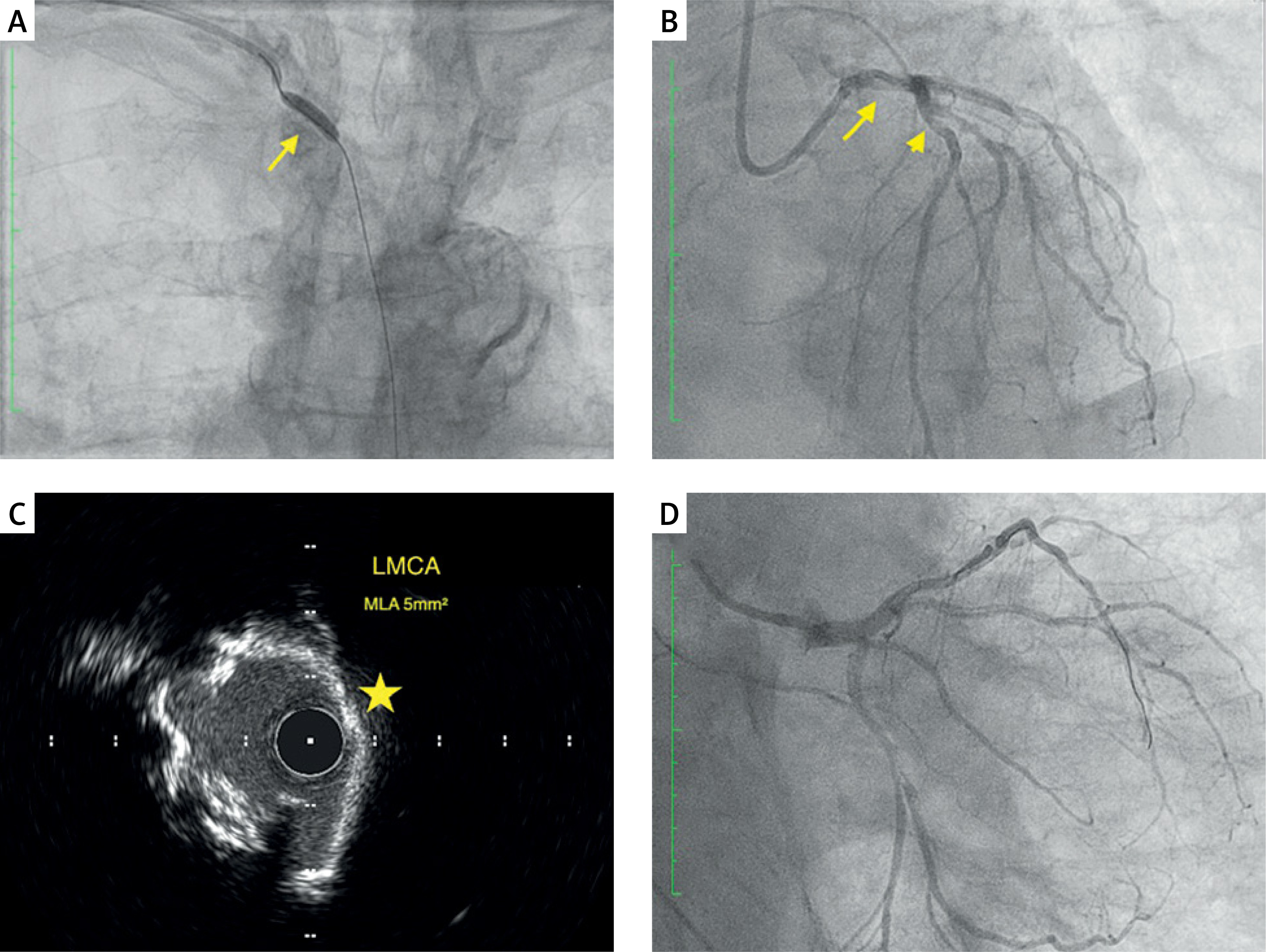
Angiography revealed a heavily calcified 60% eccentric stenosis of the left main coronary artery and an 80% stenosis of the mid left anterior descending artery (Figure 1 B). Intravascular ultrasound (IVUS) demonstrated extensive circumferential calcification (> 270°) of the left main coronary artery with a minimal lumen area of 5 mm2 [2, 3] (Figure 1 C). Rotational atherectomy of the left main was performed using a 1.75 mm burr (Rotawire Floppy), followed by lesion preparation with non-compliant balloon inflations (LAD: 3.5 × 15 mm at 15 atm; LM: 4.0 × 15 mm at 20 atm) [4].
Subsequently, two drug-eluting stents (Ultimaster Nagomi) were implanted from the left main to the left anterior descending artery (LAD: 3.0 × 18 mm; LM–LAD: 4.0 × 18 mm), followed by proximal optimization technique (POT) using a 5.0 × 12 mm balloon and final kissing balloon inflation (LAD: 4.0 mm; LCx: 3.5 mm). Final re-POT was performed to optimize stent expansion.
IVUS confirmed adequate stent expansion and apposition (minimal stent area in the left main 17 mm2) without edge dissection, and final angiography demonstrated an optimal result with preserved TIMI 3 flow (Figure 1 D). The procedure, including brachiocephalic trunk angioplasty, was performed by an experienced interventional cardiologist with expertise in peripheral interventions and complex vascular access. Periprocedural anticoagulation was achieved with unfractionated heparin, and dual antiplatelet therapy was initiated following the procedure in accordance with standard practice.
Following right brachial access, a localized pseudoaneurysm developed and was successfully treated with ultrasound-guided compression, without the need for invasive intervention. No bleeding or ischemic complications or neurological deficits were observed during hospitalization. The patient was discharged in stable condition 2 days after the procedure with planned cardiology follow-up. Further evaluation by vascular surgery was arranged to consider definitive management of the brachiocephalic trunk lesion.
This case illustrates an uncommon but clinically relevant strategy in which angioplasty of a severely stenotic supra-aortic vessel enabled coronary angiography and complex IVUS-guided left main PCI in a patient with generalized atherosclerosis and absence of conventional vascular access routes. In carefully selected patients, endovascular restoration of access vessels may represent the only feasible pathway to definitive coronary revascularization.










