Introduction
Occupational exposure to ionizing radiation remains an inherent risk in the cardiac catheterization laboratory. Despite advances in imaging technology and procedural techniques, interventional cardiologists, scrub nurses, and the remaining staff are continuously exposed to scattered radiation, which has been associated with an increased risk of several occupational diseases (e.g., cataracts and malignancies) [1]. Conventional radiation protection strategies, including ceiling-mounted shields and personal aprons, may not provide adequate shielding [2].
Furthermore, the cumulative physical burden of anti-radiation apparel has been linked to a high prevalence of musculoskeletal disorders among catheterization laboratory personnel [3]. Recently, novel mobile radiation shielding systems (RSS) have been introduced to enhance protection while improving operator ergonomics. The RAMPART Defender system is a mobile shielding solution designed to reduce scatter radiation exposure while maintaining procedural access and visibility. Early studies have demonstrated significant reduction in operator and staff radiation dose without compromising procedural workflow or efficiency. However, real-world data on its implementation remain limited, particularly in European centers [4].
Therefore, we aimed to evaluate the feasibility and effectiveness of the RAMPART Defender RSS in routine clinical practice. This study presents the initial Polish experience from a high-volume center, focusing on radiation exposure among catheterization laboratory staff during a variety of fluoroscopy-guided cardiovascular procedures.
Methods
This retrospective, single-center observational study was conducted at the Upper Silesian Medical Center in Katowice and reflects our initial real-world experience during the first week after implementation of the RAMPART Defender System. The analysis included 17 consecutive fluoroscopy-guided cardiovascular procedures performed with the RSS, including coronary angiography (CAG) with assessment of coronary physiology, percutaneous coronary interventions (PCI), patent foramen ovale (PFO) closure, and transcatheter aortic valve implantation (TAVI).
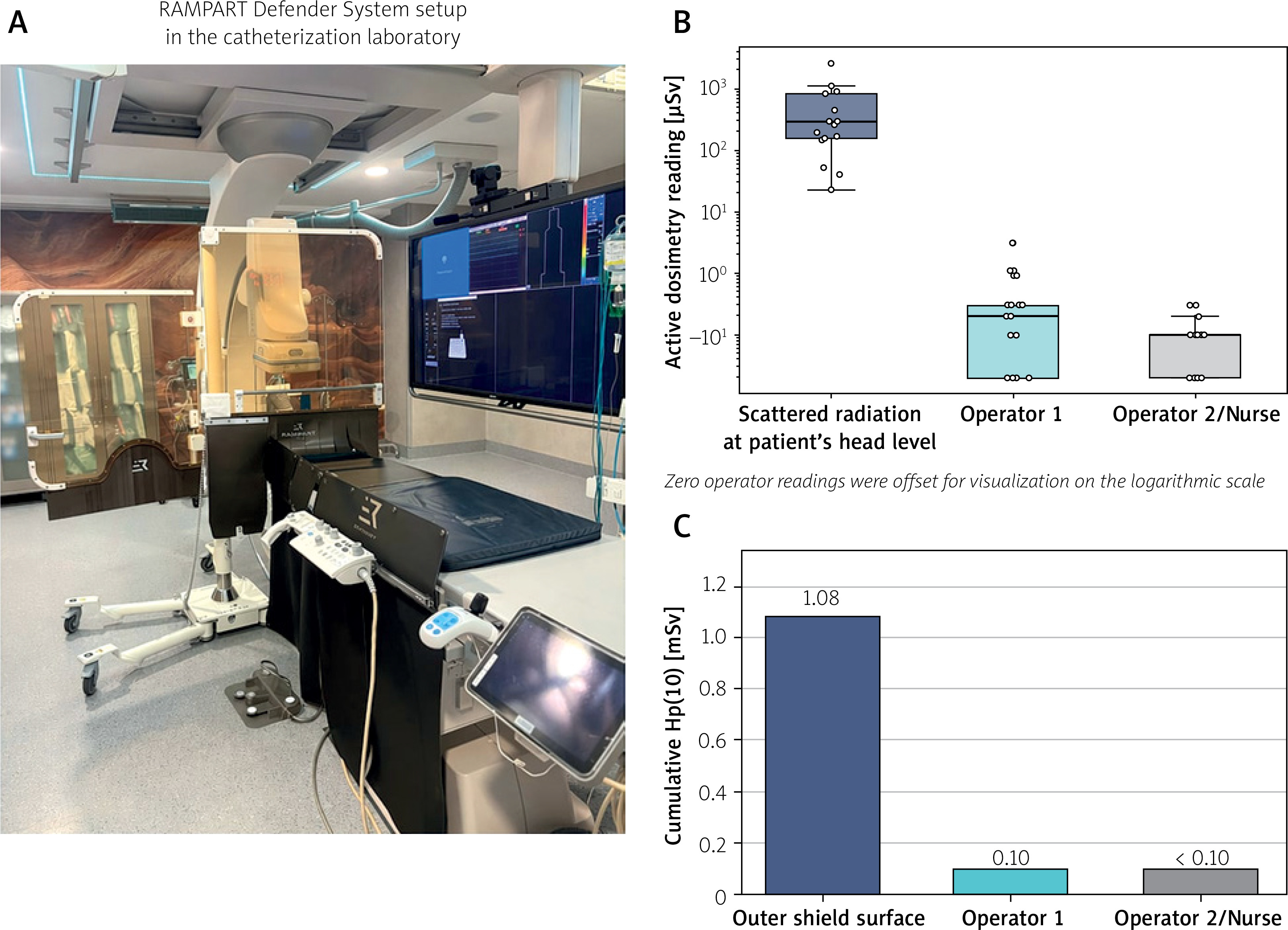
The RAMPART Defender is a mobile, lead-free RSS composed of two bismuth-infused acrylic panels (1 mm lead-equivalent) with additional soft shielding layers (0.5 mm) and table-mounted shields (1 mm lead-equivalent) (Figure 1 A). The system is designed to reduce scatter radiation and can be positioned to allow both radial and femoral access while maintaining full protection.
Figure 1
A – RAMPART Defender system setup in the catheterization laboratory. B – Individual procedural active dosimetry readings during the first week after implementation of the RAMPART Defender system, recorded at the level of scattered radiation at the patient’s head and at the positions of Operator 1 and Operator 2/Nurse. The y-axis is shown on a logarithmic scale. C – Cumulative Hp(10) readings obtained with thermoluminescent dosimeters during the same observation period, shown for the outer surface of the protective shield, Operator 1 and Operator 2/Nurse
Radiation exposure was assessed using the RaySafe i3 real-time dosimetry system with three active personal dosimeters (detection limit < 30 µSv/h). One dosimeter was positioned beneath the patient’s head to quantify scattered radiation from the primary beam (SRH), whereas two dosimeters were worn at chest level by the primary and secondary operators (or assisting nurse, depending on the procedure). The RaySafe i3 system was used in accordance with the manufacturer’s recommendations and routine quality assurance procedures. During the study period, no procedure required temporary repositioning of the RAMPART Defender system that interrupted radiation protection. Additional cumulative measurements were obtained using thermoluminescent dosimeters (TLDs, detection limit < 0.1 mSv) placed on the shield surface and operators’ chests, analyzed by an accredited laboratory. Both dosimetry systems were used according to the manufacturer’s guidelines, and a calibration process was conducted for TLDs, as the RaySafe i3 does not require calibration according to the manufacturer’s instructions. Clinical and procedural data were collected from the angiography system and procedural records. These included procedure type, vascular access site, fluoroscopy time, cumulative air kerma at patient entrance reference point, dose-area product (DAP), and dosimeter readings obtained from the SRH position and individual staff positions. Fluoroscopic projections and C-arm angulations were selected according to procedural requirements and operator preference, reflecting routine clinical practice. No predefined protocol regarding X-ray tube positioning was applied.
Owing to the retrospective nature of the analysis and the use of anonymized clinical and procedural data, the requirement for written informed consent was waived.
Statistical analysis was performed using Statistica v. 13 software (Tibco Software Inc., Palo Alto, CA, US). Continuous variables are presented as median (Q1–Q3) and categorical variables as counts (%). Normality was assessed with the Shapiro–Wilk test, and analyses were primarily descriptive given the small, exploratory sample.
Results
The analysis included 17 consecutive fluoroscopy-guided cardiovascular procedures performed with the RAMPART Defender system during the first week after implementation. Coronary procedures predominated and included CAG with physiology assessment in 2 (12%) cases, CAG alone in 5 (29%), and PCI in 7 (41%). The latter included PCI in chronic coronary syndromes, non-ST-segment elevation myocardial infarction, and left main intervention. Structural procedures included PFO closure in 2 (12%) cases and TAVI in 1 (6%) case. Radial access was used in 14 (82%) procedures, femoral access in 2 (12%), and mixed access in 1 TAVI (6%). Procedure-specific procedural characteristics and radiation exposure measurements are summarized in Table I.
Table I
Procedure-specific procedural characteristics and radiation exposure according to procedure type
| Procedure | n | DAP [Gy·cm²] | Air kerma [mGy] | Fluoroscopy time [min] | Scatter radiation SRH [µSv] | First operator [µSv] | Second operator [µSv] |
|---|---|---|---|---|---|---|---|
| CAG* | 7 | 9.19 (8.42–12.16) | 82.67 (77.72–130.02) | 5.80 (4.55–6.50) | 261.2 (172.8–295.8) | 0.30 (0.25–0.30) | 0.10 (0.05–0.10) |
| PCI | 7 | 31.85 (13.73–52.56) | 385.45 (179.96–549.76) | 14.90 (10.58–28.08) | 839.4 (235.2–1090.6) | 0.10 (0.0–0.65) | 0.10 (0.0–0.10) |
| PFO closure | 2 | 1.08 (0.98–1.18) | 4.89 (4.02–5.76) | 2.15 (1.80–2.50) | 31.8 (23.2–40.4) | 0.05 (0.0–0.1) | 0.0 (0.0–0.0) |
| TAVI | 1 | 20.80 | 136.0 | 13.10 | 454.8 | 0.10 | 0.10 |
Active dosimetry readings at operator positions remained very low throughout the observation (Figure 1 B). Median active dosimetry reading was 0.20 µSv (Q1–Q3, 0–0.30 µSv) for the first operator, and 0.10 µSv (Q1–Q3, 0–0.10 µSv) for the second operator. In contrast, the SRH dosimeter showed a median reading of 293.60 µSv (Q1–Q3, 157.50–839.40 µSv), indicating persistent scatter radiation in the procedural field. Median procedural parameters were 126.20 mGy (Q1–Q3, 77.90–268.40 mGy) for air kerma, 11.06 Gy·cm² (Q1–Q3, 8.68–18.93 Gy·cm²) for DAP, and 6.95 min (Q1–Q3, 4.83–13.28 min) for fluoroscopy time.
The median active-dosimetry-based reduction was 99.92% (Q1–Q3, 99.85–100.00%) for the first operator and 99.99% (Q1–Q3, 99.97–100.00%) for the second operator/nurse in the overall cohort. In the PCI cases, the corresponding median reductions were 99.93% (Q1–Q3, 99.86–100.00%) and 99.99% (Q1–Q3, 99.97–100.00%), respectively.
Very low operator readings were also observed in procedures associated with higher procedural radiation burden. In the left main PCI, despite a DAP of 56.83 Gy·cm², air kerma of 777.79 mGy, fluoroscopy time of 48.40 min, and an SRH reading of 2632.50 µSv, active dosimetry readings were 3.10 µSv for the first operator and 0 µSv for the second operator. Likewise, in the NSTEMI-related PCI case, with a DAP of 54.95 Gy·cm², air kerma of 513.05 mGy, fluoroscopy time of 32.10 min, and an SRH reading of 1120 µSv, both operator readings were 0.10 µSv. In the TAVI procedure, the SRH reading was 454.80 µSv, whereas both operator positions recorded 0.10 µSv. Similarly, in the two PFO closure procedures, SRH readings were 23.20 and 40.40 µSv, while first-operator readings were 0.10 and 0 µSv and second-operator readings were 0 and 0 µSv. Supplementary cumulative thermoluminescent dosimeter data are shown in Figure 1 C. Over the same first-week observation period, cumulative personal dose equivalent at 10 mm depth (Hp(10)) was 1.08 mSv at the outer side of the protective shield, 0.10 mSv for operator 1, and < 0.10 mSv for operator 2.
Discussion
In this single-center study, we present the initial Polish experience with the RAMPART Defender RSS. The main finding is that the use of this mobile shielding solution was associated with very low levels of radiation exposure among catheterization laboratory staff during fluoroscopy-guided cardiovascular procedures, without workflow disruption.
These findings align with prior studies showing marked reductions in operator radiation exposure with advanced shielding. Randomized data demonstrated reductions up to 86% at head level with radiation exposure reduced below the measurable threshold at selected detector locations [5]. Likewise, prospective data have shown significantly lower radiation exposure at the neck level among operators and assisting staff when using the RAMPART system compared with conventional protection [6, 7]. In our study, the use of RSS was associated with relative reductions in active dosimetry readings of 99.92% and 99.99% at the first and second monitored staff positions, respectively. During the most frequent procedure, PCI, the corresponding reductions were 99.93% and 99.99%, indicating that the effect remained pronounced even in procedures associated with a higher radiation burden. Novel shielding systems that effectively reduce radiation burden while potentially decreasing reliance on heavy lead garments may therefore improve long-term occupational safety [4].
Another potential advantage of this system is a reduction of the other occupational hazard related to work in the catheterization laboratory, namely spine and joint diseases, such as spondyloarthritis, hip and knee arthritis, and other musculoskeletal disorders [3]. A standard lead apron may weigh approximately 7 kg, and repeated exposure to this physical load over many years of practice may contribute to cumulative spinal and joint strain. In this context, RSS may offer a potential ergonomic advantage [4].
Importantly, RSS did not interfere with workflow, allowing both radial and femoral access without limiting catheter manipulation or visualization. This is consistent with prior reports showing improved radiation protection without compromising procedural efficiency [8].
This study is limited by its retrospective, single-center design, small sample size, and restricted dosimeter placement, which may not reflect total body radiation exposure. In addition, because this study reflects the initial implementation of the RAMPART Defender system, an observer (Hawthorne) effect related to increased operator awareness of radiation protection cannot be completely excluded.
From a practical perspective, successful implementation of the RAMPART Defender system requires appropriate procedural planning and familiarization with the device. Although no workflow disruptions were observed during our initial experience, operators should anticipate a short learning curve related to optimal positioning of the shielding system and adaptation of catheterization laboratory logistics. In addition, anesthesiology personnel and operators performing transesophageal echocardiography remain outside the protected workspace and therefore require separate radiation shielding. Finally, predefined bailout strategies should be established before each procedure to ensure rapid access to the patient during emergency situations, such as cardiopulmonary resuscitation or urgent fluoroscopy-guided pericardiocentesis, when temporary removal or repositioning of the shielding system may be necessary.
The full video coverage of a procedure performed using RAMPART Defender is available on Zabrze Cathlab TV [9].
Our findings should also be considered in the context of other emerging lead-free radiation protection strategies. In addition to mobile shielding systems such as RAMPART, alternative approaches including suspended protection systems (e.g., Zero-Gravity), barrier systems (e.g., PROTEGO), and robotic-assisted PCI have demonstrated substantial reductions in operator radiation exposure, each with distinct advantages and workflow limitations. Consequently, the optimal radiation protection strategy may depend on catheterization laboratory infrastructure, procedural complexity, and institutional experience [10].
In conclusion, our initial experience suggests that the RAMPART Defender system is a feasible radiation-shielding solution in routine catheterization laboratory practice and may represent an important step toward a new approach to occupational radiation protection.