
A 53-year-old male with conventional cardiovascular risk factors, a history of transient ischemic attack, and polymyositis was referred from an outside center for percutaneous coronary intervention (PCI) of chronic total occlusions (CTOs) of the left anterior descending artery (LAD) and right coronary artery (RCA) after a prior unsuccessful PCI attempt targeting the RCA CTO. The previous attempt had failed owing to false lumen wiring. Transthoracic echocardiography revealed a mildly reduced left ventricular ejection fraction (LVEF) of 45% with akinesis of the basal inferior wall segment and hypokinesis of the mid and apical anterior wall and septal segments. The patient reported CCS class II symptoms. Diagnostic coronary angiography revealed CTOs of the mid LAD and proximal RCA (Figure 1 A). The LAD CTO was characterized by a J-CTO score of 1 with good retrograde filling from the left circumflex artery. The RCA CTO had a J-CTO score of 3 with adequate retrograde filling via collateral vessels from the left coronary system.
Figure 1
Multimodality imaging and stepwise percutaneous revascularization of double chronic total occlusions. A – Baseline coronary angiography. From left to right: left anterior descending artery occlusion (RAO); left anterior descending artery occlusion (AP); right coronary artery occlusion (LAO) with simultaneous contralateral contrast injection into the LCA (dual injection). B – CCTA images. From left to right: 3D reconstruction of the left anterior descending artery and right coronary artery (arrows); occlusion of the right coronary artery with massive calcifications; occlusion of the left anterior descending artery. C – PCI of the LAD. From left to right: antegrade wiring with Mamba microcatheter and Judo guidewire (Boston Scientific); final angiography after successful stent implantation; IVUS cross-section showing optimal stent expansion and apposition. D – PCI of the RCA. From left to right: unsuccessful antegrade wiring with wire passage into the subintimal space; successful retrograde wiring using the reverse CART technique; final angiography after successful stent implantation
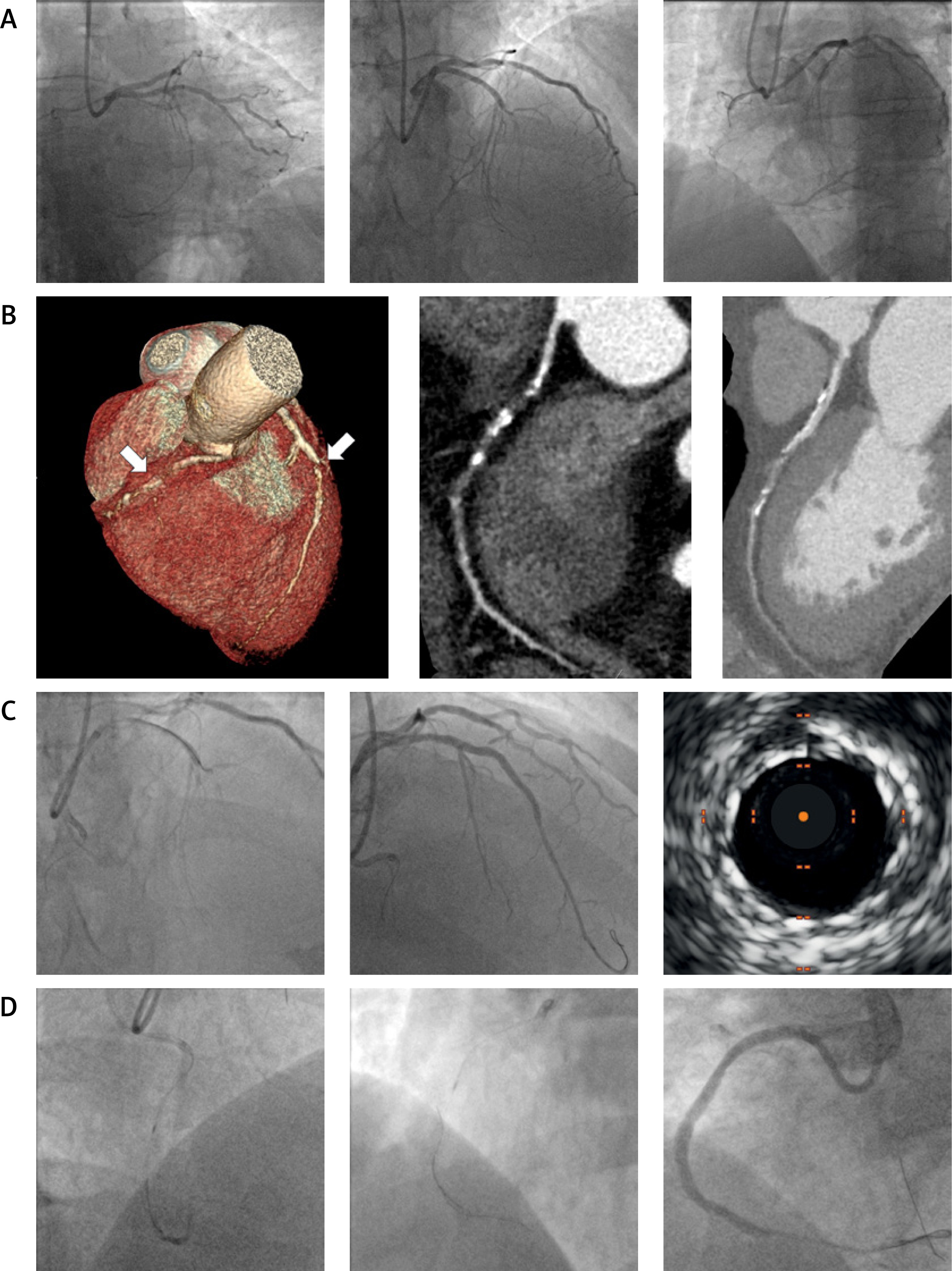
Cardiac magnetic resonance imaging (CMRI) and coronary computed tomography angiography (CCTA) were performed for anatomical, ischemia, and myocardial viability assessment. CCTA revealed massive calcifications at the distal cap of the RCA CTO with an occlusion length of approximately 34 mm. In contrast, the LAD CTO was short with a calcified proximal cap (Figure 1 B). CMRI confirmed ischemia in both the LAD and RCA territories, demonstrating subendocardial perfusion defects. Left ventricular systolic function was preserved, with an LVEF of 53%. Late gadolinium enhancement (LGE) showed no evidence of scarring, confirming preserved myocardial viability. The patient was therefore qualified for PCI of both CTOs.
The procedure was performed via bilateral radial access using 7F introducer sheaths. PCI of the LAD CTO was performed first, using a 7F EBU3.75 guiding catheter. An antegrade wire escalation strategy was employed with a Mamba microcatheter and sequential use of Judo (Boston Scientific, Marlborough, Massachusetts, USA) and Gaia 3 Next (Asahi Intecc Co., Nagoya, Japan) guidewires (Figure 1 C). The Gaia 3 Next guidewire successfully crossed into the distal segment of the vessel, followed by predilatation with a 2.0 × 20 mm semi-compliant balloon at 14 atm. Following intravascular ultrasound (IVUS) assessment, a 3.0 × 38 mm Synergy drug-eluting stent (DES) was deployed at 12 atm (Boston Scientific) and post-dilated using non-compliant balloons (3.0 × 20 mm and 3.5 × 15 mm at 20 atm). IVUS confirmed adequate stent expansion and apposition, with restoration of TIMI grade-3 flow (Figure 1 C).
Subsequently, PCI of the RCA CTO was attempted. An antegrade approach using a 7F AL1.0 guiding catheter, Mamba microcatheter, and Judo and Gladius guidewires was unsuccessful because of subintimal wire entry (Figure 1 D). A retrograde approach was therefore chosen. A Sion Black (Asahi Intecc Co., Nagoya, Japan) guidewire supported by a Turnpike LP microcatheter was advanced to the distal RCA through septal collateral channels. The reverse controlled antegrade and retrograde tracking (reverse CART) technique was then applied, with balloon inflations using semi-compliant balloons (2.5 × 20 mm and 3.0 × 20 mm at 16 atm). The tip-in technique facilitated introduction of the antegrade Mamba microcatheter into the distal RCA, establishing connection between the distal and proximal segments. Following IVUS assessment, two DES were implanted (Synergy 3.0 × 38 mm at 16–18 atm distally and Synergy 3.5 × 48 mm at 20 atm proximally; Boston Scientific). An optimal result was confirmed by IVUS and TIMI grade-3 flow (Figure 1 D). The procedure was completed without complications, and the patient remained hemodynamically stable. Sheaths were removed, and dual antiplatelet therapy with acetylsalicylic acid (ASA) and prasugrel was initiated in view of high thrombotic and low bleeding risk (PRECISE-DAPT score 18 points). The total radiation dose was 1.5 Gy, fluoroscopy time was 56 min, and total procedure time was 90 min. The contrast volume was 350 ml. At 6-month follow-up, the patient was free of anginal symptoms.
This case illustrates that percutaneous revascularization of double CTOs can be performed safely and effectively in carefully selected patients and underscores the value of preprocedural non-invasive imaging [1]. CCTA is crucial for complex CTO evaluation: it enables assessment of complex anatomy, guides technique selection, and may help reduce contrast utilization. In this case, distal cap calcifications of the RCA prompted an earlier switch to the retrograde approach, which helped shorten the procedure time. Conversely, CCTA also identified the LAD occlusion as short and relatively straightforward to cross. CMRI is valuable for confirming PCI indications in CTO candidates. Using LGE and stress perfusion (e.g., regadenoson administration), both viability and ischemia in the respective territories can be evaluated – particularly in double CTO cases, where the extent of ischemia is more difficult to assess [2–4]. Intravascular imaging is critical not only for vessel sizing, stent optimization, and reducing the risk of restenosis, but also for confirming true-lumen position in complex CTO procedures [5–7].










