
A 54-year-old male patient had a HeartMate 3 left ventricular assist device (HM3 LVAD, Abbott Laboratories, Abbott Park, IL, USA) implanted for chronic ischemic left ventricular heart failure. Ten months after surgery he presented with symptoms of congestion and lowflow alarms. Left ventricular assist device (LVAD) flow was consistently below 3 l/min (baseline 4.8–5 l/min) at a pump speed of 5200 rpm. The clinical course, infection workup, and decision to stent rather than operate in this patient were previously reported in Kardiologia Polska (Polish Heart Journal) in 2020 [1]. The present report focuses on the invasive pressure tracings obtained during the same procedure, which were not included in the earlier publication.
Neither echocardiography nor 384-slice computed tomography angiography was conclusive. Urgent left heart and HM3 graft catheterization was performed. The catheters were introduced from the femoral artery retrogradely to the ascending aorta and HM3 outflow graft. Contrast injection to the proximal outflow graft (with the pump stopped temporarily to prevent catheter entrapment) and end hole catheter pressure tracings revealed significant stenosis of the proximal graft (Figure 1). Transcatheter stenting of the stenosis was successfully performed, resulting in immediate restoration of optimal flow.
Figure 1
A, B – Pressure tracings of the patient with HM3 outflow graft stenosis. A – Catheter tip placed between pump rotor and graft-pump connection (proximal to the stenosis): heart-diastolic (a) and heart-systolic (b) with the continuous flow from the stenotic LVAD, and LVAD artificial diastolic (c) and LVAD artificial systolic (d); b/a: 190/140, d/c 266/73, mean 155 mm Hg. Note a very high peak LVAD artificial systolic pressure and high b/d difference. B – Catheter tip in the graft distal to the stenosis, b/a 61/49 mm Hg, d/c 80/30, mean 54 mm Hg (similar tracing was noted in the aorta). C – Pump outflow graft angiography – stenosis of the proximal part of the graft caused by graft tilting (arrows). D – Invasive blood pressure tracing from proximal graft after successful stenting: b/a 92/78, d/c 105/52 mean 84 mm Hg (similar tracings were noted in the distal graft and aorta). Note – same pressure standards on A, B and D panels
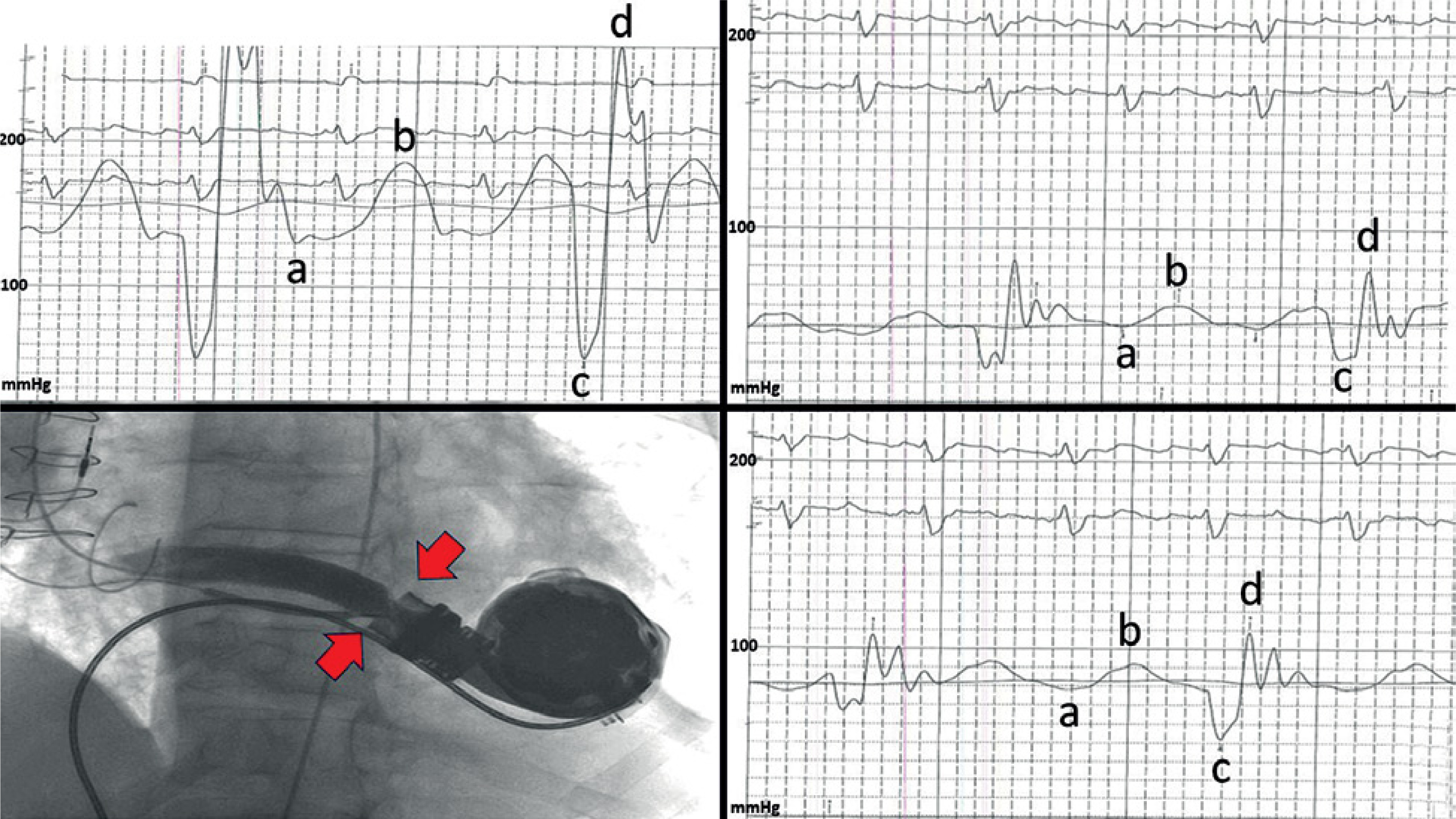
HM3 is a centrifugal design, continuous flow LVAD that creates a constant outflow resulting in a specific pressure tracing with a narrow heart pulse pressure and superimposed pump pulse created by a periodic rotor speed change (±2000 rpm) every 2 s. The pump pulse waves are not synchronized with the native heart beat waves (Figure 1) [2].
The HM3 is nowadays a commonly used implantable LVAD in patients with end stage left ventricular failure as permanent support, bridge to cardiac recovery, or bridge to transplant. Device malfunction of various etiologies is a possible complication that requires prompt diagnosis and treatment. Incidence of outflow graft stenosis up to 2.9% has been reported [3]. Percutaneous stenting is an accepted alternative to reoperation [4].
The purpose of presenting these images was to show catheterization results and analyze specific pressure tracings in a patient with HM3 outflow graft stenosis before and after successful stenting. Of note is a very high systolic pump wave, exceeding 200 mm Hg (at rotor speed of 5200 rpm), high pump/heart systolic pressure difference (76 mm Hg) and high mean pressure (155 mm Hg) proximal to the stenosis. Invasive “pump catheterization” enabled precise diagnosis of graft stenosis-related pump malfunction, followed by successful treatment in a case in which other diagnostic tools were inconclusive.










