
Introduction
Extravasation of cytostatic agents is an iatrogenic complication in oncology, occurring with an estimated incidence of 0.1–6% of intravenous chemotherapy infusions [1]. In paediatrics, precise rates are difficult to establish, but children are considered at heightened risk owing to small-calibre veins, fragile tissues, limited ability to report early symptoms, and the need for repeated venous access over prolonged therapy [2]. Although relatively infrequent, it is associated with a potentially life-threatening condition resulting from pressure buildup due to internal bleeding or tissue swelling [1, 3]. The severity of injury depends on multiple factors, including the type and concentration of the extravasated drug, the volume, and the anatomical site of leakage. The anatomical site of leakage plays a decisive role in both the extent of injury and the therapeutic approach. Extravasation from peripheral veins, particularly on the dorsum of the hand or near large joints, often results in superficial but functionally significant injuries due to the limited subcutaneous space and proximity to tendons or nerves. In contrast, extravasation from central venous access devices or implanted ports may involve deep tissue compartments, the mediastinum, or pleural space, requiring urgent imaging and often surgical intervention such as drainage or thoracoscopic exploration. Understanding the anatomical location is therefore essential to guide the choice between conservative and surgical management [3, 4]. Vesicant agents such as anthracyclines or vinca alkaloids are particularly hazardous. They may cause severe and irreversible local injuries and can also induce progressive tissue necrosis, ulceration, and permanent functional deficits [4].
Moreover, extravasation can have long-term psychological and cosmetic repercussions, which may affect quality of life well beyond cancer treatment [2, 5].
Despite its clinical importance, the approach to extravasation management remains heterogeneous worldwide. Numerous guidelines have been published by oncology societies, national health authorities, and hospital-based expert groups, but they differ with respect to definitions, classification of cytostatic agents, and recommended interventions. Furthermore, the availability of specific antidotes such as dexrazoxane varies considerably between institutions and countries, which complicates the standardization of care [6].
The absence of unified, paediatric-focused recommendations highlights the need for clear and practical instructions tailored to this vulnerable patient group. Interdisciplinary collaboration among oncologists, surgeons, nurses, and pharmacists is crucial to ensure prompt recognition and appropriate intervention [1–6].
This publication addresses this gap by presenting structured, evidence--based guidelines for the management of cytostatic drug extravasation in children, including possible surgical interventions. The protocol integrates international recommendations with institutional experience and emphasises preventive strategies, immediate bedside management, and escalation to surgical treatment when indicated. The document is intended as a practical tool to improve patient safety and to support consistent standards of care in paediatric oncology.
The content integrates evidence from published recommendations, systematic reviews, and institutional practice, and therefore represents a narrative, expert-based review rather than a systematic analysis.
Step-by-step procedure
Immediate actions upon noticing extravasation (apply to all cases)
stop the infusion immediately if extravasation is suspected,
do not remove the intravenous needle/central line/needle from the vascular access device, they may be needed for further treatment (e.g., drug aspiration),
secure the infusion set – disconnect the drug bag or syringe but keep it (to verify the type of drug and its concentration),
determine and record the name of the extravasated drug,
mark the site of the incident and perform a clinical assessment,
carefully examine the puncture site: assess the extent of swelling, erythema, pain, presence of blisters, and necrosis,
mark the boundaries of the skin lesion with a pen,
take a photo of the extravasation site (if permitted by the patient’s parents and hospital rules),
assess the patient’s general condition – pain, anxiety, systemic reactions [7–16].
Aspiration of extravasated medication
Extravasation from a peripheral injection:
Extravasation from the port, central catheter, midline:
– stop administering the drug and leave the needle in the port,
–do not flush the port,
–attempt to aspirate the cytostatic agent through the vascular port,
–perform chest imaging (X-ray or computed tomography [CT]) urgently,
– call a surgeon for consultation to consider pleural drainage (if there is fluid in the pleural space), thoracoscopy/thoracotomy (if there is fluid in the mediastinum), or subcutaneous tissue drainage (if there is a subcutaneous fluid collection) [7–16] (Table 1).
Table 1
Step-by-step management of cytostatic extravasation according to the site of leakage
Apply local treatment:
Table 2
[i] DMSO – dimethyl sulfoxide, NAB – nanoparticle albumin-bound
* Cold compress – dry (moist compresses cause skin maceration) causes vasoconstriction, reduces the area of damage, reduces pain, reduces inflammation; used together with dimethyl sulfoxide; must not be used in cases of extravasation of drugs containing platinum salts, vinca alkaloids, and taxanes. How to use: apply immediately after extravasation 4 times a day for 15–20 min, for a maximum of 48 h.
** Warm compress – dry causes vasodilation, which accelerates the flow and absorption of the extravasated drug and reduces the concentration of the cytotoxic drug outside the vascular bed. How to use: apply 4 times a day for 15–20 min, for a maximum of 48 h.
*** Dimethyl sulfoxide (DMSO) is a preparation with anti-inflammatory and vasodilatory properties. It has a high capacity to remove free radicals. Its side effects include intense local burning and reddening of the skin. It is available in liquid and ointment/gel form (RIMSO-50®, CryoPur – DMSO, Dolobene®). Method of use: immediately after extravasation, locally every 6–8 h for 7–14 days – 4 drops of DMSO per 10 cm^2 of skin surface (on an area twice the size of the area of extravasation), wait until the medication dries without applying a dressing, then apply a dry, cold compress. Apply DMSO every 8 h for at least 7 to 14 days, but do not use if blisters appear, and keep the area of extravasation as exposed to air as possible.
**** Hyaluronidase – This is an enzyme that breaks down hyaluronic acid, increases tissue permeability, and accelerates the absorption of extravasated substances. The maximum dose of hyaluronidase used is 900–1500 IU. Available preparation: Hylase Dessau 150I.E; 10 vials of 150 IU. Method of use: dissolve 150 IU in 1 ml of 0.9% NaCl and administer directly into the vessel, then another 1 ml (150 IU in 1 ml of 0.9% NaCl) should be used for subcutaneous injection into the area of extravasation.
***** Dexrazoxane – It is a catalytic inhibitor of topoisomerase II that binds iron, displacing it from anthracycline-Fe complexes, thereby preventing the formation of free oxygen radicals (Savene™ – prepare in accordance with the Product Safety Data Sheet). Method of use: do not use a single dose greater than 2000 mg, start administration of the drug as soon as possible (within 6 h of anthracycline extravasation), administer the drug in infusions on 3 consecutive days, at the same time: day 1 – 1000 mg/m^2, day 2 – 1000 mg/m^2 and day 3 – 500 mg/m^2.
Further management includes administration of pain relief and fluids intravenously. Oxygen therapy and antibiotics (in case of port extravasation) should be considered. Ongoing monitoring of the patient should be provided, and, if there is no improvement, follow-up CT scan (if there is leakage from the port) and another surgical consultation should be performed. The affected limb should be kept elevated for at least 48 h, after which gradual mobilization is recommended. In cases of extensive tissue damage or necrosis, surgical intervention may be required, sometimes in multiple stages. Every episode of cytostatic drug extravasation must be carefully documented in both the medical and nursing records.
It is essential to clearly distinguish extravasation from a local irritant reaction, because some cytostatic drugs may cause a local skin reaction (asparaginase, cisplatin, daunorubicin, doxorubicin, epirubicin, fludarabine, mechlorethamine, melphalan) or phlebitis (amsacrine, carmustine, cisplatin, dacarbazine, epirubicin, 5-fluorouracil, gemcitabine, mechlorethamine, vinorelbine). A detailed classification according to their local effect at the site of extravasation/administration is presented in Table 3 [7–16].
Table 3
List of cytostatic drugs and their classification according to their action at the site of extravasation/administration [7–16]
A designated container should be available in every unit where cytostatic drugs are administered. Suggested contents of the kit for managing cytostatic drug extravasation:
instructions for action in case of extravasation;
sterile disposable gloves – 5 pairs;
sterile compresses (5 × 5 cm; 10 × 10 cm) – 5 packs;
syringes (1 ml; 5 ml) – 5 each;
needles (0.5; 0.6; 0.8; 1.2) – 5 pieces each;
cold compresses/cooling pads (in the freezer) – 2 pieces;
warm compresses/heating pads – 2 pieces;
0.9% NaCl 10 ml – 5 ampoules;
set for marking the site of extravasation (e.g., skin marker);
antidote:
Healthcare staff should be advised when extravasating cytotoxic drugs, wear gloves and a protective apron resistant to the action/penetration of cytotoxic substances, as well as a cap, medical mask and goggles [7–16].
To reduce the risk of cytostatic extravasation, attention should be paid during administration of cytostatic agents. The following guidelines should be followed during the administration of cytostatic agents:
select the appropriate vessel:
– avoid administering cytostatics into veins on the back of the hand and around large joints (extravasation in such areas may lead to limited joint mobility),
– do not administer cytostatics into veins with thrombotic changes, in limbs with slow blood flow (e.g., in the vicinity of excised lymph nodes), swollen or with paresis/paralysis;
cytostatics that are highly damaging or used in continuous infusion, especially in planned long-term chemotherapy, should preferably be administered into large veins via a catheter inserted into a central vein or vascular port;
avoid administering cytostatic drugs into vessels that have been punctured cephalad to the currently planned puncture site within the last 48 h;
check the puncture site:
proper administration of cytostatic drugs:
– strictly follow the manufacturer’s instructions for administering cytostatic drugs, including drug concentration and administration time,
– administer highly irritating drugs first. Each time before and after administering a cytostatic drug, flush the puncture site with ≥10 ml of 0.9% NaCl,
– avoid administering the drug in a bolus,
– avoid high infusion rates/high flow pressures;
monitor the puncture sites: keep a puncture observation chart [7–16].
Surgical management in case of cytostatic drug extravasation
Surgical management of cytostatic drug extravasation is reserved for selected cases where conservative measures are insufficient or when local tissue damage is progressing despite early interventions. The primary goal is to minimise necrosis, preserve tissue viability, and reduce long-term functional and aesthetic complications. The choice of surgical technique depends on several factors: the type and volume of extravasated drug, time elapsed since the incident, depth of tissue involvement, and the presence of evolving necrosis or ulceration [6, 15, 17–19].
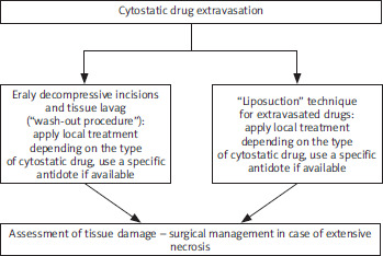
Early decompressive incisions and tissue lavage (“wash-out procedure”)
This approach, originally described by Gault, is the most recommended early surgical intervention, particularly when highly vesicant cytostatics (e.g., anthracyclines, vinca alkaloids) are involved. It should ideally be performed within the first 6 h after extravasation for optimal effectiveness. The procedure is as follows: Several small longitudinal incisions (typically 3–5, 2–3 mm each) are made radially around the extravasation site under local anaesthesia. A blunt-tipped cannula is inserted into the subcutaneous space, and a large volume (500–1000 ml) of isotonic solution (commonly 0.9% NaCl) is gently flushed through the tissue planes to dilute and remove the cytostatic agent [20, 21]. Hyaluronidase may be added to facilitate drug dispersion and reduce tissue injury. It is indicated for early extravasation of vesicant agents, especially anthracyclines, epipodophyllotoxins, and alkylating agents. This procedure minimises progression to ulceration and necrosis, often preventing the need for later extensive surgery, but requires early recognition of extravasation and immediate surgical availability [17, 18]. After the intervention, the incisions are left open, and a dressing is applied. The limb is elevated for at least 24 h [22].
The “liposuction” technique for extravasated drugs
This minimally invasive method is mainly used in the very early phase of extravasation (preferably within the first 1–4 h). It is particularly effective for agents that spread rapidly through the subcutaneous tissue, such as taxanes or anthracyclines. After local anaesthesia, a small incision (approximately 3–5 mm) is made, and a fine suction cannula is introduced into the subcutaneous space. Continuous gentle suction is applied to aspirate the extravasated fluid before significant tissue binding occurs. This procedure is less traumatic than multiple wash-out incisions, allows for removal of a significant amount of the cytostatic agent, and can be combined with lavage for improved efficacy, but not effective if performed late (> 6–8 h) or when extensive necrosis has already developed [20, 21].
Surgical excision of altered tissues
When extravasation is diagnosed late or conservative and early surgical approaches fail, progressive necrosis, ulceration, or secondary infection may develop. In such cases, wide surgical excision is indicated to remove devitalised tissues and prevent further spread of injury. It is usually performed after clear demarcation of necrotic areas, which may take several days to weeks. The technique is as follows: full-thickness excision of the affected skin and subcutaneous tissues with an adequate safety margin (commonly 0.5–1 cm of visually unaffected tissue) [19]. Negative pressure wound therapy (NPWT) (vacuum- assisted closure [VAC] therapy) is also helpful. Vacuum-assisted closure, also known as NPWT, is frequently used postoperatively to optimise wound healing and prepare the defect for secondary closure or reconstruction [23, 24]. Shortly a special open-pore foam dressing is placed into the wound cavity, sealed with an occlusive adhesive film, and connected to a controlled negative pressure device via tubing. Continuous or intermittent negative pressure is applied (usually –75 to –125 mm Hg), which promotes wound contraction and accelerates healing.
Clinical benefits include enhanced granulation tissue formation – stimulates angiogenesis and fibroblast proliferation, accelerating preparation of the wound bed; exudate management – continuously removes fluid, debris, and pro-inflammatory mediators, maintaining an optimal moist environment, reduction of oedema and local inflammation – decreases interstitial pressure and improves perfusion of surrounding tissues. Vacuum-assisted closure therapy optimises conditions for skin grafts and local flap survival by enhancing perfusion and adherence, is related to a lower risk of infection – by decreasing bacterial load and isolating the wound from the external environment. Typical settings and duration: continuous suction: –75 to –125 mm Hg for highly exudative wounds; intermittent cycles: 5 min on/2 min off for ischemic or marginally perfused tissues; dressing changes: usually every 48–72 h (can be longer) or more frequently if infection is suspected.
Complications and precautions during VAC therapy are minor bleeding due to increased capillary perfusion, especially in anticoagulated patients, pain during dressing changes; requires adequate analgesia or local anaesthesia, risk of foam adherence if dressing is left too long without changes. VAC therapy is contraindicated in patients with untreated osteomyelitis, uncontrolled sepsis, and malignant wounds [23, 24]. Secondary infections, delayed healing, and functional impairment may still occur, especially if deeper structures such as tendons, joints, or neurovascular bundles are exposed. Surgical excision combined with VAC therapy represents a salvage approach when early interventions (e.g., wash-out procedures, liposuction) are no longer feasible or have failed. By accelerating wound bed preparation and reducing the risk of infection, VAC therapy significantly improves the success rate of subsequent skin grafts and reconstructive techniques [23, 24].
Local plastic surgery or skin grafts
Following wide excision, tissue defects often require coverage to restore skin continuity and promote healing. The choice between local plastic procedures and skin grafts depends on the size and depth of the defect, as well as the involvement of critical structures [19]. Split- thickness skin grafts are preferred for larger superficial defects; donor sites typically heal rapidly [25]. Full-thickness skin grafts are indicated for areas requiring superior cosmetic outcomes, such as the face, neck, or dorsum of the hand [26]. Local advancement or rotation flaps are utilised when adjacent tissue is viable and sufficiently mobile to cover the defect [27]. These approaches can shorten recovery time, reduce scarring, and improve both aesthetic and functional outcomes [19, 25–27].
Reconstructive techniques
These procedures are indicated for extensive, deep tissue damage, particularly when tendons, nerves, or underlying muscles are exposed and conventional grafts are inadequate. Techniques include fasciocutaneous flaps (e.g., radial forearm flap), myocutaneous flaps (e.g., latissimus dorsi flap), and, in severe cases, free tissue transfer with microvascular anastomosis. They are typically employed in situations of massive necrosis, late-diagnosed extravasation, or if prior reconstructive attempts failed. Although effective, these interventions are technically demanding, involve longer recovery periods, and carry an increased risk of complications such as flap failure or infection [28, 29].
Practical considerations and recommendations
Early detection is critical, as most surgical interventions are effective only if performed promptly, ideally within the first few hours after extravasation. A multidisciplinary approach, involving collaboration between oncologists, plastic surgeons, and wound care specialists, has been shown to improve patient outcomes. Prevention remains the most effective strategy; proper intravenous access, timely administration of antidotes, and patient education can significantly reduce the need for surgical intervention. Treatment should be individualised, with the choice of the surgical technique guided by the type of extravasated drug, the extent of tissue involvement, and patient-specific factors such as comorbidities and healing potential (Figure 1) [17, 18, 29].
Conclusions
Cytostatic extravasation in children requires immediate, structured, and interdisciplinary management. Early intervention with specific antidotes and supportive measures may prevent progression to irreversible tissue damage. While surgical treatment remains a last resort, preventive strategies, vigilant monitoring, and standardised institutional protocols are essential to ensuring optimal paediatric oncology care.










