Determining the optimal treatment strategy in patients with severe aortic stenosis, concomitant ascending aortic dilatation, and a history of multiple thoracic surgical and percutaneous interventions remains challenging and is associated with an increased procedural risk [1, 2]. In patients undergoing aortic valve surgery, a borderline ascending aortic diameter must be interpreted together with valve morphology, aortic phenotype, technical feasibility, and the incremental risk of extending the operation.
A 52-year-old man with symptomatic severe bicuspid aortic stenosis and a complex medical and surgical history was admitted after syncope caused by ventricular tachycardia, which was successfully cardioverted by the emergency medical services. He was subsequently stabilized in the intensive care unit.
The patient’s history included major complications during the diagnostic work-up of a right lung mass causing tracheal compression in 2023. Following biopsy, he developed mediastinitis requiring bilateral pleural drainage via right thoracotomy. Subsequently, an esophagopleural fistula was treated endoscopically. Histopathological examination did not confirm malignancy. Computed tomography (CT) revealed an aneurysm of the right second intercostal artery, which was initially managed conservatively.
One month later, the patient was readmitted with respiratory failure caused by aneurysm progression and airway compression. The aneurysm measured 67 × 58 × 59 mm. Endovascular embolization of the aneurysm and stent-graft implantation for concomitant aortic coarctation were performed. Because of persistent airway compression, surgical resection via sternotomy was attempted but was unsuccessful. Ongoing minor bleeding into the aneurysm sac required repeat embolization, which was effective. Despite suspected microfistulization from a small bronchus, conservative management was continued.
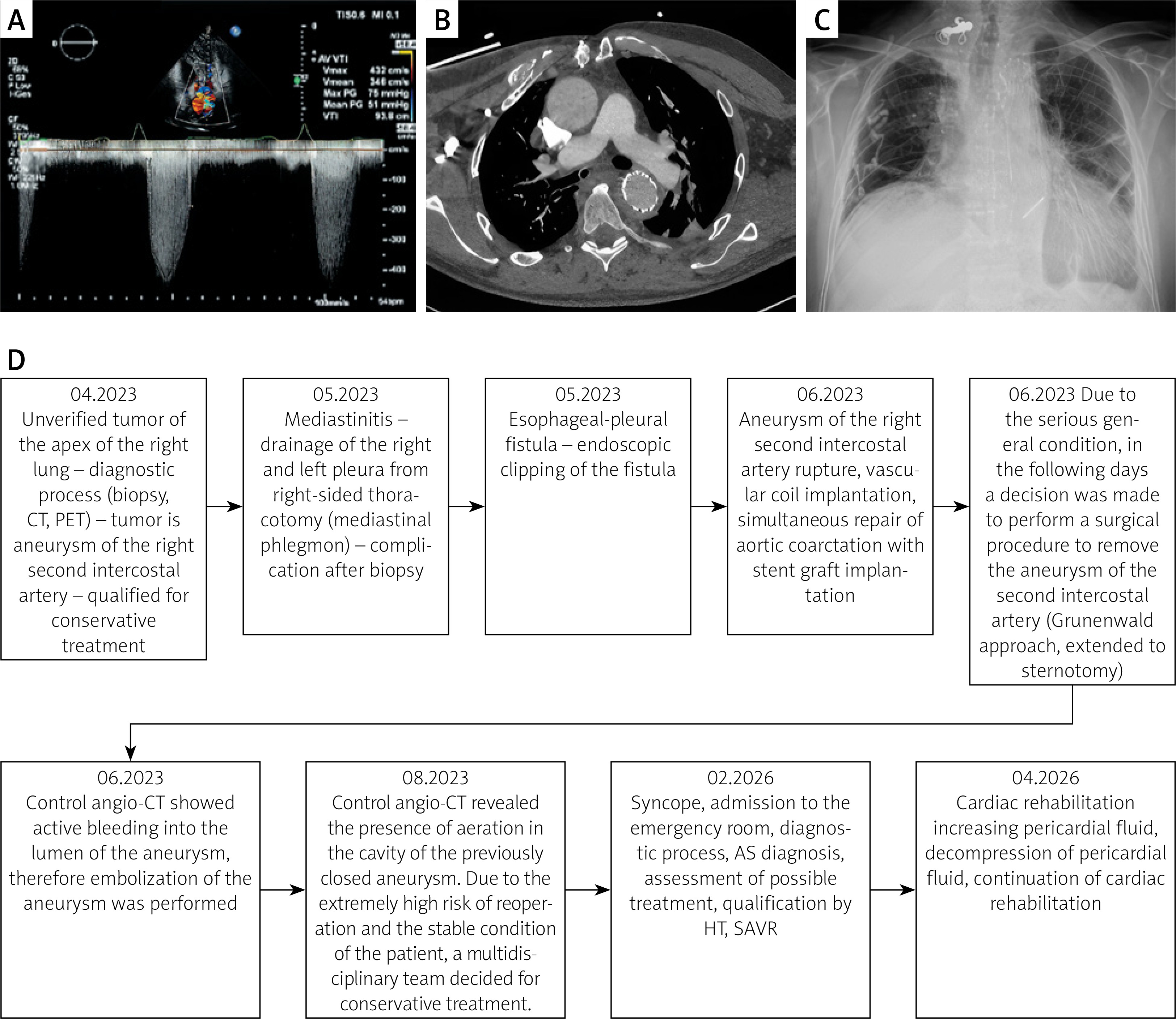
During the current admission in 2026, transthoracic echocardiography showed severe bicuspid aortic stenosis, with peak and mean transvalvular gradients of 75 and 51 mm Hg, respectively, an aortic valve area of 1.0 cm², and ascending aortic dilatation of 48 mm (Figure 1 A). CT confirmed bicuspid aortic valve morphology with fusion of the right and left coronary cusps, leaflet calcifications, ascending aortic dimensions of approximately 48 × 47 mm, and correct position of the descending aortic stent-graft (Figure 1 B).
Figure 1
A – Echocardiography with parameters of severe aortic stenosis; B – Angio-CT with dilation of the ascending aorta and stent graft in the descending aorta; C – Post-surgical chest X-ray with inflammatory-atelectatic changes, paralysis of the right diaphragmatic dome, descending aortic stent-graft, prominent right internal thoracic artery, metal sutures of the sternum, implanted mechanical aortic valve and temporary electrode; D – Flowchart of the patient’s medical history
Potential vascular accesses were analyzed before the initially considered transcatheter aortic valve implantation (TAVI). Right femoral access appeared feasible, with the right common iliac artery measuring approximately 10 × 11 mm at its narrowest point, the right external iliac artery 9 × 8 mm, and the right common femoral artery 10 × 10.5 mm. In contrast, left femoral access was unfavorable because of a 6 × 7 mm left external iliac artery and a 15-mm proximal occlusion of the left common femoral artery. Alternative subclavian access was also considered technically challenging because of imaging artifacts on the right side and marked caliber variation of the left subclavian artery.
The Heart Team considered both TAVI and surgical aortic valve replacement (SAVR). TAVI could have avoided redo sternotomy and reduced the invasiveness of the valve intervention. However, its disadvantages were substantial in this 52-year-old patient: bicuspid anatomy with calcified fused cusps, uncertain long-term durability in a young patient, inability to treat the concomitant ascending aortic dilatation, prior aortic coarctation stent-grafting, and limited alternative or bail-out access routes. SAVR offered durable valve treatment, direct intraoperative assessment of the ascending aorta, and the option of concomitant ascending aortic replacement or Bentall procedure if the operative findings justified the additional risk. Its principal disadvantages were the hostile mediastinum, adhesions, bleeding risk, previous mediastinal infection, and prior thoracic and endovascular procedures.
Because preoperative CT showed an ascending aorta of approximately 48 mm, the initial strategy was not simply isolated SAVR. The patient was referred for high-risk SAVR with readiness to perform concomitant ascending-aortic replacement or a Bentall procedure if intraoperative anatomy and safety conditions supported it. Intraoperatively, the ascending aorta measured approximately 45 mm, and the aortic wall thickness was approximately 2 mm around its circumference. The surgical team judged that extending the procedure to ascending aortic or root replacement would require substantially more extensive dissection, longer cardiopulmonary bypass and cross-clamp times, and potentially more complex distal control in a previously infected and surgically manipulated mediastinum. In this context, the incremental risk of bleeding and perioperative complications was considered disproportionate to the expected benefit of prophylactic aortic replacement. The final decision was therefore to perform SAVR with implantation of a mechanical prosthesis without replacement of the ascending aorta, followed by strict imaging surveillance.
Postoperative chest X-ray showed inflammatory and atelectatic changes (Figure 1 C), and effective antibiotic therapy was initiated. Transthoracic echocardiography demonstrated a hematoma posterior to the right atrium with signs of gradual resorption. The patient was discharged in good clinical condition and referred for standard post-cardiac surgery rehabilitation. During rehabilitation, however, cardiac tamponade occurred and urgent pericardial decompression was required, after which rehabilitation was continued. Although the exact source could not be proven retrospectively, the most plausible mechanism was delayed bleeding or oozing from the technically demanding aortotomy suture line in fragile tissue, potentially facilitated by postoperative anticoagulation after mechanical valve implantation and local inflammatory adhesions. This complication reinforced the need for close echocardiographic surveillance after high-risk valve surgery in hostile mediastinal conditions.
This case illustrates that the educational value lies not in a novel prosthesis or unconventional valve technique, but in the structured escalation and de-escalation of treatment strategy. In borderline ascending aortic dilatation accompanying bicuspid aortic stenosis, guideline thresholds should be balanced against patient-specific operative hazards. For patients with prior thoracic infection, failed sternotomy, endovascular aortic repair, and limited vascular access, the safest definitive treatment may be a staged, individualized strategy in which the final operative extent is determined by intraoperative findings and the anticipated incremental risk of adding aortic replacement [3, 4].