
Summary
The present study investigates the predictive capacity of the endothelial stress ratio, a novel multi-component biomarker, regarding the occurrence of the no-reflow phenomenon in 1256 patients undergoing coronary intervention for acute coronary syndrome. By integrating fibrinogen, albumin, C-reactive protein, and lymphocyte counts, this ratio provides a comprehensive assessment of the systemic inflammatory and endothelial burden. Our analysis demonstrates that the endothelial stress ratio is a powerful independent predictor of microvascular reperfusion failure, even after rigorous adjustment for high-risk clinical factors such as Killip class, ST-elevation myocardial infarction presentation, and pre-procedural Thrombolysis in Myocardial Infarction flow. To ensure statistical validity and prevent multicollinearity with its constituent parts, the ratio was evaluated in a dedicated multivariate model where it outperformed traditional indices such as the fibrinogen-to-albumin ratio, yielding an odds ratio of 1.48 and a high diagnostic accuracy with an area under the curve of 0.82. In conclusion, this study establishes the endothelial stress ratio as a practical and robust bedside tool for risk stratification. Its clinical application may facilitate the early identification of patients at high risk for no-reflow, potentially allowing clinicians to implement personalized pharmacological or procedural strategies to optimize myocardial perfusion.
Introduction
Restoration of epicardial vessel patency through percutaneous coronary intervention (PCI) is a cornerstone in the management of acute coronary syndrome (ACS). However, despite successful reopening of the occluded artery, inadequate myocardial reperfusion may occur, a condition known as no-reflow. The presence of no-reflow has been associated with larger infarct size, impaired left ventricular recovery, and increased short- and long-term mortality [1, 2].
The pathophysiology of no-reflow is multifactorial, involving distal thromboembolic obstruction, ischemia–reperfusion injury, microvascular spasm, endothelial swelling, oxidative stress, and inflammatory cell infiltration [3, 4]. Among these mechanisms, endothelial dysfunction plays a pivotal role by contributing to microvascular obstruction, impaired vasomotor regulation, and reduced capillary perfusion.
Several circulating biomarkers have been proposed to reflect systemic inflammation and endothelial injury. The fibrinogen-to-albumin ratio and the C-reactive protein-to-albumin ratio are established indicators of vascular inflammation and thrombotic activation [5, 6] while the neutrophil-to-lymphocyte ratio reflects oxidative stress and immune imbalance [7]. These markers have individually shown associations with no-reflow; however, their predictive performance remains modest when used alone.
To provide a more comprehensive reflection of endothelial stress, combining these parameters may yield superior predictive power. Therefore, in the present study, we introduce a new composite index, the endothelial stress ratio (ESO2), calculated as (fibrinogen/albumin) × (C-reactive protein/lymphocyte). We aimed to investigate the ability of this ratio to predict no-reflow in patients with ACS undergoing PCI and to evaluate its incremental prognostic value when added to established clinical and angiographic models.
Aim
The aim of this study was to investigate the predictive value of the endothelial stress ratio, a novel composite biomarker derived from routine laboratory parameters, for the occurrence of the no-reflow phenomenon in patients with acute coronary syndrome undergoing percutaneous coronary intervention. Additionally, we aimed to evaluate whether the endothelial stress ratio provides incremental prognostic value beyond established clinical, laboratory, and angiographic risk factors.
Material and methods
Study design and population
This retrospective observational study included patients diagnosed with acute coronary syndrome who underwent invasive treatment between January 2021 and October 2025 at our angiography center. Patients aged over 18 years who underwent coronary angiography and PCI with complete clinical, laboratory, and angiographic data were included. Exclusion criteria were prior coronary artery bypass graft surgery, active infection, malignancy, chronic inflammatory or autoimmune disease, and missing laboratory parameters.
Biochemical measurements and calculation of the endothelial stress ratio
All laboratory measurements were obtained from venous blood samples collected before coronary intervention. Hematologic parameters (white blood cells, neutrophils, lymphocytes, platelets) and biochemical parameters (glucose, creatinine, uric acid, total cholesterol, triglycerides, low- and high-density lipoprotein cholesterol, fibrinogen, albumin, and C-reactive protein) were recorded.
ESO2 was calculated to represent the combined effects of endothelial dysfunction and systemic inflammation using the following formula [6, 7]: ESO2 = (fibrinogen/albumin) × (C-reactive protein/lymphocyte).
All parameters were standardized to consistent measurement units prior to analysis: fibrinogen in mg/dl, albumin in g/dl, C-reactive protein in mg/l, and lymphocyte count in ×109/l. Conversion factors (g/l ×100 = mg/dl; g/l ÷ 10 = g/dl; mg/dl × 10 = mg/l; cells/µl ÷ 1000 = ×109/l) were applied when necessary. Because of a right-skewed distribution, the log10-transformed ESO2 value was used in regression analyses.
Angiographic evaluation
All patients underwent conventional coronary angiography according to standard protocols. The no-reflow phenomenon was defined as a post-procedural TIMI flow grade ≤ 2 or TIMI 3 flow with a myocardial blush grade ≤ 1.
Angiographic images were evaluated by two experienced interventional cardiologists who were blinded to laboratory and clinical results. In case of disagreement, a third observer’s assessment was considered final.
Statistical analysis
All analyses were performed using IBM SPSS Statistics version 25.0 (IBM Corp., Armonk, NY, USA). The normality of continuous variables was tested using the Kolmogorov–Smirnov test. Normally distributed variables were expressed as mean ± standard deviation, while non-normally distributed variables were presented as median (interquartile range). Group comparisons were made using Student’s t-test or the Mann–Whitney U test, as appropriate. Categorical variables were expressed as counts and percentages and compared using the χ2 test.
To avoid potential multicollinearity between the endothelial stress ratio (ESO2) and its constituent components (such as the fibrinogen-to-albumin ratio), these variables were evaluated in two separate multivariate logistic regression models. Model 1 included the fibrinogen-to-albumin ratio along with other significant clinical and laboratory variables, while Model 2 specifically evaluated the independent predictive value of the ESO2 index. The variance inflation factor (VIF) was used to assess for multicollinearity among the variables, and a VIF < 5 was considered acceptable. Variables associated with no-reflow in univariate analysis (p < 0.10) were entered into the models. Model discrimination was evaluated using receiver operating characteristic (ROC) curve analysis, and calibration was tested with the Hosmer–Lemeshow goodness-of-fit test. A two-tailed p-value < 0.05 was considered statistically significant.
Results
A total of 1256 patients were included in the study, with a mean age of 61.8 ±10.7 years. The no-reflow phenomenon occurred in 73 (5.8%) patients. Baseline demographic, clinical, and laboratory characteristics of the study population are presented in Table I. Patients who developed no-reflow had a higher incidence of STEMI presentation, higher Killip class on admission, and more frequent multivessel disease (all p < 0.05). In the laboratory analysis, patients in the no-reflow group had significantly higher levels of fibrinogen, C-reactive protein, and uric acid, while albumin and lymphocyte levels were lower (all p < 0.05).
Table I
Baseline demographic, clinical, and laboratory characteristics of patients
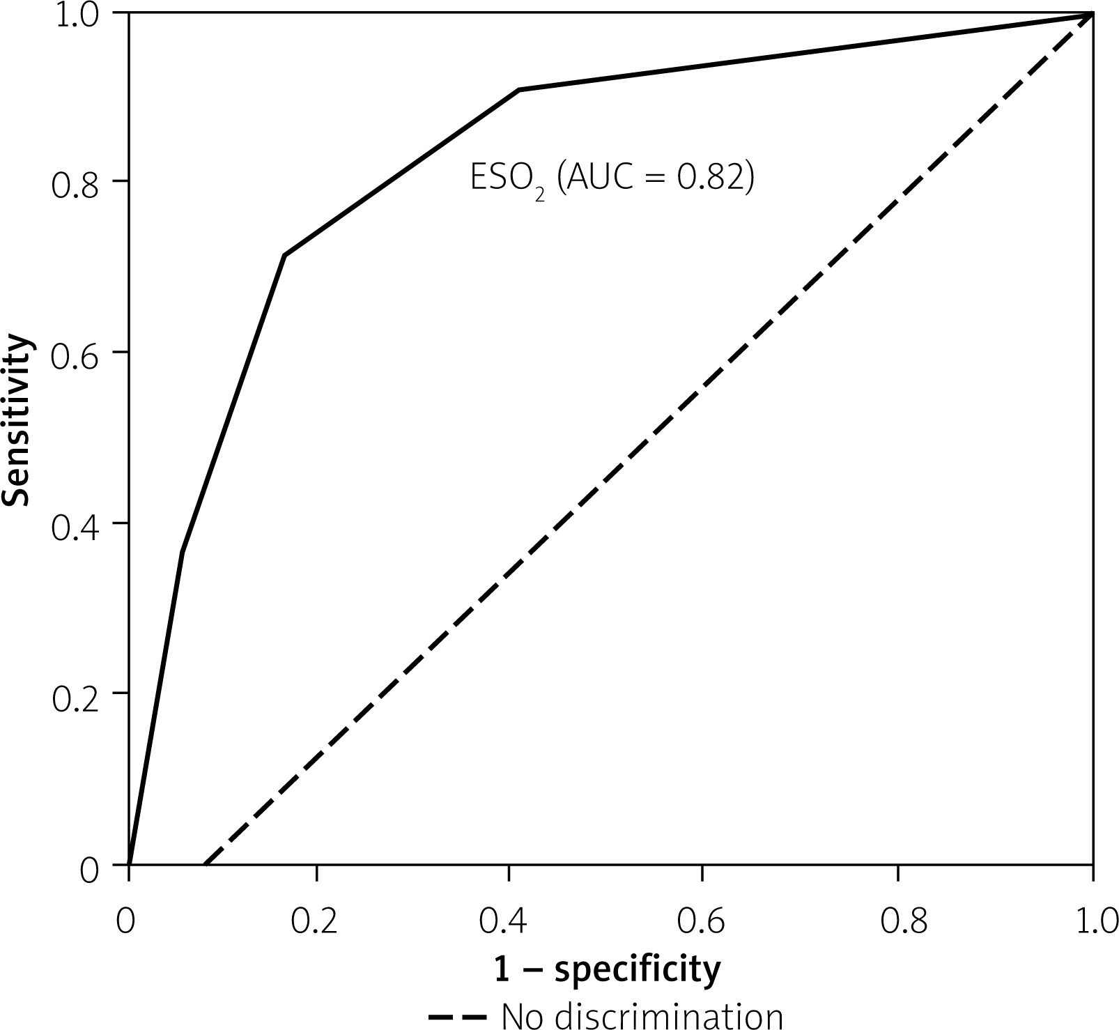
In the univariate logistic regression analysis, hypertension, diabetes mellitus, uric acid, fibrinogen-to-albumin ratio, and ESO2 were significantly associated with the occurrence of no-reflow. To avoid multicollinearity between the composite indices, two separate multivariate models were constructed (Table II). In Model 1, hypertension, uric acid, and the fibrinogen-to-albumin ratio (OR = 1.37, 95% CI: 1.11–1.68, p = 0.002) remained independent predictors. In Model 2, ESO2 demonstrated the strongest independent association with the no-reflow phenomenon (OR = 1.48, 95% CI: 1.21–1.82, p < 0.001). Receiver operating characteristic (ROC) analysis demonstrated that ESO2 had strong discriminative performance, with an area under the curve (AUC) of 0.82 (p < 0.001) (Figure 1).
Table II
Multivariate logistic regression models for predictors of no-reflow
Figure 1
Receiver operating characteristic (ROC) curve demonstrating the diagnostic performance of the endothelial stress ratio (ESO2) for predicting the no-reflow phenomenon in patients with acute coronary syndrome. The ESO2 index showed strong discriminative ability with an area under the curve (AUC) of 0.82. The diagonal dashed line represents the reference line of no discrimination
Discussion
In this study involving 1256 patients with ACS who underwent PCI, we found that the no-reflow phenomenon occurred in 5.8% of cases. Patients with no-reflow had significantly higher fibrinogen, CRP, and uric acid levels, while albumin and lymphocyte counts were lower. The variables identified in univariate analysis, including hypertension, diabetes mellitus, uric acid, fibrinogen-to-albumin ratio, and ESO2, were consistent with the baseline differences observed between the no-reflow and non-no-reflow groups. ESO2 calculated as (fibrinogen/albumin) × (CRP/lymphocyte), was markedly elevated in the no-reflow group and remained an independent predictor in multivariate analysis. The strong discriminative capacity (AUC = 0.82) and the improvement in predictive accuracy after incorporating ESO2 into the clinical-angiographic model (∆AUC = 0.07, p = 0.01) emphasize its value as a practical and powerful biomarker for identifying patients at risk of microvascular obstruction.
The observed no-reflow rate in our cohort aligns with previous large-scale analyses reporting an incidence of approximately 5–10% after PCI [8]. Although successful epicardial recanalization restores coronary patency, microvascular dysfunction remains a major determinant of infarct size, ventricular recovery, and mortality [9]. Mechanistic studies have demonstrated that microvascular obstruction results from endothelial swelling, oxidative stress, platelet and leukocyte activation, and distal embolization [3]. Among these mechanisms, endothelial dysfunction serves as a central link between inflammation and impaired reperfusion [10].
Numerous studies have evaluated inflammatory and oxidative biomarkers such as the neutrophil-to-lymphocyte ratio (NLR), fibrinogen-to-albumin ratio (FAR), and C-reactive protein-to-albumin ratio (CAR) as predictors of adverse outcomes in ACS [6]. However, their predictive performance is modest when used alone because each reflects only one dimension of vascular stress. The ESO2 index proposed in this study integrates both endothelial and inflammatory components, offering a broader pathophysiological perspective. Elevated fibrinogen and CRP promote leukocyte adhesion and platelet aggregation within the microcirculation, whereas low albumin and lymphocyte levels indicate reduced antioxidant defense and immune modulation [11]. This multidimensional representation may explain why ESO2 outperformed other ratios in predicting no-reflow.
The present study demonstrated that the ESO2 is a potent and independent predictor of the no-reflow phenomenon in patients with ACS. In this revised analysis, we incorporated a more comprehensive set of baseline characteristics, including Killip class, ACS presentation type, and detailed angiographic findings. Our findings revealed that patients experiencing no-reflow presented with a higher risk profile, characterized by a higher prevalence of STEMI and multivessel disease. Despite the presence of these established clinical and procedural risk factors, ESO2 remained a robust independent predictor in multivariate models. To ensure statistical rigor and address potential multicollinearity, we evaluated ESO2 and the FAR in separate models. While FAR is a recognized marker of vascular inflammation, the ESO2 index provided a higher odds ratio (OR: 1.48 vs. 1.37), suggesting that the integration of both inflammatory and endothelial components yields superior predictive power. This multidimensional representation captures the cumulative burden of microvascular stress more effectively than single or double ratios. Furthermore, the higher ESO2 levels observed in the no-reflow group may reflect an aggressive systemic inflammatory milieu that predisposes patients to microvascular obstruction even after successful epicardial recanalization. From a clinical perspective, identifying high-risk patients through ESO2 before intervention may allow for the early implementation of preventive strategies, such as the use of intracoronary vasodilators or distal protection devices.
Our findings also support the growing evidence linking systemic inflammation to microvascular reperfusion failure [12]. Inflammation triggers cytokine release, endothelial activation, and reactive oxygen species generation, which further aggravate vascular permeability and microvascular plugging [13]. Elevated ESO2 values in our study likely reflect this interaction between inflammatory stress and endothelial dysfunction. Moreover, patients with high ESO2 may represent a phenotype with a more aggressive systemic inflammatory milieu, predisposing them to microvascular obstruction even after technically successful PCI. From a clinical standpoint, ESO2 offers clear advantages: it is easily calculated from routine laboratory parameters, cost-effective, and rapidly available before intervention. Early identification of patients with elevated ESO2 could help tailor preventive measures such as distal embolization control, intracoronary vasodilators, or intensified anti-inflammatory therapy [14]. Integration of ESO2 into existing risk models could further refine individualized patient management and guide interventional strategies.
At the mechanistic level, endothelial stress plays a central role in the pathogenesis of no-reflow. Endothelial injury leads to impaired nitric oxide bioavailability, microvascular constriction, and capillary plugging, ultimately reducing myocardial perfusion despite an open epicardial artery. Inflammatory activation, characterized by cytokine surge and leukocyte–platelet interactions, perpetuates this process and contributes to reperfusion injury. Experimental data have confirmed that oxidative stress and endothelial edema are major drivers of microvascular obstruction following reperfusion [15]. These mechanisms are also consistent with the anti-inflammatory benefits observed in clinical trials targeting cytokine pathways, which have reduced cardiovascular events by attenuating endothelial inflammation [16]. In this context, ESO2 may serve as a surrogate for global endothelial stress, capturing the cumulative inflammatory burden responsible for no-reflow.
Finally, the interplay between inflammation, oxidative stress, and endothelial dysfunction forms a self-sustaining cycle that worsens microvascular perfusion [17]. The strong correlation between high ESO2 and the no-reflow phenomenon observed in this study reinforces the concept that systemic inflammatory activation parallels coronary microvascular injury. Future multicenter prospective studies should explore whether lowering ESO2 through targeted anti-inflammatory or endothelial-protective therapies can translate into improved myocardial reperfusion and outcomes [3, 18].
This study has several limitations that should be acknowledged. First, its retrospective and single-center design limits the ability to establish a causal relationship between endothelial stress ratio and the no-reflow phenomenon. Second, laboratory measurements were obtained only once before PCI; thus, dynamic changes in inflammatory or endothelial parameters over time were not evaluated. Third, we did not perform advanced imaging modalities such as cardiac magnetic resonance imaging to directly quantify microvascular obstruction. Importantly, key procedural/angiographic determinants of no-reflow – particularly baseline (pre-PCI) TIMI flow and thrombus burden (e.g., thrombus grade) – were not systematically recorded for the entire cohort and therefore could not be incorporated into the multivariable regression models. This may have resulted in residual confounding and should be considered when interpreting the independent association between endothelial stress ratio and the no-reflow phenomenon. Finally, the absence of external validation or comparison with other inflammatory indices in an independent population may restrict the generalizability of our findings. Future multicenter prospective studies are warranted to confirm the predictive value of ESO2 and to determine optimal cut-off values for clinical implementation.
Conclusions
ESO2, derived from routine laboratory parameters, is an independent and strong predictor of the no-reflow phenomenon in patients with acute coronary syndrome undergoing percutaneous coronary intervention. ESO2 effectively integrates inflammatory and endothelial components, providing a practical and cost-efficient biomarker for early risk stratification. Incorporating ESO2 into pre-procedural assessment may help clinicians identify high-risk patients and guide personalized therapeutic strategies aimed at preventing microvascular reperfusion failure.










