Coarctation of the aorta (CoA) is a congenital narrowing of the thoracic aorta, occurring in approximately 3 per 10,000 live births and accounting for 5–8% of congenital heart defects [1, 2]. Recoarctation remains a recognized late complication after childhood repair and may present in adulthood with persistent or resistant hypertension [1, 2]. In contemporary practice, transcatheter stent implantation is an established treatment option for native and recurrent coarctation in adolescents and adults, providing high procedural success and favorable hemodynamic outcomes [1–4].
A 27-year-old man with resistant arterial hypertension treated with five antihypertensive agents (an angiotensin II receptor blocker, a β-blocker, a thiazide diuretic, a calcium channel blocker, and eplerenone), with ambulatory blood pressure persistently exceeding 150/80 mm Hg, was referred for elective treatment of recurrent aortic obstruction. In infancy (1998), he had undergone an arterial switch operation for transposition of the great arteries, ventricular septal defect closure, resection of aortic coarctation, and aortic arch reconstruction using a pulmonary homograft patch. Computed tomography (CT) performed during the diagnostic work-up for secondary hypertension demonstrated aneurysmal dilatation of the aortic root (48 × 45 × 46 mm) and ascending aorta (40 mm at the tubular ascending aorta, 45 mm at the sinotubular junction), as well as a narrowing of the distal aortic arch to 11 mm over a 15-mm segment, distal to the left subclavian artery. Doppler echocardiography showed accelerated flow in the descending aorta with a peak velocity of approximately 4.0 m/s, corresponding to a peak gradient of about 65 mm Hg, and a tardus-parvus pattern in the abdominal aorta, consistent with hemodynamically significant recoarctation (Figure 1 A).
Figure 1
A – Accelerated flow at the site of aortic recoarctation, with an increased peak gradient. B – Patient-specific 3D-printed model of the aorta, created from CT data, showing the stenotic segment (arrow). C – Final angiographic result after the procedure showing the stent position (arrow). D – Follow-up Doppler echocardiography showing reduction of peak velocity and pressure gradient
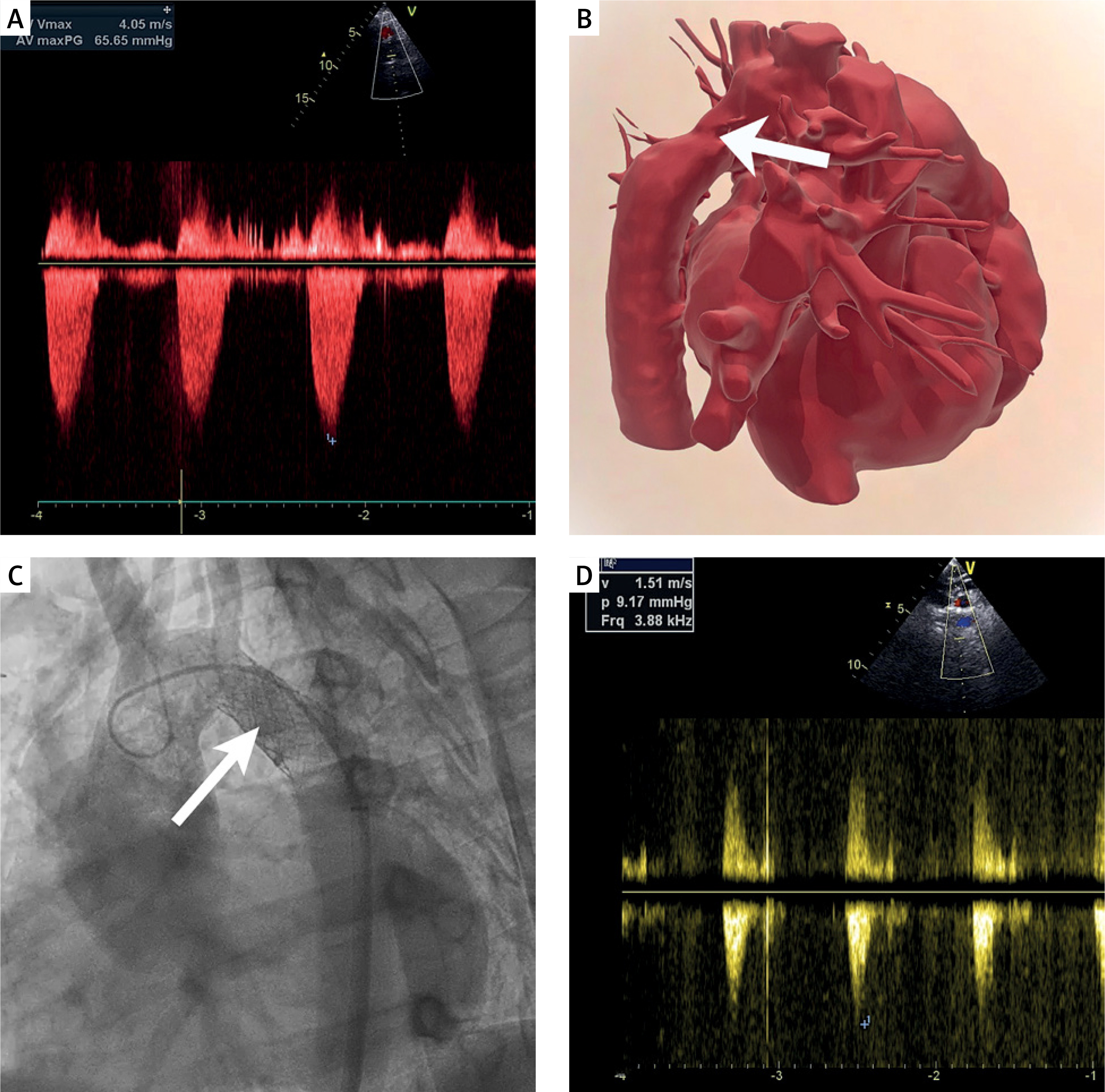
Because of the complex postsurgical anatomy, a patient-specific 3D-printed model of the aorta was created from CT data to improve anatomical understanding and procedural planning (Figure 1 B). The model was particularly helpful in assessing the spatial relationship between the recoarctation segment and the ostium of the left subclavian artery, facilitating procedural planning and optimal stent positioning. The transcatheter repair was performed using dual vascular access. Aortography obtained from radial access confirmed the aortic narrowing, while pull-back pressure measurements performed via femoral access demonstrated a peak systolic gradient > 20 mm Hg, supporting intervention in the setting of resistant hypertension [1]. After exchange for a 12-F sheath, an AndraStent Optimus Co-Cr XL 33-mm stent was implanted using a balloon-in-balloon catheter and expanded to 20 mm at the site of recoarctation, immediately distal to the origin of the left subclavian artery. Final angiography demonstrated complete expansion of the stenotic segment without residual waist (Figure 1 C). Post-deployment invasive assessment demonstrated complete abolition of the systolic pressure gradient (0 mm Hg). The procedure and hospital stay were uneventful.
Follow-up Doppler echocardiography demonstrated a reduction in peak velocity within the descending aorta to approximately 1.5 m/s (peak gradient 9 mm Hg), normalization of the abdominal aortic Doppler flow profile (Figure 1 D), and normalization of arterial blood pressure. During ambulatory follow-up, antihypertensive treatment was progressively de-escalated. Initially, the doses of antihypertensive medications were reduced, and subsequently the regimen was simplified from five to three agents, with sustained normalization of blood pressure.
This case highlights that recurrent aortic obstruction should be actively considered in young adults presenting with resistant hypertension after childhood coarctation repair [1, 2]. In appropriately selected patients, endovascular stent implantation represents an effective alternative to redo surgery, providing immediate gradient relief and durable restoration of aortic lumen diameter [3, 4]. Advanced imaging combined with patient-specific 3D printing may be particularly valuable in complex congenital and postsurgical anatomy, facilitating spatial understanding of the lesion, optimizing procedural planning, and assisting with device selection and stent positioning. In this young adult with complex congenital heart disease and distal arch recoarctation after infant surgery, CT-guided, 3D model-assisted transcatheter stent implantation was safe and effective, resulting in immediate elimination of the invasive pressure gradient and early normalization of Doppler flow parameters.