
Carotid atherosclerotic plaque calcification is common but calcification intensity and location patterns are variable [1, 2]. By today’s criteria, at least one in every twenty patients referred for carotid revascularisation is considered ineligible for carotid artery stenting (CAS – transfemoral/transradial, or transcarotid stent-assisted revascularization – TCAR) due to the index lesion severe calcifications [3]. Traditionally, calcification was believed to constitute a process stabilising the atherosclerotic plaque into a “lower-risk lesion”, but evidence is increasing that not all calcified lesions are benign [4–6]. Symptomatic carotid disease shows double peaks at calcium scores around 200–400 and 600–800 with a dip at 400–600 Agatson units [6]. Calcified nodular protrusions into the carotid artery lumen (projecting calcific nodules) and larger calcific intraluminal protrusions (exophytic calcifications) provide anatomic basis for thrombus formation with the risk of spontaneous distal embolisation (or luminal occlusion) and clinical symptoms of cerebral tissue injury such as stroke or transient ischaemic attack [5, 7]. Furthermore, bulky calcifications and positive ‘rim sign’ calcifications (defined as calcification < 2 mm in thickness with adjacent soft plaque > 2 mm in thickness) are significantly associated with ischaemic events (χ2 = 4.552, p = 0.033; χ2 = 10.448, p = 0.001 respectively) [1]. Recent analysis of the relationship between calcification and vulnerability of carotid plaques demonstrates a significantly higher incidence of ipsilateral spontaneous cerebral ischaemic lesions at preoperative computed tomography in patients with high-level calcifications (56% vs. 32%, p = 0.01), indicating that calcific carotid stenosis may be indeed associated with increased risk of spontaneous embolism [4]. Highly-calcified carotid stenoses (HCCS) belong to acute carotid-related stroke presentations that require emergency revascularisation as ‘tandem’ (extracranial carotid + intracranial occlusion) or ‘non-tandem’ lesions, posing important challenges to interventional management [2, 8].
In a series of conventional carotid artery stenting (CAS) procedures in carotid artery stenosis involving plaque calcification of more than 75% of the vessel circumference (mean arc of calcifications 320.1 ±24.5 degrees; range: 278–360 degrees), Tsutsumi et al. [9, 10] reported, with the routine use of non-compliant balloons, effective multiple fragmentations of the calcifications on computed tomography imaging in 94.4% of lesions (cracks in the calcified plaque with lack of effective fragmentation in only 5.6%), and excellent stent expansion achieved and maintained in all lesions. This, however, has not been the experience of other groups [11–16], possibly because of the relevance of not only the circumference of calcification but also its other chatacteristics such as, for instance, the thickness. Highly calcified carotid stenoses (HCCS) are known to pose significant hurdles to stent-assisted revascularisation using conventional lesion preparation techniques and conventional carotid stents [11, 12, 14, 17, 18]. Heavy, concentric calcification is an important predictor of complications with CAS [11–13]. With heavy calcification, it can be technically difficult or even impossible to advance the embolic protection device beyond the lesion [11, 19]. Heavy calcification causes difficulties in tracking devices, lesion dilation, and achieving adequate stent expansion and apposition [11, 18, 19]. Consequently, there may be suboptimal luminal gain, increased risk of perforation and embolic events, and reduced long-term patency [19, 20]. Severely calcified carotid lesions are prone to embolism, technical failure of the CAS attempt, stent compression/collapse and restenosis, markedly increasing the risk of peri-procedural and long-term clinical complications [3, 11, 13, 14, 17, 19]. Calcification circumference, area, and its location (in particular when severe circumferential or eccentric/exophytic) are significantly associated with carotid stent underexpansion and stent fracture and restenosis [3, 15, 16, 19]. In a series of 140 consecutive carotid revascularisation procedures (81 carotid endarterectomy, CEA; 59 CAS), high calcium score was a predictor of reduced stent expansion (Wallstent or Precise single-layer stents; routine predilatation and postdilatation; distal and/or proximal cerebral protection; residual stenosis with CAS: 9.7 ±13.3% vs. 1.7 ±6.1%, p < 0.001). Multiple logistic regression analysis revealed that calcium score was an independent pre-CAS predictor of residual stenosis [21]. Furthermore, on multivariate analysis, carotid arteries with high-grade calcification were clearly prone to develop restenosis after CAS (aOR = 6.04, 95% CI: 1.29–28.36, p = 0.02) [15]. Immediate post-procedural minimum stent area correlates negatively (r = –0.37; p < 0.01) with the risk of in-stent restenosis [22]. In a study by Chang et al. [23], presence of calcified plaque on cine imaging was significantly associated with an increased rate of stent fracture or deformation (p = 0.0006). A high calcification grade and more thick calcifications are independent predictors of bad clinical outcome with CAS over a follow-up of up to 11 years [13].
In a recent large-scale analysis from the Vascular Quality Initiative, 35% of patients had 51% to 99% arc of calcification, and 3% had 100% circumferential calcification/intraluminal calcium protrusion [24]. In transfemoral CAS patients, severe circumferential calcification/protrusion was associated with higher in-hospital stroke/death odds (OR = 2.0; 95% CI: 1.2–3.4; p < 0.013), whereas in TCAR patients, 51% to 99% (circumference) calcification was associated with increased odds of in-hospital stroke/death (OR = 1.5; 95% CI: 1.1–2.2; p < 0.025) [24]. At 1 year, circumferential calcification/protrusion was associated with higher odds of ipsilateral stroke/death (12.4% vs. 6.6%; hazard ratio, 1.64; p < 0.002) [24].
Recent analysis of stent occlusions in 12,143 patients showed that lesion calcification with 100% circumference and calcification protruding into lumen increased the risk for stent occlusion (2.3-fold and 4.7-fold, p < 0.05 for both) [25]. In a study by Katano et al. [21], in patients with HCCS, restenosis at 6 months was frequent and similar after both CAS and CEA (18.8% and 20.0%, respectively) [21]. After CAS, cerebral new ischaemic ipsilateral lesion(s) on post-operative diffusion-weighted imaging were more frequent in HCCS than non-HCCS after CAS (33.3% vs. 20.0%) [21]. Multiple logistic regression analysis revealed HCCS as an independent predictor of residual stenosis after CAS (OR = 13.7, p = 0.025; for diabetes OR = 3.5) [21]. However, criteria for the choice of a particular revascularisation mode (CAS or CEA) in a particular lesion were unclear [21], and all-comer (or randomised) series are needed for bias-free comparisons. Recent experimental analysis of carotid plaques acquired from standard endarterectomy procedures, with quantitative assessment of calcification performed using high-resolution computed tomography, documented a strong negative correlation between calcification and the plaque stretch ratio [26]. Only plaques containing concentric calcifications with < 20% calcification volume successfully reached the minimum required stretch, avoiding plaque rupture [26]. Index lesion calcification was identified as an independent predictor of stroke within 30 days from CAS (OR = 2.47, p < 0.001) [12]. Severe calcification (circumferential or exophytic) has been considered a contraindication to classic CAS [27, 28].
Conventionally, carotid calcification is subjectively graded as “mild”, “moderate” or “severe” [29] (or none-to-minimal versus moderate-to-severe [3]), raising signifcant concerns about comparability of the assessment by different investigators or core laboratories (calcification that is “moderate” to one observer or one core laboratory may be “mild” or “severe” to another). The apparent “differences” in outcomes reported for HCCS by different group might thus arise, at least in part, from poorly discriminative definitions, leading to implementations of observer-dependent judgement. Recently, a highly reproducible carotid calcification angiographic severity scoring system has been developed in collaboration with an angiographic core laboratory and introduced into clinical practice [30]. Evaluated characteristics include the calcific segment length in relation to lesion length, minimal calcification thickness, calcification circularity index (number of quadrants around the lumen), and calcification severity according to a five-grade ranking system (G0-G4; carotid calcification severity scoring system, taking into consideration calcification intensity on still frames and cine images as well as the calcification pattern) [30]. HCCS angiographic criteria include calcific segment length to lesion length ratio ≥ 2/3, calcification circularity index ≥ 3, calcification thickness ≥ 3.0 mm, and calcification severity grade ≥ 3 [30]; all four criteria are needed for the “HCCS” label (for carotid calcification severity scoring see Table II in Reference [30]).
During development of the PARADIGM-IVL study protocol (ISRCTN94131858) we identified several scenarios relevant to endovascular management of HCCS; those are summarised in Figure 1 and 2 [30–44], whereas reference [30] provides our prior data on HCCS percutaneous management limited to lesion preparation with ‘step-up’ diameter non-compliant balloons followed by non-compliant balloon(s) post-dilatation to optimise the stent. Lesion dilation before stenting (predilatation) has been strongly recommended in highly-calcific stenoses [11] and it has been implemented in routine clinical practice (Figures 1, 2). Scoring balloons (Figure 2, Scenario #2) employ wire-like elements on their surface that create incisions in the plaque during inflation, allowing more efficient dilatation [45]. Scoring/cutting balloon plaque modification has been reported to minimise conventional carotid stent underexpansion and structural damage, to enhance stent adhesion to the vessel wall, and to improve outcomes in percutaneous treatment of HCCS [45]. However, in a series of patients with heavily calcified lesions and baseline lesional median arc of calcification of 311° (294–334°), treated with single-layer stent CAS after plaque modification using scoring balloon angioplasty, still 70% of cases (first-generation stent in all) showed post-procedural residual stenosis of ≥ 29% [45]; a critical parameter of the suboptimal result of endovascular management and the risk of in-stent restenosis [46, 47]. Indeed, high-pressure balloon dilatation with non-compliant or specialty balloons may have insufficient force to fracture calcium and achieve vessel expansion and can lead to barotrauma-related arterial wall dissections or perforation [20, 36] (example in Figure 2, Scenario #1). This, taken together with the unmet clinical need for optimizing percutaneous HCCC emergency management in acute carotid-related stroke presentations (examples in Figure 2 scenario #1 and scenario #2) and in elective patients at high risk of surgery in the context of presently suboptimal outcomes of HCCS management using conventional CAS techniques, indicates a need for improved endovascular HCCS preparation and improved stent-assisted lumen reconstruction.
Figure 1
Endovascular management of a highly-calcified carotid stenosis using multiple “step-up diameter” non-compliant balloon predilatations and a dual metallic layer (dual-braided) closed-cell stent. A – Baseline intracranial angiogram demonstrating suboptimal supply to the left haemispheric vessels from the left internal carotid artery (ICA). B – Tight, highly calcified (Grade 4/4 [30]; red arrowheads) circumferential carotid bifurcation stenosis with luminal irregularities and lesion ulcerations. C – Initial predilatation with Ø2.0 mm semi-compliant balloon to enable (after initial crossing attempt) filter delivery; purple arrow denotes distal filter. D – Step-up predilatation with Ø3.0 mm non-compliant balloon at 18 atm. E – Final predilatation with Ø4.0 mm non-compliant balloon at 20 atm. F – Dual metallic layer, dual-braided closed-cell stent (Casper/Roadsaver; inner nitinol mesh, nominal device size 8.0 × 20 mm) [31, 34, 35, 37] implanted and now optimised with Ø5.0 mm non-compliant balloon at 18–20 atm. G – Final angiography showing a largely malapposed (red stars) closed-cell stent with angiographic 53% residual stenosis due to the stent collapse on a non-modified (cf., Figure 2, Scenario #3 and #4) ‘stone’ of calcium (inset; contrast-free image, yellow arrows); residual minimal lumen diameter (MLD) of 2.43 mm. Use of the dual metallic layer, dual-braided closed-cell stent has been identified as an independent risk factor for in-stent restenosis [34, 44] that is particularly relevant when in-stent MLD is ≤ 3 mm [41]). H – Final intracranial angiogram demonstrating a normalised supply to the left haemispheric vessels from the left ICA in absence of signs of embolisation. The procedure was neurologically uncomplicated but transient haemodynamic instability required noradrenaline infusion. I – 30-day duplex ultrasound follow-up showing stent patency with turbulent in-stent flow and increased velocities consistent with the stent focal underexpansion/compression, in absence of any neurologic complications.
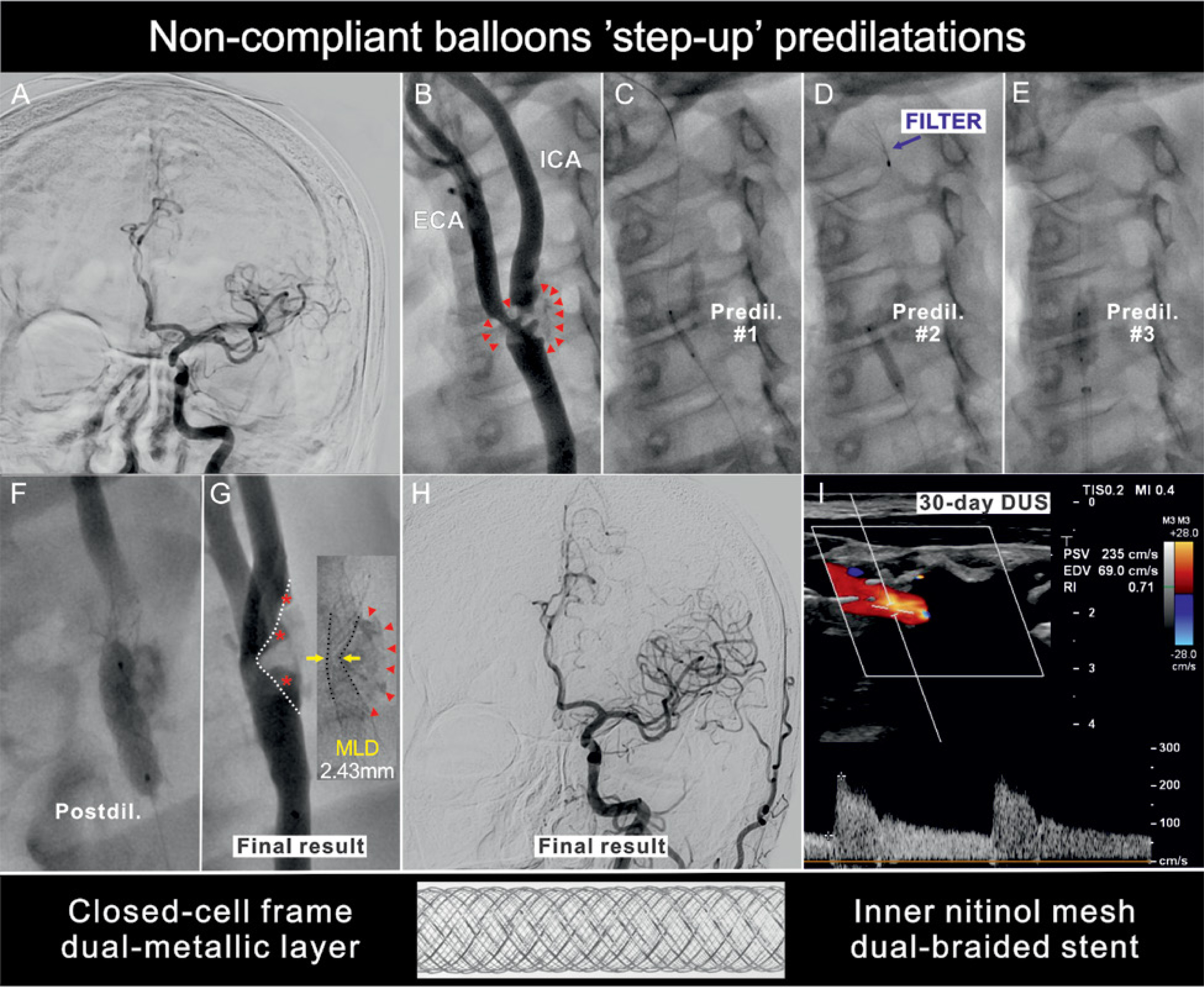
Figure 2
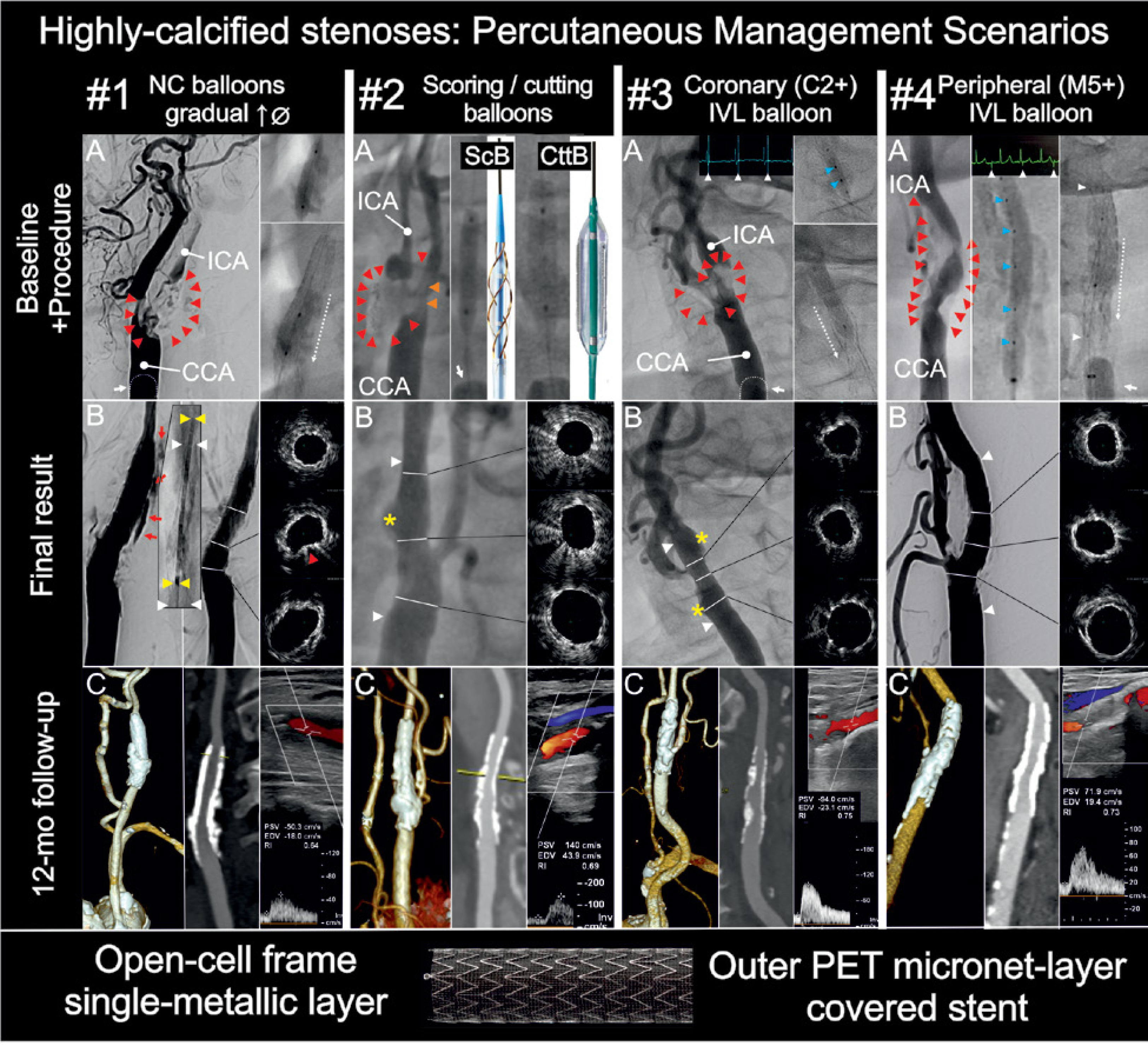
Endovascular management of highly-calcific carotid stenoses using a high radial force open-cell (frame), MicroNET-covered anti-embolic stent.
Scenario #1. Multiple ‘step-up’ diameter non-compliant balloon predilatations for lesion preparation. A – A tight, highly-calcified lesion (red arrowheads, Grade 4/4 [30]) underwent ‘step-up’ preparation with non-compliant balloons of increasing diameter (high-pressure inflations), followed by implantation of a high radial force open-cell (frame), MicroNET-covered stent (9.0 × 40 mm) [32, 39]. The stent was optimised by stepup pressure postdilatation with Ø5.0 mm non-compliant balloon (up to 20 atm). Proximal cerebral protection (mono-balloon/MonoBalloon catheter) was employed throughout the procedure (white arrow, common carotid artery, CCA, balloon denoted with a dotted line). The patient presented for emergency endovascular recanalization of a stuttering stroke, with contraindications to surgery. B – Left – control angiography showing optimal stent expansion and lumen gain; however, there was an increasing segmental extravasation of dye, indicating perforation; this was initially controlled (but not resolved) with prolonged balloon inflations. Sealing was achieved with implantation of another MicroNET-covered stent (8.0 × 30 mm; stent-in-stent technique [36]), taking advantage of the sealing properties of MicroNET [42, 43]. Intravascular ultrasound imaging showed overall acceptable expansion of the stents (note focal deformation/indentation, red arrow, on suboptimal-only fragmentation of calcification) and optimal apposition of the stent-in-stent to the arterial wall (cf., Figure 1). External carotid artery occlusion (MicroNET-covered stent-in-stent for perforation rescue) was asymptomatic. The procedure was uncomplicated neurologically and uncomplicated haemodynamically. C – 12-month follow-up imaging with computed tomography angiography and duplex ultrasound showed a fully maintained lumen (absence of in-stent restenosis/stent-in-stent) and normal in-stent velocities. Scenario #2. Scoring/cutting balloon balloon–assisted ‘step-up diameter’ predilatations for lesion preparation. A – A tight, highly calcific lesion (Grade 4/4 [30], red arrowheads) was initially prepared with scoring and cutting balloons (ScB, 2.0 × 15 mm; CttB, 3.0 × 10 mm) in a patient presenting with acute carotid-related stroke for emergency recanalization after intravenous thrombolytic therapy. Further predilatations were performed with a Ø4.0 mm and Ø5.0 mm non-compliant balloons (not shown), and were followed by implantation of a MicroNET-covered stent (CGuard 10 × 40 mm). The stent was postdilated with a Ø5.5 mm and (proximally) Ø6.0 mm non-compliant balloon (max 20 atm). Note that, in this anatomy, our initial attempt to insert a dual-balloon proximal cerebral protection system [33, 46] had failed due to severe calcifications in carotid bifurcation and proximal external carotid artery (orange arrowheads) – and thus a common carotid artery (CCA) mono-balloon proximal neuroprotection system (white arrow in the middle image) was used. B – Control angiography and intravascular ultrasound showed optimal stent expansion and apposition (except a short focal malapposition in the post-stenotic aneurysmatic segment, orange star), with a fully patent external carotid artery. C – 12-month follow-up imaging with computed tomography angiography and duplex ultrasound demonstrated a fully maintained lumen (absence of in-stent restenosis) and normal in-stent velocities (note higher-range compared to those in non-calcified carotid arteries post stent implantation). Also note full healing (MicroNET flow-diverting characteristics) of the focal stent malapposition in the aneurysmatic post-stenotic segment (normalised lumen; full, optimal, durable endovascular revascularisation).
Scenario #3. Intravascular lithotripsy (IVL) – assisted lesion preparation: coronary IVL balloon, 2-emitter Rx system. A – Initial preparation of a highly-calcified (Grade 4/4 [30], red arrowheads) carotid stenosis, in a clinically asymptomatic patient with (so far clinically silent) ipsilateral cerebral infarcts, was performed with Ø3.5 mm non-compliant balloon predilatation (not shown; note that the upfront-introduced IVL balloon ruptured upon gentle inflation, prompting initial predilatation with a non-compliant balloon to enable IVL therapy); this was followed by delivery of 120 pulses of calcium-cracking ultrasound energy (top right, emitters are marked with blue arrowheads, 4.0 × 12 mm balloon coronary IVL system). The IVL balloon was repeatedly repositioned for maximised delivery of the calcium-cracking energy throughout the entire lesion length, as the greatest acoustic pressure is exerted directly perpendicular to emitters [40]. Electrocardiographic demonstration of spikes of calcium-cracking energy delivery is shown on the ECG trace (inset, white arrowheads). A MicroNET-covered stent (9.0 × 30 mm) was implanted and optimised with Ø5.0 mm and Ø5.5 mm non-compliant balloon postdilatations up to 24 atm throughout the stent length from top (bottom-right image) to bottom (dotted arrow). Proximal cerebral protection was achieved throughout the procedure (documented flow reversal in the internal carotid artery, ICA) with a mono-balloon common carotid artery catheter (CCA, balloon denoted with a dotted line, white arrow). B – Control angiography showed optimal stent expansion and lumen gain, with two focal malapposition spots (yellow stars) that were minor on intravascular ultrasound imaging (open-cell frame, high conformability) and which spontaneously healed due to the stent (MicroNET) flow-diverting properties (cf., 3C below). C – 12-month follow-up imaging with computed tomography angiography and duplex ultrasound showed a fully maintained lumen (absence of in-stent restenosis) and normal in-stent velocities (note higher-range compared to those in non-calcified carotid arteries post stenting). Full, durable optimal endovascular revascularization. Rx – rapid exchange.
Scenario #4. Intravascular lithotripsy (IVL) – assisted lesion preparation: peripheral IVL balloon, 5-emitter over-thewire (OTW) system. A – Long, highly calcified carotid tandem stenosis (Grade 4/4 [30]; red arrowheads]) in a symptomatic patient at a high risk for surgery was initially prepared with non-compliant balloon (Ø2.0 mm, Ø4.0 mm and Ø5.0 mm) predilatations (not shown), followed by delivery of 300 pulses of calcium-cracking ultrasound energy (5.5 × 60 mm, peripheral IVL system) and implantation of a 10 × 40 mm MicroNET-covered stent (CGuard, white arrowheads in B). The stent was optimised with Ø6.0 mm non-compliant balloon postdilatations along its length (up to 20 atm). Severe external carotid artery stenosis as part of the highly-calcific carotid bifurcation disease prohibited use of a dual-balloon proximal cerebral protection system [33]; thus a mono-balloon common carotid artery catheter was employed (CCA, balloon denoted with a white arrow). The ‘back’ pressure was low (24/20 mm Hg) and initial test demonstrated a short-lived (ca. 2 min) clinical tolerance. Therefore a distal filter compatible with the OTW IVL system (Spider FX 6.0 mm 320/190 cm) was added (not shown); filter delivery (under proximal protection) required initial predilatation of the distal aspect of the lesion with Ø2.0 mm. Due to limited tolerance, the proximal protection had to be interrupted throughout the procedure (each time after aspiration) to allow for the IVL system multiple charging requirement (10 cycles) and ultrasonic energy bursts of 30 per series as well as balloon repositioning to optimise calcium cracking along the lesion (as the greatest acoustic pressure is exerted directly perpendicular to emitters [40]). After the IVL phase of the procedure (emitters marked with blue arrowheads), the filter system was shortened to 190 cm and the procedure was continued in a less cumbersome Rx mode, minimising the risk of filter movement with non-compliant balloon. B – Final angiography showing optimal stent expansion and apposition (both confirmed on intravascular ultrasound imaging) with no signs of contrast extravasation. Full, durable, optimal endovascular revascularization. C – 12-month follow-up imaging with computed tomography angiography and duplex ultrasound showed a fully maintained lumen (absence of in-stent restenosis) and normal in-stent velocities (in higher-range compared to those in non-calcified carotid arteries post stenting)
Intravascular lithotripsy (IVL) is a technique, recently adapted (Food and Drug Administration Breakthrough Designation) from lithotripsy technology used for treatment of ureterorenal calculi, to manage severely calcific vascular stenotic lesions using ultrasonic shockwaves generated in a balloon-based delivery system [40, 48]. Shockwaves induce calcium micro-fractures (microcracks), which facilitate lesion preparation, stent expansion and luminal gain [40, 48]. Currently coronary (such as C2+; example of the procedure in Figure 2, Scenario #3)– and peripheral–dedicated peripheral (such as M5+; example of the procedure in Figure 2, Scenario #4) IVL systems are available [40, 48]. The C2+ system has 2 emitters that simultaneously deliver up to 120 pulses of ultrasonic shockwave energy (10 s emission for each 10-pulse series; total emission time: 2 min) [49]. The M5+ system has 5 emitters that simultaneously deliver up to 300 pulses of ultrasonic shockwave energy (10 s emission for each 10-pulse series; total emission time: 5 min) [49]. The M5+ system (through the larger number of pulses of calcium-cracking energy) appears more powerful in cracking carotid heavy calcifications but the available “peripheral” IVL balloon lengths are excessive for carotid stenoses, require long wires (OTW system), and thus their navigation to avoid filter movement(s) is difficult (Figure 2, Scenario #4). Because the greatest acoustic pressure is exerted directly perpendicular to emitters [40], IVL balloon repositioning along the vessel length, between the series of emissions, may be advantageous. Both systems have been introduced into carotid use (Figure 2) [50–54].
Although disruption of calcium by IVL may obviate the need for very aggressive balloon dilatations, the risk of distal atherothrombotic embolisation still remains despite the use of distal cerebral protection devices [52]. It has been hypothesised that proximal embolic protection might be more effective in transfemoral CAS employing IVL but clinical data are presently lacking. So far, either only distal or only proximal intraprocedural cerebral protection has been employed for carotid artery IVL-assisted modification of heavily calcific stenoses [50–54]. There are distinct limitations of distal (unprotected lesion crossing, filter apposition, filter capacity, landing zone requirements) and proximal (duration-dependent tolerance, risk of intolerance, risk of suboptimal efficacy in case of poor/absent flow reversal with low ‘back’ pressures) [46, 47]. These limitations and potentially large amounts of the embolic material with IVL calcium-cracking, the PARADIGM-IVL study protocol mandates use of dual-protection (proximal dual– or mono-balloon system as per anatomic criteria + distal filter). Notably, a high rate of ISR (5% at a mean of 4 months; criteria: peak systolic velocity > 300 cm/s, end diastolic velocity > 90 cm/s, or an ICA to CCA ratio > 4) has been recently reported with the use of IVL with a first-generation (single-layer) carotid stent [53]. This, taken together with a rather high rate of neurologic complications (5.8–6.8% by 30 days) [53, 54] suggests important room for further improvement. Our pilot data suggest that a high-radial force (yet highly conformable [32, 38]) MicroNET-covered stent may play an important role in optimising HCCS endovascular management. Independent bench evaluation demonstrated that the CGuard MicroNET-covered carotid stent (Figure 2) has a 5-fold higher radial force than the double metallic layer Casper/Roadsaver stent illustrated in Figure 1 (0.055 vs. 0.011 N/mm) [31, 32]. Furthermore, open-cell design is crucial in adaptation to the diseased arterial wall (‘self-tapering’/SmartFil characteristics with minimised malapposition [38, 39, 53–55]) while the anti-embolic properties of MicroNET [56, 57] may be particularly important during the stent post-dilatation optimisation and for post-procedural protection against plaque-related embolism [46, 57, 58]. The carotid stent mechanical properties relate to the risk of restenosis and late cerebral events [37, 38, 44, 59].
In conclusion, lesion-level heavy calcifications significantly increase the procedural risks of CAS, reduce the endoluminal quality of conventional stenting (stent underexpansion, compression, fractures), and impair long-term efficacy and durability of endovascular revascularisation of carotid stenoses needed in emergency stroke management and in primary and secondary prevention of carotid-related stroke [8, 60, 61]. IVL delivers localised pulses of shockwave ultrasonic energy that fractures calcifications (formation of microcracks) and modifies non-compliant ‘stone-like’ calcifications (traditionally prohibitive for endovascular management) into more compliant lesions, facilitating safer angioplasty and stent deployment and post-dilatation optimisation. Today, IVL is widely used in the coronary and peripheral arterial bed but it has not yet been systematically evaluated in the treatment of severely calcific carotid disease. PARADIGM-IVL study (ISRCTN94131858; note routine use of dual, proximal + distal, cerebral protection and anti-embolic MicroNET-covered, high radial force, highly-conformable stents) is evaluating the role of CAS with IVL plaque modification in managing consecutive patients with severely calcifed concentric/exophytic carotid stenosis referred for carotid revascularisation in primary and secondary stroke prevention.










