
Introduction
Cardiac hypertrophy, a fundamental cellular process involving the enlargement of cardiac myocytes, is a crucial adaptive mechanism in response to increased workload or stress on the cardiac tissue [1, 2]. In mammals, the normal proliferative capacity of cardiomyocytes declines sharply after birth [1], and continued growth of cardiomyocytes in response to increased workload on the heart occurs without dividing [2]. Generally, cardiac hypertrophy is an adaptive response of cardiac tissue to various stimulations. The stimulators include physiological factors like physical training and pathological factors such as valvular insufficiency, hypertension, aortic stenosis, and myocardial infarction [2–4]. Hence, the hypertrophic response of the cardiomyocyte is a complex process orchestrated by a multitude of regulatory pathways. These pathways integrate mechanical, hormonal, and metabolic signals to elicit a coordinated increase in myocyte size, protein synthesis, and organelle biogenesis [1–3]. The physiological pathway of cardiac hypertrophy, commonly observed in athletes and individuals engaging in regular physical activity, is characterized by a coordinated increase in myocyte size, mitochondrial content, and capillary density. This adaptive remodeling typically results in improved contractility, enhanced diastolic filling, and increased resistance to ischemic stress, ultimately contributing to improved cardiovascular health and performance [1, 2]. This condition, known as physiological hypertrophy, is accompanied by maintained or improved cardiac function and progressive changes in the ventricle [5].
The primary type of physiological cardiac hypertrophy is exercise-induced, which occurs in response to physical exercise [6]. In other words, physiological hypertrophy refers to the sustained increase in cardiac dimensions (10–20%) and cardiac output following physical exercise [7]. This adaptive remodeling of cardiac tissue mostly requires a balanced increase in ventricular muscle mass and chamber volume [8]. Studies indicate that mechanical loads of the blood influence physiological hypertrophy. Specifically, physiological hypertrophy is associated with increased blood return to the heart, driven by increased muscle pump function and vasodilation. This leads to improved myocardial contractility and reduced myocardial ischemia [9–11]. Scientists studied the precise mechanism of stroke volume increase following heightened cardiac workload for decades. However, recent studies suggest that elevated diastolic volume, increased diastolic filling velocity, and improved systolic contractility are the primary factors contributing to the higher stroke volume [12]. Given the significant impact of physical exercise on cardiac tissue, various training regimens, including resistance, endurance, circuit, and high-intensity interval training, can lead to beneficial adaptations [13]. For the first time, Galanti et al. described two morphological cardiac patterns following various exercise types, including predominant augmentation of wall thickness and major cavity size in chamber dimensions, depending on the prevalence of static or dynamic components [14].
To understand the various effects of physical exercise on cardiac tissue, different exercise training categories should be described, with emphasis on intensity and volume (duration, distance, number of repetitions or sets). Resistance exercise (strength exercise) enhances muscular fitness by contracting skeletal muscles against an overload produced by free weights, weight machines, resistance ropes, chains, balls, and bands, or the individual’s own body weight [15, 16]. High-intensity interval training (HIIT) is characterized by intermittent activity-to-rest with short time workouts (20–60 s) of high-intensity exercise (commonly more than 80% of maximum heart rate). This exercise method primarily activates anaerobic energy systems [17, 18]. Endurance exercise, known as aerobic training, is usually performed at 50–80% of an individual’s maximum heart rate and is generally sustained for extended durations compared to anaerobic activities (resistance and high-intensity interval activities) [19]. Concurrent exercise is defined as the systematic integration of resistance and endurance workouts within a single exercise schedule [20].
As mentioned by Galanti et al. (2016), studies have reported that structural adaptations in cardiac tissue following various types of physical exercise may manifest through changes in the size of the heart chamber and dimensions of cavities [8, 14, 21], or functionally, including improvement of stroke volume and cardiac output [22]. Physiological hypertrophy of cardiac tissue preserves or increases systolic function without causing interstitial fibrosis or cell death [23]. Interestingly, resistance training involves external loads that impose pressure overload on the cardiac muscles, resulting in concentric hypertrophy. High-intensity interval training (HIIT) predominantly induces concentric cardiac hypertrophy due to the pressure overload imposed by intense exercise bouts, characterized by increased left ventricular wall thickness without significant chamber dilation. However, studies also reported modest chamber enlargement and atrial dilation, indicators of eccentric remodeling, particularly during prolonged or repeated high-volume intervals, resulting in a mixed (concentric–eccentric) hypertrophy [24].
This process results in a significant increase in cardiomyocyte width and left ventricular wall thickness. Importantly, it does not reduce the size of the internal cavity during diastole, leading to concentric left ventricular hypertrophy [16]. The increase in wall thickness resulting from pressure overload is primarily due to an expansion of the cardiomyocyte cross-sectional area [25]. In contrast, eccentric hypertrophy induced by endurance training is characterized mainly by the addition of sarcomeres in series due to volume overload. This leads to an increase in myocyte cell length and a durable increase in cardiac mass, ultimately resulting in the enlargement of the ventricular cavity [25]. Nevertheless, conflicting reports exist regarding morphological and functional changes following different exercise training programs, and the dose-dependent responses of the heart to exercise duration and intensity remain unclear. A recent study by Wang et al. found that both endurance and resistance training led to an expansion of the heart cavity and improved contractile ability of the left ventricle. This expansion reduces the cardiac workload under equal pressure compared to a normal heart [26]. Another study by Barauna et al. found that resistance exercise induces concentric hypertrophy without reducing ventricular cavity size or causing systolic dysfunction [24]. De Souza et al. found that while endurance training, resistance training, and a combination of these training regimens significantly improve cardiac hypertrophy, 8 weeks of daily resistance training results in greater increases in wall thickness and left ventricular weight relative to body weight compared to combined training (concurrent training) [27]. In general, concentric hypertrophy arises in response to pressure overload, characterized by an increase in cardiomyocyte width and ventricular wall thickness without significant enlargement of the diastolic cavity. In contrast, eccentric hypertrophy occurs due to volume overload, resulting from the addition of sarcomeres in series, which leads to elongated myocytes and dilation of the ventricular chamber. Despite these distinct structural adaptations, both forms represent physiological remodeling processes that enhance cardiac performance under their hemodynamic conditions. Table I illustrates the structural changes in cardiac hypertrophy following different exercise training regimens.
Table I
Structural changes in cardiac tissue following exercise training regimens
| Type of intervention | Subject | Intensity and duration | Findings | Conclusion | Reference |
|---|---|---|---|---|---|
| Endurance running | Elite male runners | 11 years/12 h per week/6 days per wk | Increased LV mass (17%); LVEDV 34% increase than controls | Increased physiological hypertrophy | [34] Utomi et al., (2014) |
| Endurance running training | Male/female mice with metabolic syndrome | 0.9 km/h running/45 min/5 days per wk/7 months | Increased LVEF in female mice, decreased cardiac performance in male mice | Decreased cardiac function in spite of increased physiological hypertrophy in male mice | [36] Tóth et al., (2022) |
| Endurance swimming training | Male Wistar rat | 60 min with 5% body weight/5 days per wk/8 wk | Increase in LV weight (13%) and myocyte dimension (21%) | Increased physiological hypertrophy | [35] Medeiros et al., (2011) |
| Endurance swimming training | FoxO1 knockout (cKO) mice | 10–90 min/2 times per day/7 days per wk/4 wk | Increase in heart weight/tibia length ratio, Contribution of FoxO1 in exercise-induced cardiac hypertrophy | Increase in heart weight (21%) in FoxO1-mice compared to controls | [29] Weeks et al., (2021) |
| Endurance (ET); high intensity interval (HIIT) swimming training | Male Wistar rat | ET: 30–75 min/5 days per wk/12 wk HIIT: 20 s–1 min/5–16 set/2–16% body weight/4 days per wk/12 wk | Increased LVEDV, LVESV, SV, EF%, heart and LV weight | Increases in physiological hypertrophy in ET and HIIT | [8] Gharaat, et al., (2018) |
| Endurance (ET) and high intensity interval resistance (HIIT) swimming training | Male Wistar rat | ET: 75 min/5 days per wk/12 wk HIIT: 5–16 set/interval 20–60 s/5–16% body weight/4 days per wk/12 wk | Increased heart weight: ET (15%); HIIT (29%) | Increases in physiological hypertrophy in ET and HIIT | [51] Gharaat et al., (2023) |
| Endurance training; resistance training; combined resistance and endurance training | Male Wistar rat | Endurance: run 1 h/day for 5 days/8 wk/30–60 min/60% of the maximum effort test (MET) on the treadmill at 15% gradient Resistance: climbing a ladder with load (50% of body weight)/every day/8 wk Combined: both protocols | Increased LVW/BW 11.4% in endurance, 35% in resistance, and 18% in combined training. Increased LV wall thickness 6% in endurance, 17% in resistance, and 10% in combined training | Increased physiological hypertrophy | [27] De Souza et al., (2014) |
| Combined endurance (75–80%) and resistance (20–25%) training | Elite soccer and rugby player | Soccer: 5 years/11 months/5 sessions running with 70–80% VO2max/1 session lower body weightlifting Rugby: 5 years/11 months/more resistance training (weight lifting) and less sub-maximal running | Increased LV mass 22% in soccer and 3% in rugby compared to sedentary; increased 18% LV mass in soccer than rugby | Increased physiological hypertrophy in soccer, no significant change in rugby | [14] Galanti et al., (2016) |
| Resistance training | Male Wistar rat | 4 sets/12 reps/65–75% of 1RM/5 days per week (wk)/4 wk or 8 wk or 12 wk | 8% (4 wk), 12% (8 wk), and 16% (12 wk) increase in LV mass of RT | Increased physiological hypertrophy and function | [37] Barauna, et al., (2008) |
| Resistance training (squat with canvas jacket) | Male Wistar rat | 4 sets/12 reps/65–75% of 1RM/5 days per wk/4 wk and 12 wk | Increases in LV mass 11.7% after 8 wk and 16.7% after 12 wk | Increased LV hypertrophy after 4, 8 and 12 wk training; increased LV mass after 8 and 12 wk | [24] Barauna, et al., (2007) |
| Resistance training (squat with canvas jacket) | Male Wistar rat | 4 sets/10–12 reps/65–75% of 1RM/5 days per wk/4 wk | Increased LV weight 12% after 4 wk | Increased physiological hypertrophy | [38] Barauna, et al., (2005) |
| Resistance (weightlifting and Aikido) | Elite male weightlifter | 12 years/11 h per week/6 days per week | No significant change in LV mass and LV end-diastolic volume compared to control | No significant change in physiological hypertrophy | [34] Utomi et al., (2014) |
While reviewing the literature on physiological hypertrophy, the extensive investigation of the cellular and molecular mechanisms underlying it in the context of various physical exercise regimens is remarkable [28, 29]. This is crucial because evaluating intracellular signaling pathways is essential for understanding how cardiomyocytes respond to physiological hypertrophy. From a mechanistic perspective, exercise training acts as a hemodynamic (volume and pressure) overload, which activates cardiac transcription factors. Several gene products, including β-myosin heavy chain [30], atrial natriuretic factor (ANF) [31], α-skeletal actin [32], and atrial myosin light chain 1 [33], are re-expressed during exercise training that are typically expressed only during cardiac development. It seems that some elements of the embryonic cardiac transcriptional program may be reactivated in response to the hemodynamic loads induced by physical activity.
According to the inevitable role of physical activity on cardiac structure and function which is related to physiological hypertrophy and results in a healthier heart with improved blood pumping capacity, as evidenced by higher maximal VO2 and increased cardiac output during physical activity, a comprehensive review of the cellular and molecular mechanisms involved in physiological hypertrophy is essential for identifying the hypertrophic pathways and understanding the interconnected effects that characterize its progression. This study aims to briefly explore the impact of exercise-induced cellular and molecular pathways on cardiac physiological hypertrophy and to classify reports on the types, intensities, and volumes of physical activities that may lead to a better understanding and scheduling of physical exercise training regimens for a healthier heart.
Comparison between exercise types and intensity on physiological hypertrophy
Numerous studies suggest that different forms of exercise – including endurance, resistance, combined training (e.g., circuit training), and high-intensity interval training – contribute to cardiac remodeling, also known as physiological hypertrophy. However, inconsistencies remain in the existing literature. Regarding endurance training, Gharaat et al. observed an 11% increase in heart weight following 12 weeks of endurance swimming [8], while Utomi et al. reported a 17% increase in left ventricular (LV) mass after 11 years of professional running in male athletes [34]. Similarly, Medeiros et al. found a 13% increase in heart weight after 8 weeks of endurance swimming [35], and de Souza et al. documented an 11.4% increase in left ventricular weight following 8 weeks of endurance running [27]. Interestingly, Tóth et al. reported that 28 weeks of endurance running led to a 28% increase in heart weight in male rats compared to females, despite a decline in cardiac function in the males [36]. This suggests that the duration of training plays a critical role, as most adaptations occur early in the training period, with prolonged training potentially leading to a plateau or even a reduction in physiological hypertrophy.
Based on the available endurance training literature, the most effective protocol for inducing physiological hypertrophy appears to involve 30 to 75 min of running, cycling, or swimming at an intensity of 60–80% of VO2max. Research on resistance training and cardiac remodeling has yielded varying results. Barauna et al. reported progressive increases in heart weight, with an 8% gain after 4 weeks, 12% after 8 weeks, and 16% after 12 weeks of resistance training [37]. Similarly, Barauna et al. documented a 12% increase in heart weight within just 4 weeks [38]. De Souza et al. observed an even more pronounced effect, reporting a 35% increase in left ventricular weight following 8 weeks of resistance training [27]. While these findings suggest that resistance training induces physiological hypertrophy, other studies have presented contrasting results. Utomi et al. found no significant changes in left ventricular weight or end-diastolic volume among elite weightlifters and Aikido practitioners after 12 years of professional training, compared with sedentary individuals [34]. This suggests that cardiac adaptations to resistance training may diminish over time, with improvements in cardiac function potentially occurring through increased left ventricular cavity size and flexibility rather than hypertrophy alone. Based on the available evidence, the most effective resistance training protocols for promoting cardiac adaptations typically involve three to four weekly sessions of free weight training at intensities ranging from 50% to 75% of one-repetition maximum (1RM).
Several studies have examined the impact of interval training on physiological hypertrophy, revealing positive adaptations in cardiac tissue. Gharaat et al. reported an 11% increase in heart weight following high-intensity interval swimming [8], while Gharaat et al. found a 9% increase in left ventricular weight after 12 weeks of similar training [9]. Naderi et al. observed varying degrees of cardiac hypertrophy depending on intensity, with an 18% increase in heart weight following low-intensity interval training (55–60% VO2max), a 17% increase with moderate-intensity interval training (65–70% VO2max), and a striking 35% increase after high-intensity interval training (85–90% VO2max) in rats [39].
Due to differences in training duration and intensity, as well as additional variables such as external loads (e.g., free weights and incline exercises), identifying a single optimal protocol for maximizing cardiac hypertrophy remains challenging.
Significant inconsistencies exist in the literature on combined training regimens. De Souza et al. reported an 18% increase in left ventricular mass following 8 weeks of concurrent endurance and resistance training [27]. In contrast, Galanti et al. observed only minimal increases in left ventricular mass after 5 years of rugby training, which combined weightlifting and endurance running, compared with sedentary individuals [14]. This discrepancy suggests that combined training may favor increases in left ventricular dimensions and flexibility rather than solely promoting left ventricular mass hypertrophy.
Overall, endurance training has been shown to increase heart weight and left ventricular mass, with adaptations primarily occurring during the early phases of training. Resistance training also promotes hypertrophy, but long-term studies suggest adaptations may shift toward improved flexibility and cavity size rather than increased mass. Interval training appears highly effective, with hypertrophic responses varying based on intensity. However, variations in protocols make it difficult to determine an optimal approach. Combined training studies show mixed results, with some indicating significant hypertrophy and others minimal effects, suggesting adaptations may favor structural flexibility over mass gain.
Methods
This review synthesized evidence on cardiac physiological remodeling by reviewing findings from experimental, clinical, and observational studies. A comprehensive literature search was performed across major scientific databases, including PubMed, Scopus, Web of Science, and ISC, covering publications from 1975 to 2025. The search strategy combined relevant keywords and Medical Subject Headings (MeSH) terms, such as “physical exercise”, “cardiac hypertrophy”, “gene expression”, and related terms.
Studies that met the following criteria were included: (1) articles published in peer-reviewed journals; (2) articles written in the English language; and (3) articles addressed molecular mechanisms of cardiac physiological hypertrophy and physical activity related to the review topic. Editorials, commentaries, conference abstracts, and non-scholarly publications were excluded. Given the nature of narrative reviews, no formal risk-of-bias assessment or meta-analysis was conducted; however, methodological quality and relevance were considered when interpreting the study findings.
Key signaling pathways associated with exercise-induced cardiac hypertrophy
The potential adaptive mechanisms within the body have spurred numerous studies exploring signaling pathways associated with exercise-induced cardiac hypertrophy. These investigations focus on how cardiac cells adapt to external loads, converting these stimuli into intracellular signals. Several pathways, including the renin-angiotensin pathway, the IGF1/PI3K/AKT pathway, the downstream mediators of vascular endothelial growth factor, hepatocyte growth factor, and platelet-derived growth factor, neuregulin-1, and microRNAs (miRNAs), play important roles in physiological hypertrophy. These pathways are primarily evoked following various exercise training programs [40–42]. The upcoming paragraphs delve into the potential pathways that play a significant role in cardiac remodeling. The discussion also explores the structural and functional impacts of different training approaches on cardiac hypertrophy (Table I).
IGF1-centered signaling pathways (IGF1/IGF1-R/PI3K/AKT axis)
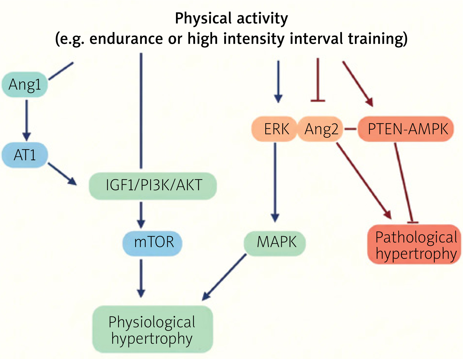
As shown in Figure 1, the IGF1/PI3K/AKT axis emerges as the core signaling pathway that drives cardiac adaptation to physical exercise, leading to physiological cardiac hypertrophy [15, 26, 43, 44]. IGF1 exerts its effects primarily through binding to its high-affinity receptor, IGF1-R, whose intrinsic tyrosine kinase activity triggers downstream intracellular signaling cascades essential for cardiomyocyte growth, survival, and functional enhancement [45–47]. Activation of this axis has been consistently associated with increased heart mass, improved systolic performance, and adaptive hypertrophic remodeling in both experimental models and human populations exposed to exercise training [21, 48–50]. The coordinated upregulation of IGF1, IGF1-R, PI3K, and AKT following endurance, resistance, and interval training underscores the central role of this pathway in mediating physiological myocardial growth [29, 51–56].
AKT-dependent hypertrophic signaling and mTOR activation
One of the main downstream factors of PI3K activation, AKT, functions as a critical effector that links IGF1 signaling to myocardial hypertrophy by activating mTOR, a principal regulator of protein synthesis and cardiomyocyte growth [28, 56]. Exercise-induced stimulation of the AKT/mTOR pathway promotes physiological hypertrophy by enhancing myocardial protein accretion in response to both endurance and resistance training stimuli [56]. Modulation of this pathway by reduced PTEN expression further amplifies PI3K/AKT/mTOR signaling, reinforcing its contribution to adaptive cardiac growth [56]. Following prolonged exercise training, signaling through this pathway appears to support structural remodeling that favors improved ventricular compliance rather than continuous increases in myocardial mass [34].
Mitogen-activated protein kinase signaling (Raf/MEK/ERK pathway)
In parallel with PI3K/AKT signaling, IGF1-R activation also stimulates the Raf/MEK/ERK signaling cascade [46, 47]. This pathway has been implicated in the development of physiological hypertrophy, particularly in response to swimming-based exercise protocols. Evidence indicates that exercise-induced cardiac adaptation involves the coordinated regulation of both AKT and ERK signaling pathways, highlighting their complementary roles in supporting myocardial growth and functional adaptation [57].
Vascular and endothelial signaling mediated by IGF1
In addition to the mentioned effects of IGF1 on cardiomyocytes, the IGF1 signaling axis contributes to cardiovascular adaptation through pathways involved in vascular and endothelial regulation. These contributions include stimulation of nitric oxide (NO) production in endothelial cells and vascular smooth muscle cells (VSMC), regulation of blood pressure through moderation of eNOS activity, stimulation of NO production in endothelial cells and VSMC, and promotion of vascular smooth muscle cell proliferation and migration [45]. Although these signaling mechanisms are not directly responsible for hypertrophic growth, they provide essential support for cardiovascular function and hemodynamic regulation during exercise-induced cardiac remodeling.
Regulatory modulation of IGF1 signaling by PTEN
Activation of the IGF1/PI3K/AKT axis is tightly controlled by intracellular regulatory mechanisms, among which PTEN plays a key inhibitory role (Figure 2). Exercise-induced reductions in PTEN expression enhance PI3K/AKT/mTOR signaling, thereby facilitating physiological cardiac hypertrophy [56]. This regulatory interaction highlights the importance of maintaining signaling balance to achieve effective and sustainable myocardial adaptation to training stimuli.
Figure 2
Molecular crosstalk related to cardiac hypertrophy following physical exercise via the RAF/MEK/ERK signaling pathway
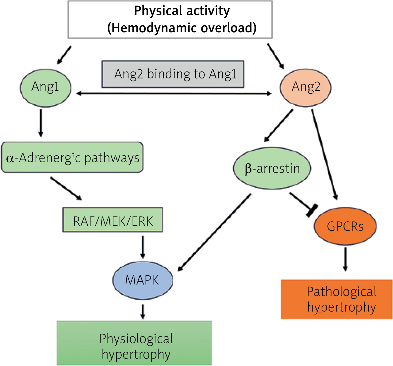
Modulation of IGF1-centered signaling, AKT/mTOR-mediated hypertrophic pathways, MAPK/ERK signaling and vascular NO-related pathways following exercise training
As seen in Table II, the IGF1/PI3K/AKT axis generally shows a dose-dependent relationship with exercise, with higher intensity and frequency driving stronger cardiac adaptations. However, future studies are needed to find the optimal exercise prescription to maximize the effects of these pathways. Previous studies have shown that in humans, the IGF1/PI3K/AKT axis is overexpressed in the heart following training at 40–60% of VO2max, leading to physiological hypertrophy [48]. Similarly, another study found that IGF1-R and AKT levels increased significantly after 8 weeks of swimming [49]. Additionally, Cheng et al. found that components of the physiological hypertrophy pathway, such as IGF1, IGF1-R, PI3K, and AKT, were elevated in endurance-trained rats with type 2 diabetes after 10 weeks of training at 48–66% VO2max [50]. Other studies have also suggested that voluntary exercise training (e.g., running and swimming) may help prevent cardiovascular disease by improving heart function through the IGF1/PI3K/AKT pathway [29, 51].
Table II
Molecular and cellular changes in cardiac tissue following different exercise training regimens
| Type of intervention | Subject | Intensity and duration | Findings | Conclusion | Reference |
|---|---|---|---|---|---|
| Endurance swimming training | C57/BL6 mice | Ramp protocol/incremental 10 to 90 min/2 times per day | mRNA level of GATA4 increased, no significant change in mRNA levels of ANP and BNP | Activation of cardiac progenitor cells and physiological hypertrophy | [65] Xiao et al., (2014) |
| Endurance swimming training | Male C57BL/6 mice | 10–90 min swimming/2 times per day/7 days per wk/3 wk or voluntarily wheel running/2 times per day/3 wk | 1.5-fold increase in miR-17-3p following swimming and 1.4 fold increase following voluntary wheel running | Induced cardiac growth | [130] Shi et al., (2017) |
| Endurance ramp swimming or running training | B6 mice for swimming, rat for running | Swimming: 10–90 min/2 sessions a day/7 days per wk/4 wk; running: 40 min/maximal speed 20 m per min/5 days/4 wk | Increased CITED4 expression with exercise; negative regulation of CITED4 by c/EBPβ expression | Increased physiological hypertrophy following CITED4 expression | [87] Boström et al., (2010) |
| Endurance exercise, ramp swimming or voluntary wheel running | Male C57BL6/J mice | 10–90 min/2 sessions a day/7 days per wk/4 wk | miR-222 enhanced following both training protocols | Necessity of miR-222 for exercise-induced cardiomyocyte growth and proliferation | [92] Liu et al., (2015) |
| Endurance running training with α2A/α2CARKO knockout | C57BL6/J mice | Incremental time-intensity up to 60% maximal speed/5 days per wk/8 wk | Decrease in the calcineurin mediated signaling pathway in α2A/α2CARKO mice after exercise; no significant change in NFAT, GATA4, and heart weight in trained mice | Increased cardiac function and exercise tolerance; anti-remodeling effects of α2A/α2CARKO knockout by training | [79] Oliveira et al., (2009) |
| Endurance running training | Mice | Running exercise: voluntary wheel running/7 days per wk/4 wk | Increased miR-222 expression | Inhibition of miR-222 blocks cardiomyogenic response to training | [11] Vujic et al., (2018) |
| Endurance running training | Wistar rat | Running at 15° inclination/1 h per day/6 days per wk/8 wk | Decreased expressions of LATS and MAP4K after training | Preventing pathological hypertrophy, increased physiological hypertrophy | [132] Tabrizi et al., (2019) |
| Endurance running training with α2A/α2CARKO knockout | Male C57BL6/J mice | Incremental time-intensity up to 60% maximal speed/5 days per wk/8 wk | No significant change in expression of AKT and mTOR in heart of wild type and α2A/α2CARKO mice | Insignificant relation of exercise training to AKT/mTOR signaling activation | [79] Oliveira et al., (2009) |
| Endurance swimming training | Female rat | Swimming exercise: ramp protocol followed by 60 min sessions with 5% body overload, twice per day/5 days per wk/8 wk | Increase in PIK3/AKT/mTOR pathway gene expression, decrease in PTEN gene expression | Increased physiological hypertrophy | [56] Ma et al., (2013) |
| Endurance swimming training | FoxO1 knockout (cKO) mice | 10–90 min/2 times per day/7 days per wk/4 wk | Increased AKT phosphorylation and IGF1-R | Increased physiological hypertrophy (21%) in FoxO1-mice | [29] Weeks et al., (2021) |
| Endurance swimming exercise | Male mice | Duration not mentioned/5 days per wk/10–11 wk | Increased PI3K | Exercise and PI3K increase attenuated pathological heart growth | [21] McMullen et al., (2004) |
| Endurance swimming training | Male CIGF1RKO and CIRKO mice | 10–90 min/2 bouts per day/5 wk | Increase in AKT phosphorylation hearts of exercise-trained, CIRKO and CIGF1RKO mice; increase in IGF1 expression. Decreased activation of AMPK in trained mice | No data | [53] Kim et al., (2008) |
| Endurance running training + Nigella sativa (N.S) Supplementation | Wistar rat | 18 meters per min at 32° inclination/2 h per day/8 wk | Increased GH, IGF1, AT-1, and ET-1 in the exercise group; decreased Erk-1 in the exercise group | Increased physiological hypertrophy in the exercise group | [132] Tabrizi et al., (2019) |
| Endurance (ET) and high intensity interval resistance (HIIT) swimming training | Male Wistar rat | HIIT: 5–16 sets/interval 20–60 s/5–16% body weight/4 days per wk/12 wk ET: 75 min/5 days per wk/12 wk | Increased heart weight in ET (15%) and HIIT (29%); IGF1 increased in ET and HIIT | Higher levels of IGF1 confirmed by physiological hypertrophy in training groups | [51] Gharaat et al., (2023) |
| Moderate- and high-volume swimming training | Female Wistar rat | Moderate (T1): 60 min/5% body weight/5 days per wk/10 wk; high (T2): 60 min/5% body weight/5 days per wk/10 wk/in 9th wk 2 times and in 10th wk 3 times per day | miRNA-126 increased 26% in T1 and 42% T2; PI3KR2 reduced 39% in T1 and 78% in T2 | Regulation of PI3K/AKT pathway and physiological hypertrophy | [133] Da Silva et al., (2012) |
| Running training (low intensity (LIT), moderate intensity (MIT) and high intensity (HIT)) | Wistar rat | 10 sets/4 min/5 days per wk/6 wk- LIT: 55–60% VO2max, MIT: 65–70% VO2max, HIT: 85–90% VO2max | Increased GATA4, NKX2.5 and TBX5 mRNA after all training protocols; GATA4, NKX2.5 in HIT higher than LIT and MIT | Higher cardiac weight and function following training, increased physiological hypertrophy | [39] Naderi et al., (2019) |
| Cardiac NKX2-5 knockout | Mice | Did not utilize any exercise intervention | Interfering effects of CSX/NKX2.5 null cells on cardiac development | Early embryonic lethality and defects in cardiac ring morphogenesis | [111] Tanaka et al., (1999) |
| Intensive intermittent running training | Wistar rat | 30 min intermittent running/50–60% VO2max or 85–90% VO2max/3 days per wk/8 wk | Increased TGF-b1 and TIMP gene expression in the heart | Increased molecular factors involved in physiological hypertrophy | [134] Xiong et al., (2023) |
| Resistance training | Wistar rat | 4 sets/12 reps/65–75% of 1RM/5 days per wk/8 wk | Increased expression of AT1 receptor; insignificant change in AT II | Increased physiological hypertrophy | [37] Barauna et al., (2008) |
The hypertrophic response of cardiac tissue to exercise, mediated by the IGF1/PI3K/AKT axis, may depend on the frequency, intensity, and duration of the training. Numerous studies on rats and mice that have involved endurance treadmill running [52] or swimming training [9, 52, 53] have consistently supported these findings. For instance, Neri Serneri et al. demonstrated that increased IGF1 expression in cardiomyocytes among elite soccer players confirms the positive effects of exercise training on physiological hypertrophy. The underlying mechanism involves increased stretching of cardiac myocytes due to higher stroke volume during exercise [54]. Another study by Kim et al. found that cardiac hypertrophy in normal, non-transgenic PI3K mice was more pronounced after 5 weeks of chronic incremental swimming training than in age- and weight-matched dnPI3K mice. This study highlighted the crucial role of myocardial IGF1 signaling in cardiac hypertrophy [53]. Another well-designed study demonstrated that while AKT signaling can induce cardiac hypertrophy, it is not fully effective without the presence of IGF1 [54]. It has been demonstrated that swimming training induces physiological hypertrophy by regulating both the AKT and ERK signaling pathways [57]. Ma et al. found that 8 weeks of swimming training, combined with an additional 5% body weight, increased PI3K/AKT/mTOR signaling by reducing PTEN expression, leading to physiological hypertrophy [56].
The IGF1/PI3K/AKT axis plays a critical role in regulating physiological cardiac hypertrophy, with its activation closely linked to exercise duration and intensity. Evidence suggests a dose-dependent response: moderate to high-intensity exercise increases IGF1 and its downstream signaling components, promoting myocardial adaptation.
Endurance training at 40–70% VO2max has been shown to significantly upregulate IGF1, IGF1-R, PI3K, and AKT, leading to physiological hypertrophy [48, 52]. Similarly, chronic swimming and treadmill running have been shown to enhance IGF1 signaling, particularly in endurance-trained rodents and elite athletes [45, 54]. However, prolonged training may lead to a plateau in hypertrophic adaptations, suggesting that the heart’s response diminishes over time. Resistance training also activates the IGF1/PI3K/AKT pathway, thereby increasing myocardial protein synthesis and promoting structural remodeling via mTOR activation [56]. However, long-term adaptations appear to shift toward enhanced left ventricular compliance rather than mass enlargement [34]. High-intensity interval training (HIIT) elicits the strongest IGF1/PI3K/AKT activation, with intensities of 85–90% VO2max producing significant cardiac hypertrophy [39]. This response may result from both mechanical and metabolic stimuli that amplify IGF1 signaling, indicating a potent stimulus for cardiac remodeling.
In summary, the signaling pathways addressed in the text (and shown in Table II) are coherently classified into IGF1-centered signaling, AKT/mTOR-mediated hypertrophic pathways, MAPK/ERK signaling, vascular NO-related pathways, and regulatory modulators such as PTEN. Together, these interconnected pathways form a coordinated molecular network that underlies physiological cardiac hypertrophy in response to exercise, with activation patterns closely influenced by training intensity, duration, and modality [39, 48, 52, 54, 56].
G-protein-coupled receptor and angiotensin contribution with ERK
Myocardial G-protein-coupled receptors (GPCRs), including adrenergic, angiotensin, and endothelin (ET1) receptors, are transmembrane receptors that regulate both physiological processes in the cardiac tissue. These receptors play a crucial role in modulating cardiac hypertrophy and function [58]. Stimulation of GPCRs by angiotensin II (AT-II) activates the RAF-MEK-ERK pathway through the recruitment of β-arrestin [59]. Additionally, α-adrenergic (αAR) receptors positively regulate cardiac contractility and hypertrophic responses [60]. Studies have shown that activation of ERK signaling via the α-adrenergic pathway plays a stimulatory role in the development of cardiac physiological hypertrophy [60]. Hemodynamic overload on the heart activates the local renin-angiotensin system, which in turn engages the angiotensin II type 1 receptor pathway and induces the α-adrenergic pathway. Previous studies have shown that angiotensin II binds to the Ang1 receptor, which serves as a key regulator of fluid volume, blood pressure, and cardiovascular change. The role and mechanism of the AT2 receptor, along with the AT1 receptor, in cardiac hypertrophy following exercise training are crucial, particularly in relation to the increased expression of key marker genes of cardiac hypertrophy [18]. This local activation contributes to the adaptive remodeling seen in athletes, characterized by increased cardiomyocyte size and improved cardiac function without the adverse effects typically associated with AngII [18]. The signaling pathways following AngII signaling through the AT1 receptor include the mitogen-activated protein kinase (MAPK) pathway and the phosphoinositide 3-kinase (PI3K)/Akt pathway [48]. However, there is a significant lack of academic studies specifically addressing this pathway in the context of exercise-induced physiological hypertrophy. Limited studies in this area indicate that exercise training increases Ang1 expression without altering Ang2/II levels, ultimately leading to cardiac hypertrophy [37, 60]. Exercise training potentially decreased Ang2/II, a vasoconstrictor that causes hypertension, and increased angiotensin (1-7), a vasodilator that decreases blood pressure, contributing to protective effects that prevent cardiac pathological hypertrophy [61]. Therefore, the activity of the Ang1 receptor may be involved in the cardiac hypertrophy induced by exercise training by inhibiting Ang2/II, a pathological excitatory factor of cardiac hypertrophy.
Overall, Ang1 and Ang2/II play distinct roles in cardiac hypertrophy, particularly in response to exercise training. Ang1 primarily contributes to physiological cardiac remodeling by activating the Ang1 receptor, which has been associated with adaptive hypertrophy, increased cardiomyocyte size, and improved cardiac function without pathological consequences [18]. This receptor is engaged during exercise-induced hemodynamic overload and stimulates signaling pathways such as PI3K/Akt and MAPK, which are crucial for physiological hypertrophy [48].
In contrast, Ang2/II, which binds to the Ang1 receptor, is more commonly associated with pathological cardiac remodeling, fluid volume regulation, and blood pressure control [59]. While Ang2/II can contribute to hypertrophy, its effects are often linked to maladaptive cardiac changes. Interestingly, studies suggest that exercise training may selectively upregulate Ang1 expression while maintaining or even reducing Ang2/II levels, thereby promoting beneficial cardiac adaptations while preventing pathological hypertrophy [36, 61]. Additionally, exercise training appears to increase angiotensin (1-7), a vasodilator that counteracts the hypertensive effects of Ang2/II, further supporting the cardioprotective nature of Ang1-mediated physiological hypertrophy [61]. Thus, the balance between Ang1 and Ang2/II activity plays a critical role in determining whether cardiac hypertrophy remains within physiological limits or progresses toward pathological remodeling. More research is needed to clarify the precise mechanisms through which exercise modulates this system, particularly in the context of long-term cardiac adaptations [62].
MAPK/ERK signaling
The extracellular signal-regulated kinase (ERK) signaling pathway, a member of the mitogen-activated protein kinase (MAPK) family, plays a critical role in regulating cardiac hypertrophy. However, its effects can be context-dependent, contributing to both adaptive and maladaptive remodeling. The nature of its impact is influenced by various factors, including the type and intensity of physiological stimuli [59].
Studies in transgenic models and adult rodents have demonstrated that exercise training activates the ERK cascade, promoting concentric cardiac hypertrophy characterized by increased left ventricular wall thickness and enhanced myocardial contractility [63]. The ERK pathway achieves this by phosphorylating a diverse set of intracellular targets, including transcription factors such as ELK1, GATA4, and MEF2, which play key roles in cardiac gene expression and hypertrophic remodeling. Activation of ERK has been shown to regulate the expression of genes associated with cardiomyocyte growth, extracellular matrix remodeling, and metabolic adaptation, thereby facilitating a transition toward physiological hypertrophy.
Recent evidence suggests that exercise-induced activation of ERK is closely linked to other pro-hypertrophic pathways, including the IGF1/PI3K/AKT axis. For instance, swimming training has been shown to induce physiological hypertrophy by concurrently activating ERK and AKT signaling pathways while modulating cardiac microRNAs, such as miR-21 and miR-33, in adult male Sprague-Dawley rats [57]. These molecular interactions highlight the integrative nature of ERK signaling in exercise-induced cardiac adaptations.
However, the role of ERK in cardiac hypertrophy remains controversial. While its activation is commonly associated with beneficial remodeling in response to exercise, under conditions of chronic stress or pathological stimuli such as hypertension and myocardial infarction, ERK signaling has been implicated in maladaptive hypertrophy and fibrosis. This duality suggests that the duration, intensity, and nature of ERK activation are crucial determinants of its effects on cardiac structure and function. Further research is needed to delineate the precise mechanisms by which ERK contributes to exercise-induced hypertrophy and to distinguish its beneficial effects from pathological remodeling.
Hepatocyte growth factor axis
Hepatocyte growth factor (HGF) is a paracrine growth factor secreted by mesenchymal cells which primarily targets epithelial and endothelial cells. HGF exhibits cardioprotective properties [64] and is involved in both the enlargement of existing cardiomyocytes and the generation of new cells [65]. Moreover, HGF-based gene therapy actively initiates cardiac remodeling. Its primary effects include promoting cardiomyocyte hypertrophy and preserving cardiac function [64]. Previous studies have reported significant cardiomyocyte hypertrophy with HGF gene therapy, especially when combined with angiotensin II [66]. Yasuda et al. found that high-intensity exercise increases HGF production, which is linked to elevated VO2peak in post-infarction patients, especially those with reduced exercise capacity [67]. Additionally, Wahl et al. reported a significant increase in HGF following aerobic training, which was associated with improved power output and VEGF expression [68]. Also, Kitta et al. demonstrated that HGF protects cardiac muscle cells against apoptosis via MEK/ERK-dependent phosphorylation of GATA4 [69]. Therefore, HGF plays a role in cardiac physiological hypertrophy and function. However, further research is needed to fully establish a direct connection between exercise-induced increases in HGF and cardiac hypertrophy.
Platelet-derived growth factor axis
Platelet-derived growth factor (PDGF) is a member of the human growth factor family. It is a dimeric glycoprotein composed of four polypeptide chains, encoded by four genes located on different chromosomes, which regulates cell growth and division [70]. It has been shown that essential myocardial remodeling processes, such as mitosis, migration, angiogenesis, and matrix modulation, are induced following PDGF therapy [71]. In humans, studies have reported a strong correlation between left ventricular hypertrophy and increased PDGF production in cardiac tissue. Notably, PDGF plays a crucial role in modulating cardiac physiological hypertrophy in response to increased workload following exercise training [54]. Additionally, PDGF is essential for capillary development, which enhances cardiac function and contributes to cardiac remodeling and hypertrophy [72]. Wang et al. showed that endurance exercise training increases levels of the cardioprotective PDGF-BB via PDGF-BB/PDGFR-β signaling [73]. In summary, PDGF plays a key role in cardiac tissue remodeling, which is essential for physiological hypertrophy.
Neuregulin-1 regulatory role
Neuregulin-1 (NRG1) is a member of the epidermal growth factor family, encoded by the NRG1 gene. It plays a crucial role in physiological hypertrophy by activating pathways such as the NRG1/ErbB signaling, which is involved in myocardial development and provides cardioprotective effects [74]. These pathways are stimulated in response to cardiac overload during exercise training [40–42]. A study by Ryall et al. reported that upregulation of NRG1 induces cardiac CITED4, which contributes to eccentric hypertrophy by promoting cardiomyocyte elongation and proliferation [75]. In addition, the potential impact of NRG1 on left ventricular function has also been observed [76]. Previous studies have shown that the loss of NRG1 disrupts cardiac growth and survival processes, leading to cardiac atrophy [77], while it enhances myoblast differentiation and promotes cardiac remodeling following myocardial infarction [78]. In summary, NRG1 plays an essential role in cardiac physiological hypertrophy and repair.
Calcineurin-NFAT cascade
The nuclear factor of activated T cells (NFAT) is a key molecular modulator of cardiac hypertrophy, influencing both pathological and physiological pathways. It interacts with the GATA4 transcription factor and is also linked to fibronectin-associated factors. These relationships warrant further discussion and investigation [79, 80]. Previous studies have demonstrated a direct interaction between GATA4 and NFAT, a transcription factor that calcineurin dephosphorylates in the cytoplasm. This dephosphorylation facilitates NFAT’s nuclear translocation, where it activates hypertrophic responses [79, 81, 82]. Increased calcineurin activity leads to greater NFAT translocation into the nucleus, elevated GATA4 expression, and increased fibronectin production, which is a matrix protein. This rise in fibronectin, often observed in cardiac dysfunction, appears to be a reparative and protective response to heart failure [83]. The literature on this topic yields controversial findings. Eto et al observed that in mice trained for 10 weeks at 59–70% of VO2max, there was a significant increase in calcineurin and p-ERK1/2, associated with physiological left ventricular hypertrophy and NFAT overexpression [84]. However, a study involving NFAT2-deficient mice found that physiological hypertrophy was unaffected after 4 weeks of voluntary exercise training. This suggests that NFAT plays a crucial role in the induction of cardiac hypertrophy [85]. In summary, the calcineurin/NFAT/GATA4 axis appears to be a key pathway in cardiac hypertrophy. However, further research is needed to fully clarify its role in physiological hypertrophy.
c/EBPβ diverse role in physiological hypertrophy
CCAAT-enhancer binding protein β (c/EBPβ) is a crucial factor in cardiac hypertrophy. Its reduction has been associated with enhanced cardiac proliferation and physiological hypertrophy. This effect is mediated by the downregulation of key proteins, such as αMHC, CITED4, and PGC1α (peroxisome proliferator-activated receptor gamma coactivator 1α), in response to exercise training [86, 87]. Previous research has shown that overexpression of c/EBPβ leads to the downregulation of several key transcription factors involved in physiological hypertrophy, including GATA4, T-box transcription factor 5 (Tbx5), and NKX2.5, among others [87]. c/EBPβ is one of the downstream targets of the PI3K/AKT pathway, which is a major molecular pathway involved in exercise-induced cardiac hypertrophy. This process is mediated through the c/EBPβ-CITED4 induction pathways [88]. Inhibition of c/EBPβ in cardiomyocytes leads to increased cell size and number, suggesting that c/EBPβ deficiency promotes cardiomyocyte growth and proliferation [87].
A well-designed study by Konstandin et al. reported that c/EBPβ expression decreased by 48% in control mice and 46% in knockout mice following training [83]. Additionally, another study reported significant increases in c/EBPβ expression following both moderate and high-intensity interval running training [81]. Contrary to the protective role of fibronectin in cardiac tissue growth, which is associated with c/EBPβ downregulation induced by physical training, researchers found that removing fibronectin does not affect physiological hypertrophy following running [79, 83]. Two primary axes are involved in the c/EBPβ pathway in response to exercise: the c/EBPβ-CITED4 axis and the c/EBPβ-GATA4 axis.
c/EBPβ-CITED4 axis
CITED4, a key component in the c/EBPβ–CITED4 signaling pathway, is associated with cardiac hypertrophy. Research shows that CITED4 (CBP/p300–interacting trans-activator with ED-rich carboxy-terminal domain-4) levels significantly increase in the hearts of individuals who engage in exercise [73, 75]. A study has identified CITED4 as a stimulator of the mTOR pathway, effectively promoting physiological hypertrophy [86]. Recent analyses of cardiac microRNA pathways in two distinct exercise models have highlighted the role of miR-222 in exercise-induced cardiac and cardiomyocyte growth. It was demonstrated that miR-222 negatively regulates CITED4 in neonatal cardiomyocytes [73]. Consequently, CITED4 is implicated as a downstream effector in exercise-related transcriptional and microRNA pathways in neonatal cardiomyocytes [86]. Exercise-induced CITED4 upregulation can stimulate cardiomyocyte growth under basal conditions [87]. CITED4 induces physiological hypertrophy, leading to a 25% increase in cardiac tissue size, in both male and female mice [86]. In summary, increased levels of CITED4 are beneficial for cardiac tissue, as it regulates myocyte elongation and promotes myocyte proliferation following exercise training.
GATA4 signaling pathways
GATA binding protein 4 (GATA4) is a key transcription factor involved in cardiac hypertrophy and cardiomyocyte viability [52, 89]. It interacts with several signaling pathways, including IGF1/PI3K/PKB, calcineurin/NFAT, and c/EBPβ. This modulatory factor regulates the transcription of crucial structural and cardiovascular genes such as myosin heavy chain (MHC), atrial natriuretic peptides (both A-type and B-type), and nitric oxide synthase (NOS). Additionally, GATA4 plays a critical role in modulating responses of the cardiac tissue, including myocyte survival and hypertrophy, in response to exercise [90]. Exercise training significantly increases cardiac GATA4 expression in healthy and myocardial infarction models, promoting cardioprotective effects [39, 52, 91]. It engages signaling pathways such as IGF1/PI3K/PKB, calcineurin/NFAT, and c/EBPβ, controlling genes like myosin heavy chain, atrial natriuretic peptides, and nitric oxide synthase [90]. Studies revealed that increased GATA4 expression following exercise induces physiological hypertrophy, as shown in treadmill and swimming studies [39, 65, 90]. Naderi et al. observed that 6 weeks of low (10 intervals of 4 min at 55–60% VO2max), moderate (10 intervals of 4 min at 65–70% VO2max), and high-intensity (10 intervals of 4 min at 85%–90% VO2max) running significantly upregulates the mRNA levels of GATA4 in the cardiac tissue of rats [39].
On the other hand, GATA4 deficiency triggers pro-apoptotic proteins, lowers fibroblast growth factor 1 and epidermal growth factor receptor levels, and weakens hypertrophic responses [89]. It is essential during embryonic cardiocyte differentiation and remains active in adult cardiac cells. Running at various intensities elevates GATA4 mRNA in rat cardiac tissue [39]. Further research is needed to clarify the full role of GATA4 in cardiac adaptation to physical activity.
Interaction between HMBOX1 and miR-222
HMBOX1 (Homeobox 1) is a transcription factor that plays a pivotal role in various cellular processes, including apoptosis and autophagy. It has been identified as a significant regulator of cardiac hypertrophic responses. Specifically, HMBOX1 is integral to the molecular mechanisms governing cardiac hypertrophy induced by physical exercise training. Its interaction with miR-222 and its involvement in critical signaling pathways underscore its potential as a biomarker for physiological hypertrophy [48]. Notably, the inhibition of miR-222 has been linked to the exacerbation of pathological hypertrophy and heart failure in response to stressors such as pressure overload. Recent research suggests that HMBOX1 may be part of a regulatory network modulated by miR-222 [92].
Relationship between GSK-3β and cardiac remodeling
GSK-3β (glycogen synthase kinase 3 beta) is a serine/threonine kinase that plays a significant role in numerous cellular processes, including cell survival, proliferation, and differentiation, and is critically involved in cardiac hypertrophy. Exercise training has been shown to activate the PI3K/Akt signaling pathway, which subsequently inhibits GSK-3β activity. Additionally, this cellular factor regulates Wnt signaling by phosphorylating β-catenin, leading to its degradation. Dysregulation of this pathway can result in adverse cardiac remodeling and heart failure. Regular aerobic exercise has been shown to engage protective signaling pathways that inhibit GSK-3β, thereby facilitating physiological remodeling. This includes enhanced Akt signaling, which directly inhibits GSK-3β activity, reduces apoptosis under stress conditions such as ischemia, and promotes angiogenesis, further supporting cardiac health during physical stress [93, 94]. GSK-3β is a pivotal regulator of cardiac remodeling following myocardial injury or pressure overload. Its dual function as both a promoter of fibrotic responses and an inhibitor of pathological hypertrophy highlights the complexity of the mechanisms underlying cardiac remodeling [94]. Conversely, studies show that GSK-3 activation prevents cardiac hypertrophy, underscoring its protective role against pathological remodeling [94, 95].
The role of p27 in physiological hypertrophy
p27 (cyclin-dependent kinase inhibitor 1B) is a critical molecular mediator of cell-cycle regulation, with its expression levels significantly affecting cardiac cell proliferation and hypertrophy. Research has demonstrated that p27 is upregulated by exercise, playing a vital role in regulating cardiomyocyte proliferation and survival. This upregulation correlates with improved cardiac performance and adaptation to increased workloads during physical activity [96]. Furthermore, modulation of p27 expression following exercise training affects miR-222 expression, a regulator known to inhibit pathological hypertrophy while promoting physiological adaptations [23]. Notably, p27 interacts with signaling pathways activated by IGF-1 and involved in cardiac remodeling [94].
The relationship between NELF-A and cardiac hypertrophy
NELF-A (negative elongation factor A) is a transcriptional regulator that plays a crucial role in cardiac function. Exercise-induced physiological hypertrophy is characterized by increased cardiomyocyte size and improved cardiac performance, which is partially regulated by NELF-A through its effects on gene transcription involved in muscle growth and adaptation [94]. The mechanisms by which NELF-A influences cardiac hypertrophy are likely interconnected with signaling pathways, such as the Akt/mTOR pathway, which enhance the activity of crucial protein synthesis and muscle growth pathways. NELF-A plays a role in these pathways by regulating the expression of genes responsive to mechanical stress and to metabolic demands during physical activity [97].
The role of RhoA in cardiac remodeling
RhoA (Ras homolog family member A) is a small GTPase which plays a pivotal role in various cellular processes, including cell proliferation and survival. Its involvement in cardiac remodeling in response to mechanical stress, such as exercise training, has significant implications for cardiovascular health [98]. RhoA signaling promotes cardiomyocyte hypertrophy by activating downstream effectors, including Rho-associated protein kinase (ROCK). Research indicates that eccentric exercise training activates RhoA signaling pathways, contributing to hypertrophic responses and actin biogenesis [18]. This activation is associated with transcriptional changes that enhance muscle remodeling and recovery following exercise. Specifically, eccentric exercise has been shown to increase RhoA expression within the STARS/RhoA/AP1 and NFAT/AP1 signaling pathways, which are essential for hypertrophic adaptation [18]. Additionally, studies have reported that low-intensity exercise training protects against structural and functional deterioration in hypertensive models by inhibiting RhoA/ROCK signaling, thereby improving cardiac function and reducing pathological remodeling [98, 99].
The role of CDC42 in physiological hypertrophy
CDC42 (cell division control protein 42) is a small GTPase belonging to the Rho family. The relationship between CDC42, cardiac remodeling, and physical exercise is an important area of investigation in cardiovascular research. Cardiac remodeling refers to the structural and functional changes in the heart induced by various stressors, including pressure overload, ischemia, and exercise [94, 99]. Regular training reduces the expression levels of CDC42 and its downstream effectors in models of heart failure and hypertrophy, promoting a shift from maladaptive to adaptive remodeling. CDC42 activation promotes cardiomyocyte hypertrophy through pathways such as the PI3K/Akt pathway [100].
The role of C-MYB in cardiac hypertrophy
C-MYB (MYB proto-oncogene) is a transcription factor that plays a significant role in regulating cell proliferation, differentiation, and survival. It is part of the MYB gene family, characterized by a highly conserved DNA-binding domain and involved in the expression of genes critical for cell cycle regulation and hematopoiesis. Research indicates that exercise training can alter the expression levels of C-MYB and other transcription factors involved in cardiac hypertrophy. For instance, studies have demonstrated that regular physical activity promotes physiological hypertrophy, characterized by increased cardiomyocyte size and improved cardiac performance. C-MYB partially regulates this adaptation by modulating gene transcription related to muscle growth and metabolic efficiency [101]. The PI3K/Akt/mTOR signaling pathway, which is essential for protein synthesis and muscle growth during exercise, enhances C-MYB transcriptional activity, thereby promoting adaptive changes in cardiomyocytes [48].
PHLPP signaling pathway
The PH domain leucine-rich repeat protein phosphatase (PHLPP) typically inhibits AKT phosphorylation [65]. The targets of the PHLPP isoform include AKT, protein kinase C (PKC), p70S6 kinase 1 (p70S6K), and the RAF/MEK/ERK cascade. A study found that in mice deficient in PHLPP-1, AKT phosphorylation increased after 20 days of swimming training (at 70% of VO2max), leading to physiological hypertrophy compared to wild-type mice that underwent the same training [102]. Swimming training has been shown to alleviate pathophysiological hypertrophy induced by pressure overload. This results in increased heart size, elevated cardiomyocyte levels, and higher expression of hypertrophic genes. In mice lacking PHLPP-1, this reduction in pathophysiological hypertrophy is associated with decreased fibrosis and cell death. Additionally, the absence of PHLPP-1 is associated with increased VEGF expression and increased capillary density in the myocardium, as PHLPP-1 deletion promotes endothelial tube formation [103]. In conclusion, physical training reduces PHLPP-1 levels, thereby enhancing the activity of AKT, p70S6 kinase, and ERK. This leads to increased heart size, improved angiogenesis, greater capillary density, and elevated expression of hypertrophic genes [102, 104].
Effective gene and protein expression changes in physiological hypertrophy
The genetic literature on cardiac hypertrophy identifies 175 transcription factors [87]. Among these, several key factors have been extensively studied, including HAND2, NKX2.5, GATA4, Tbx5, c/EBPβ, PHLPP, HMBOX1, GSK-3β, NELF-A, RhOA, C-MYB, CDC42, and MEF2c. Additionally, some genes negatively impact cardiac growth when down-regulated following training. Notable examples include NFAT and c/EBPβ.
HAND2 gene expression
Heart and neural crest derivatives expressed transcript 2 (HAND2) is a basic helix-loop-helix (BHLH) transcription factor that plays crucial roles in normal development, including ventricular septation and the formation of outflow tract structures in cardiac tissue [63]. In the adult heart, HAND2 is expressed in response to stimulation of the IGF1/PI3K/AKT axis [104]. In a recent study examining the effects of high-intensity interval swimming training (20–60 min intermittent exercise, 5–16% body weight over lactate threshold, 5 days per week for 12 weeks) and endurance swimming training (30–75 min, 5 days per week for 12 weeks), our team found that both regimens regulated the PI3K enzyme and HAND2 gene in the cardiac tissue of young Wistar rats. These changes resulted in clear physiological hypertrophy of the cardiac tissue, including both structural and functional development [105]. Additionally, earlier studies have shown that the HAND2 gene, in conjunction with the transcription factors GATA4 and TBX5, regulates cardiac-specific promoters. This regulation is crucial for cardiac cell proliferation and development [106, 107]. Another study found that endurance aerobic training effectively increases HAND2 gene expression [108]. Additionally, a study by Zargani et al. demonstrated that swimming training upregulates HAND2 in elderly rats, highlighting its important role in cardiac protection [109]. To further validate the role of HAND2 in cardiac hypertrophy, researchers inhibited HAND2 expression in cardiac tissue, resulting in cardiovascular developmental abnormalities [63]. Consequently, the elevation of HAND2 following exercise training is pivotal for promoting physiological cardiac hypertrophy.
GATA4 expression
GATA binding protein 4 (GATA4) is a key transcription factor involved in cardiac hypertrophy [52]. The main role of GATA4 in cardiac hypertrophy was previously described.
MEF2c expression
Myocyte enhancer factor 2 (MEF2) is a protein-coding gene involved in cardiac hypertrophy. MEF2 plays a crucial role in the MEF2C/miR-17-3p/PTEN pathway. Studies in animal models, such as rats, suggest that targeting this pathway could represent a novel therapeutic strategy to mitigate myocardial dysfunction [65, 101, 110]. Interestingly, a study in rats found that MEF2C mRNA levels were significantly elevated following moderate- and high-intensity interval training compared with the control group [39]. Furthermore, endurance exercise training was found to elevate miR-17-3p levels, a mediator associated with MEF2, thereby inducing cardiac hypertrophy in animal models [80, 92, 110]. Ma et al. demonstrated that 8 weeks of swimming training, with an added 5% body weight, led to reduced PTEN gene expression. This reduction contributed to physiological hypertrophy, as evidenced by increased heart and ventricular weight, via the MEF2/PTEN pathway [56].
NKX2.5 expression
NK2 Homeobox 5 (NKX2.5) plays a crucial role in cardiac remodeling. It has been noted that NKX2.5 significantly influences heart morphogenesis and promotes the growth of atrial, ventricular, and conduction cells during embryogenesis. This factor plays a multifaceted role in the early stages of mammalian cardiogenesis [91]. In a research study, mice with cardiac NKX2.5 knockout displayed early embryonic lethality and anomalies in cardiac ring morphogenesis [111]. Additionally, Serpooshan et al. observed that NKX2.5 cardiomyoblasts play a role in cardiomyogenesis in the neonatal heart [112]. Naderi et al. also reported a significant increase in the expression of NKX2.5 and Tbx5 following interval training at various intensities, including low intensity (55–60% VO2max), moderate intensity (65–70% VO2max), and high-intensity interval training (85–90% VO2max) [39]. Since the reduction and down-regulation of NKX2.5 are linked to heart disease phenotypes, the increase in this factor following physical training is considered a key indicator of cardiac protection through enhanced physiological hypertrophy. In conclusion, the activation of the cardiac homeobox gene product NKX2.5 during cardiogenesis plays a crucial role in regulating adult cardiac growth.
Cardiac mass atrophy following deconditioning
In general, the molecular and cellular mechanisms underlying cardiac remodeling and atrophy induced by deconditioning are complex and controversial and require further elucidation. In this review, we provide a brief overview of the most important molecular regulators of cardiac atrophy following decreases or cessation of physical activity.
Lifestyle changes, such as sedentary behavior or decreased physical activity, lead to cardiac remodeling and atrophy [7]. Atrophy is defined as a reduction in the size of a tissue or organ due to cellular shrinkage; this decrease in cell size results from the loss of organelles, cytoplasm, and proteins [113]. As described by Laplace’s law, mechanical stress alters the morphological shape and functional performance of the cardiomyocyte in accordance with intraventricular pressure and wall stress. Adversely, reduction of wall stress (same as ventricular or hemodynamic unloading) results in cardiomyocyte size atrophy [114, 115]. Cosper and Leinwand showed that reduced cardiomyocyte volume was attributed to degradation of sarcomeric proteins in parallel rather than to preferential loss of proteins [116]. Although deconditioning led to cardiac atrophy in retired elite athletes, the reversal of LV cavity dilatation was incomplete after 5 years in 80% of them [117]. From a molecular perspective, muscle mass decrease is generally associated with cardiomyocyte autophagy mediated by the Forkhead box-containing protein O subfamily (FoxO) [118]. Increased conjugation of ubiquitin to muscle proteins; increased ATP-dependent activity in proteasome; increased protein; upregulation of transcripts encoding ubiquitin, ubiquitin-conjugating enzymes (E2), and ubiquitin-protein ligases (E3) [113]. Expression of constitutively active mutants of FoxO1 and FoxO3 in cardiac muscle cells triggers muscle atrophy without activating apoptosis. Reduction of cell size, in this way, seems to be due to increased protein degradation through FoxO-mediated activation of two muscle-specific ligases, atrogin-1 (MAFbx) and MuRF-1 [119]. On the one hand, Razeghi et al. demonstrated that the ubiquitin-proteasome system (UPS) is up-regulated and responsible for protein degradation in the heart [115]. It is thought that calcineurin is a reciprocal regulator of pro-atrophic pathways, coordinating the cardiac response. In atrophic remodeling, FoxO1 and FoxO3a reduce calcineurin phosphorylation and expression of calcineurin/NFAT pathway targets, including modulatory calcineurin-interacting protein 1.4 (MCIP1.4). Indeed, studies reported that overexpression of FoxO3a and repression of the calcineurin/NFAT pathway in a hypertrophic heart reduces heart mass [120] and causes atrophic remodeling in a healthy heart [121]. In this line, previous studies have established the main role of calcium-dependent calpain proteases in UPS activity and the degradation of myofibrillar proteins during atrophic remodeling. Expression of calpains 1 and 2 is up-regulated following ventricular unloading of the rat and human heart [122].
Contribution of microRNAs in regulatory mechanisms of cardiac hypertrophy
Apart from structural and functional changes, several cellular mechanisms are crucial to hypertrophy development. Recent research has delved into the role of micro-RNAs (miRNAs) in physiological cardiac hypertrophy, examining their effects in human and animal models [112, 123, 124]. MiRNAs are short, single-stranded RNAs that contain 17-22 nucleotides and can regulate the expression of target genes. MiRNA genes cluster into families based on their sequence similarity. Each miRNA may upregulate or downregulate several transcripts, exerting excitatory or inhibitory effects on physiological hypertrophic genes that promote heart development and increase cardiac function, or on pathological hypertrophy that leads to heart defects [27]. Based on the wide range of studies related to miRNAs, downregulation of specific miRNAs can change the cellular responses of cardiomyocytes to specific signals on pathological hemodynamic overload or inhibition of physiological hypertrophy responses, leading to anti-hypertrophic signaling with or without pathological cardiac hypertrophy [125]. Also, studies have shown that exercise training increases cardiomyocyte growth and survival by regulating pro-hypertrophic miRNAs or suppressing the hypertrophic inhibitors. In this line, the scientific literature supports controversial stimulatory effects of various exercise routines (e.g., aerobic, anaerobic, high-intensity interval, or resistance training) on miRNAs.
Also, we classified the main miRNAs that contribute to various hypertrophic, suppressive, or atrophic processes in cardiac tissue.
The role of microRNAs in cardiac hypertrophy
Apart from structural and functional changes, several cellular mechanisms are crucial to hypertrophy development. Recent studies have investigated the role of microRNAs (miRNAs) in both pathological and physiological cardiac hypertrophy in human and animal models [112, 123, 124]. Here, we classified miRNAs based on their biological functions.
miRNAs that regulate myocyte growth
This group of miRNAs regulates cardiac cell growth and proliferation. The so-called miRNAs in this category include miR-1 (target genes: IGF1, IGF1-R), miR-17 (target gene: MEF2), miR-21 (target gene: PTEN), and miR-222 (target genes: HMBOX1, P27, HIPK1), which increase in response to exercise and promote physiological cardiac hypertrophy by inhibiting negative growth regulators [57, 92, 126–128]. Additionally, miR-17-3p and miR-18a-3p mediate cardiomyocyte proliferation and non-pathological growth following training [27, 57, 129, 130].
miRNAs that promote angiogenesis
These miRNAs enhance angiogenic pathways and improve blood supply, such as miR-126 targeting Spred-1, PI3KR2, and VEGF, which are upregulated with exercise training [126–128]. These enhancements involve cardiac hypertrophy, driven by increased capillary density.
miRNAs that control fibrosis
miRNAs regulating fibrotic responses include miR-29a/c (target gene: collagen) and miR-30 (target gene: CTGF), critical for modulating fibrosis in response to hemodynamic stress and pathological hypertrophy which prevent cardiac atrophy [126–128].
miRNAs that modulate anti-hypertrophic or stress response
Specific miRNAs, such as miR-21 (target genes: Spry1, PTEN), miR-23a (target gene: MuRF1), miR-27a/b (target genes: GATA4, PPAR-γ), and miR-146 (target genes: IRAK, TRAF6), upon downregulation affect cardiomyocyte responses to pathological overload or inhibit physiological hypertrophy signaling [99, 125].
miRNAs that are responsive to exercise training
Exercise training leads to upregulation of several miRNAs – miR-21, miR-23a, miR-27a/b, miR-30e, miR-98, miR-133, miR-208a/b, miR-222, and miR-499 – that support cardiomyocyte growth and survival [92, 99, 125–128]. Some miRNAs, such as miR-1 and miR-133, show variable expression changes depending on exercise type and intensity [126–128].
This classification highlights the dynamic and multifaceted roles of miRNAs in regulating myocyte growth, angiogenesis, fibrosis, and cardiac adaptation to both physiological exercise and pathological stress. Also, previous studies reported the controversial roles of other miRNAs, such as miR-26b (target genes: IGF1 and PI3K), miR-95a (target gene: β-MHC), miR-99b (target gene: IGF1-R, AKT and mTOR), miR-100 (target gene: IGF1-R, AKT and mTOR), miR-124 (target gene: PI3K), miR-143 (target gene: ECA2), miR-144 (target gene: TSC2), miR-145 (target gene: PI3K), miR-150 (target gene: GS3K-β and C-MYB), miR-199b (target gene: Dyrk1A) and miR-208b (target gene: β-MHC), in relation to cardiac physiological hypertrophy following exercise training [99, 125–128].
Conclusions
Cardiac physiological hypertrophy represents an adaptive enlargement of the heart in response to increased workload. Unlike pathological hypertrophy, this form maintains or even enhances myocardial function and improves resilience to stressors such as ischemia. It is driven by stimuli including increased venous return, elevated arterial pressure, and exercise-induced vasodilation, which collectively promote efficient cardiac remodeling. These adaptations contribute to superior cardiovascular performance, reflected in higher maximal oxygen uptake (VO2max) and increased cardiac output following training [2, 12, 14, 131–135].
A diverse network of autocrine and paracrine signaling pathways orchestrates this remodeling process. Central pathways that drive physiological hypertrophy include IGF1/PI3K/AKT, hepatocyte growth factor (HGF), platelet-derived growth factor (PDGF), the angiotensin/α-adrenergic/ERK axis, and neuregulin-1 (NRG1). Exercise training also modulates several transcription factors that promote physiological hypertrophy, including HAND2, GATA4, MEF2, NKX2.5, and TBX5, all of which contribute to pro-growth gene expression programs [39, 65, 79, 107, 109, 111]. In contrast, factors that decline with training – such as NFAT, c/EBPβ, and PHLPP – are associated with controlled, non-pathological myocardial growth [88, 97, 100, 109]. In addition, multiple micro-RNAs, including miR-17-3p, miR-18a-3p, miR-21, miR-33, and miR-222, play essential regulatory roles in signaling pathways that coordinate cardiomyocyte remodeling, adaptation, and survival during exercise [57, 73, 92, 130, 131].
Research has shown that different training modalities elicit distinct structural adaptations in the heart. Resistance training primarily induces pressure overload, promoting concentric hypertrophy characterized by parallel sarcomere addition and increased ventricular wall thickness. In contrast, endurance training produces volume overload, leading to eccentric hypertrophy marked by sarcomere elongation and an overall increase in cardiac mass. Importantly, both forms of exercise-induced hypertrophy contribute beneficially to cardiac structure and functional capacity. Growing evidence indicates that exercise-induced cardiac remodeling can be effectively leveraged through individualized exercise prescriptions, which offer a cost-effective and non-invasive approach to improve cardiovascular health. Endurance exercise is generally associated with eccentric left ventricular remodeling, which enhances stroke volume and overall cardiovascular efficiency without evidence of increased myocardial stiffness. Overall, endurance exercise conducted for approximately 30–75 min per session at 60–80% of VO2max – typically via running, cycling, or swimming – has been associated with beneficial physiological adaptations. However, these adaptations appear to reflect observational dose–response ranges rather than a definitive optimal prescription, underscoring inter-study heterogeneity and individual variability.
Other endurance and interval training protocols, including those performed at the physiological threshold, have been associated with greater enhancements in VO2max compared with traditional endurance training [136]. Nevertheless, the extent to which these protocols should be favored for promoting physiological cardiac hypertrophy has not yet been clearly established and requires further investigation. Several reports suggest that resistance training performed 3–4 times per week at intensities of about 50–75% of 1RM is associated with favorable cardiac and muscular adaptations, with greater workloads and training volumes generally producing larger adaptive responses. However, a recent review by Hansen et al. suggested that resistance training in isolation has limited effects on cardiac hypertrophy, but may serve as a valuable adjunct to aerobic exercise by improving overall cardiovascular health and muscular strength [137]. In contrast, interval training encompasses a wide range of protocols that differ in work-to-rest ratios, duration, and intensity, making it difficult to establish a standardized dose-response model for maximizing exercise-induced cardiac hypertrophy. Nevertheless, although high-intensity interval training can be beneficial in selected healthy populations, its use in clinical settings should be approached with caution, as inappropriate intensity or volume may be associated with maladaptive cardiac remodeling [137]. This review has several limitations that should be acknowledged. The included studies were characterized by substantial heterogeneity in study design, participant characteristics, exercise protocols, and outcome measures, which limits direct comparability across studies. In addition, a considerable proportion of the available evidence is derived from animal models and short-term interventions, which may restrict the generalizability of the findings to long-term adaptations and clinical or athletic populations.
Brief summary
Physiological cardiac hypertrophy represents a beneficial, non-pathological adaptation that improves cardiac efficiency in response to increased functional demands during physical activity. This form of remodeling is mediated by multiple interconnected signaling pathways, including IGF-1/PI3K/AKT, HGF, and PDGF signaling cascades. Key transcription factors such as GATA4, MEF2, and HAND2 coordinate gene expression programs that support adaptive cardiac growth. In addition, emerging evidence indicates that specific microRNAs, including miR-173p, miR-21, and miR-222, contribute to the fine-tuning of molecular responses underlying physiological hypertrophy.
Distinct exercise modalities appear to induce different patterns of cardiac remodeling. Resistance training is generally associated with concentric hypertrophy, characterized by increased ventricular wall thickness, whereas endurance training more commonly promotes eccentric hypertrophy, involving cardiomyocyte elongation and chamber enlargement. However, the extent to which these adaptations depend on training intensity, volume, duration, and individual characteristics remains incompletely understood.
Future research should aim to address several unresolved questions. In particular, well-designed longitudinal and controlled human studies are needed to clarify how specific exercise modalities and training parameters influence physiological hypertrophy and whether these adaptations translate into improved clinical outcomes in disease prevention and cardiac rehabilitation settings, such as post-myocardial infarction recovery. In addition, mechanistic studies integrating molecular, cellular, and systemic approaches are required to elucidate intercellular communication pathways, as well as the genetic and epigenetic mechanisms through which different exercise regimens modulate hypertrophic signaling. Finally, further investigation is warranted to determine how exercise-induced physiological hypertrophy interacts with pathological remodeling processes, including fibrosis, and whether targeted exercise prescriptions can attenuate maladaptive cardiac remodeling over the long term.










