
Introduction
Fibroblast growth factor receptor (FGFR) signalling is regarded as one of the key mechanisms underlying breast cancer (BC) pathophysiology. Fibroblast growth factor receptor mediated crosstalk with oestrogen receptor (ER), progesterone receptor (PR) and human epidermal growth factor receptor 2 (HER2) signalling has been implicated in tumour progression, adaptation to the microenvironment and therapy resistance [1]. There is evidence suggesting that FGFRs might also be involved in BC plasticity and acquisition of specific biological traits, enabling cell survival and colonization at secondary sites [1–3]. However, most clinical studies of the FGFR prognostic value have focused on either the mRNA/protein expression or genetic alterations at the primary tumour [1]. Much less is known about the FGFR status in paired disease sites and the relation of a possible discordance to systemic disease readouts such as circulating tumour cells (CTCs).
Circulating tumour cells are regarded as a systemic manifestation of tumour dissemination and may provide valuable information beyond tissue-based biomarkers [4, 5]. Their detection has been linked to metastatic potential, treatment resistance, and poor outcome in advanced disease [6, 7]. However, the biological significance of CTCs in locoregional progression remains less clear, particularly in studies combining peripheral blood analysis with paired assessment of primary tumours (PT) and matched lymph node metastases (LNM) for the features of progression-related phenotypic stability.
The aim of this study was to evaluate associations between potential alterations in the FGFR1, FGFR2, FGFR3, and FGFR4 status in PT and matched LNM, and CTC burden in relation to routine clinicopathological characteristics, including BC subtype.
Material and methods
Study group and clinicopathological data collection
Sixty-seven patients with invasive breast carcinoma of no special type (IBC, NST), diagnosed between 28 October 2022 and 18 March 2025, were recruited at the Department of Pathology, Chair of Oncology, and the Department of Surgical Oncology, Central Teaching Hospital of the Medical University of Lodz, and the Regional Oncologic Centre of Copernicus Memorial Hospital, Lodz, Poland. Inclusion criteria comprised treatment-naïve tumours and the availability of formalin-fixed paraffin-embedded (FFPE) tissue suitable for further histopathological analyses.
Initial diagnoses were reviewed on haematoxylin and eosin-stained sections, and clinicopathological data were collected for all patients. Relevant prognostic and predictive factors were assessed in accordance with the 5th edition of the World Health Organisation Classification of Breast Tumours, including ER and PR status, Ki-67 index, and HER2 status [8]. Oestrogen receptor and PR were evaluated by immunohistochemistry and assessed according to the Allred scoring system and ESMO guidelines [9, 10]. Human epidermal growth factor receptor 2 status was determined by immunohistochemistry using HercepTest™ (Agilent, Santa Clara, CA, USA), with fluorescence in situ hybridisation performed where applicable. The study flowchart and cohort characteristics are available in Figure 1 and Table 1, respectively.
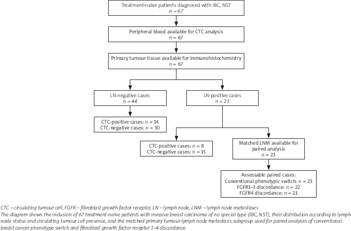
Table 1
Clinicopathological characteristics of the analysed cohort according to circulating tumour cell status
[i] BC – breast cancer, CTC – circulating tumour cell, DCIS – ductal carcinoma in situ, ER – oestrogen receptor, HER2 – human epidermal growth factor receptor 2, IHC – immunohistochemistry, IQR – interquartile range, LNM – lymph node metastases, PR – progesterone receptor
Comparison of clinicopathological features between circulating tumour cell (CTC)-negative and CTC-positive cases. Continuous variables are presented as median with interquartile range, and categorical variables as number of cases with percentages in brackets. P-values refer to comparisons between CTC-negative and CTC-positive groups. The Mann-Whitney U test was used for comparing continuous variables, and Fisher’s exact test for categorical variables.
Blood collection, circulating tumour cell enrichment, and imaging flow cytometry analysis
Peripheral blood samples (7.5 ml) were collected from a peripheral vein into EDTA tubes. To minimise potential contamination with epithelial and endothelial cells from the puncture site, the first 2–3 ml of blood were discarded. Samples were processed within 3 h of collection.
The peripheral blood mononuclear cell (PBMC) fraction was isolated by density gradient centrifugation, as described previously [11]. Briefly, blood was first centrifuged to separate platelet-rich plasma, then diluted with 1× phosphate-buffered saline (PBS), layered onto Histopaque®-1077 (Sigma-Aldrich, St. Louis, MO, USA), and centrifuged according to the manufacturer’s recommendations. The peripheral blood mononuclear cell fraction was collected, fixed in 4% formaldehyde, and stored at –80°C until further analysis.
Immunofluorescent staining of PBMC fractions containing putative CTCs was performed as described previously [11]. Briefly, thawed cells were washed with 1 ml of 1× PBS to remove formaldehyde and then incubated for 30 min at 4°C with an antibody cocktail diluted in 1× Perm/Wash Buffer (BD Biosciences, Franklin Lakes, NJ, USA). The panel included antibodies against pan-keratins (AE1/AE3 clone, AF488-conjugated, Thermo Fisher Scientific, Waltham, MA, USA, #53-9003-82; C11 clone, AF488-conjugated, Thermo Fisher Scientific, #MA5-18156) and vimentin (D21H3 clone, AF647-conjugated, Cell Signaling, Danvers, MA, USA, #9856) to identify epithelial and mesenchymal tumour cell features, as well as CD45 (REA747 clone, APC-Vio770-conjugated, Miltenyi Biotec, North Rhine-Westphalia, Germany, #130-110-635), CD31 (WM59 clone, APC-Cy7-conjugated, BioLegend, San Diego, CA, USA, #303120), αSMA and CD29 to exclude leukocytes, endothelial cells, fibroblasts and mesenchymal stromal cells, respectively. Antibodies were used at dilutions of 1 : 2500, 1 : 2500, 1 : 2500, 1 : 2500, 1 : 50, 1 : 50, and 1 : 10, respectively.
After incubation, cells were washed in 1× PBS, resuspended in 46 µL of 1× PBS, counterstained with 4′,6-diamidino-2-phenylindole (DAPI) (BD Biosciences; 1 µg/ml), and analysed immediately using the Amnis® ImageStream® X Mk II imaging flow cytometer (Cytek® Biosciences, Inc.). Before each measurement session, the instrument underwent automated internal calibration and quality control using the Amnis® ASSIST protocol to ensure consistent performance across acquisition runs. Peripheral blood mononuclear cell fractions were analysed under standardised conditions, as described previously [11]. Laser powers were set at 100 mW for pan-cytokeratin and DAPI, and at 150 mW for vimentin, CD45, and CD31. Image acquisition was performed at low flow speed using 40× magnification.
Fluorescence compensation was performed using UltraComp eBeads™ Plus compensation beads (Thermo Fisher Scientific, Waltham, MA, USA), stained individually with each antibody included in the panel. DAPI compensation was performed using cells, as recommended for DNA-binding dyes. In accordance with the ImageStream® X Mk II compensation workflow, single-colour controls were used to calculate fluorescence spillover, while instrument background and baseline performance were controlled through the Amnis® ASSIST quality control procedure. The compensation matrix was calculated in IDEAS™ 6.4 software (Cytek® Biosciences, Inc.), visually inspected, and verified before analysis.
DAPI-positive, and CD45/CD31-negative objects were identified, archived, and counted using the INSPIRE™ ImageStreamX MkII System software (Cytek® Biosciences, Inc.). Putative CTCs were defined as DAPI+/CD45–/CD31– cells with a diameter of at least 5 µm and were gated according to fluorescence intensity. Circulating tumour cell counts were normalised per 1 million PBMCs. Representative images illustrating different CTC phenotypes based on pan-keratins and vimentin presence are available in the CTC Atlas (www.ctcatlas.org).
Immunohistochemistry
Immunohistochemical (IHC) staining for FGFR1–4 was performed on 4-µm FFPE tumour sections using a standard protocol according to the manufacturers’ recommendations. The following primary antibodies were used: FGFR1 (NB100-2080, Novus Biologicals, Colorado, USA; pH 6.0, dilution 1 : 100), FGFR2 (H00002263-M01, Abnova, Taipei City, Taiwan; pH 6.0, dilution 1 : 600), FGFR3 (B-9, sc-13121, Santa Cruz Biotechnology; pH 6.0, dilution 1 : 50), and FGFR4 (M00769-2, Boster Bio, CA, USA; pH 6.0, dilution 1 : 500) (Figure 2). Omission of the primary antibody was used as a negative control.
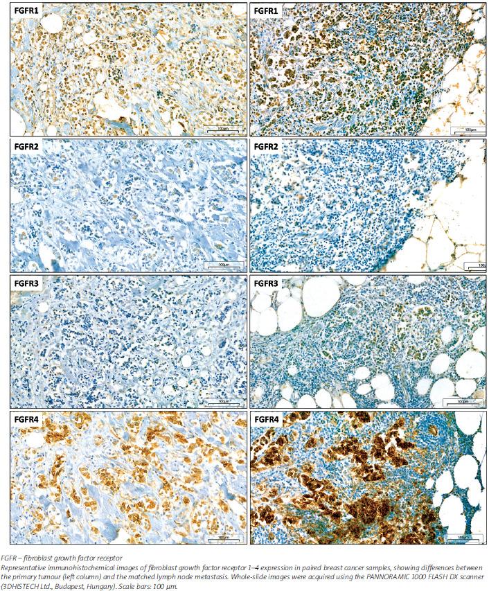
Quantitative evaluation of IHC was performed using the H-score system (range 0–300), calculated as the percentage of positively stained tumour cells (0–100) multiplied by staining intensity (0–3). In parallel, a semi- quantitative scoring approach analogous to the Allred scale (0–8) was applied. For dichotomous analyses, expression of each FGFR was classified as low or high according to terciles: cases within the first tercile of H-score were regarded as low, whereas cases within the second and third terciles were classified as high. Fibroblast growth factor receptor 1–4 protein expression was assessed independently by two evaluators, and discrepant cases were resolved by consultation with an expert reviewer, in line with previously published protocols.
Digitalisation of histological slides, imaging, IHC assessment, and figure preparation were performed using the PANNORAMIC 1000 FLASH DX, SlideCenter, and SlideViewer systems (3DHISTECH Ltd., Budapest, Hungary), as well as MedLAN Slide Viewer (MSV) v2.0b (MedLAN Sp. z o.o., Bialystok, Poland).
Definition of phenotypic switch and fibroblast growth factor receptor discordance
Immunohistochemistry-based BC subtype switch was defined as a change in the phenotype determined by routine IHC markers (ER, PR, HER2, and Ki-67) between the primary tumour and matched LNM. Fibroblast growth factor receptor discordance was assessed quantitatively using paired H-score values of the respective FGFR measured in the primary tumour and matched LNM, and qualitatively based on dichotomised FGFR status, with low-to-high or high-to-low conversion classified as FGFR discordance.
Statistical analysis
Continuous variables were presented as medians with interquartile ranges (IQR), while categorical variables were reported as numbers with corresponding percentages in brackets. The distribution of continuous variables was assessed using the Shapiro-Wilk test. Depending on data distribution and the number of groups compared, continuous variables were analysed using the Mann-Whitney U test, Kruskal-Wallis test, Student’s t-test, or analysis of variance, as appropriate. Comparisons between categorical variables were performed using Pearson’s χ2 test or Fisher’s exact test, depending on subgroup size. Correlations were analysed using Spearman’s rank correlation coefficient. Disease-free and overall survival data were summarised descriptively because of the very low number of events and were not used for formal outcome modelling. All statistical analyses were performed using STATISTICA version 13.1 (Dell Inc., Round Rock, TX, USA). A two-sided p-value < 0.05 was considered statistically significant.
Results
Study group characteristics and circulating tumour cell counts
At diagnosis, 23 of 67 patients with IBC, NST (34.3%) had regional LNM, but no patient had advanced disease; all cases were classified as stage I–IIIA according to the 8th edition of the American Joint Committee on Cancer Tumor-Node-Metastasis classification. Median age was 66.9 years; 10 patients (14.9%) were premenopausal and 57 (85.1%) were postmenopausal. Most tumours (39/67; 58.2%) were of intermediate grade. According to IHC-based markers, 31 cases (46.3%) were classified as luminal A, 28 (41.8%) as luminal B, 4 (6.0%) as HER2-enriched, and 4 (6.0%) as triple-negative BC (Table 1).
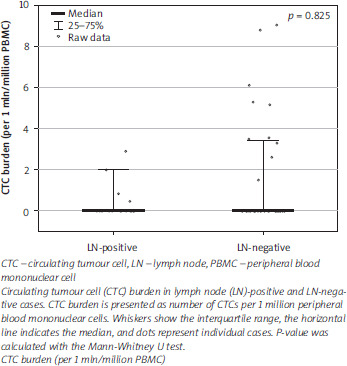
Circulating tumour cells were detected in 22/67 patients (32.8%). Median CTC count among positive cases was 5.7 CTCs per 1 million PBMC (IQR: 3.0–17.8). Circulating tumour cell presence (i.e. at least 1 detected CTC/1 million PBMC) was not associated with lymph node (LN) status and was observed in 8/23 LN-positive cases and 14/44 LN-negative cases (34.8% vs. 31.8%; p = 0.806). Circulating tumour cell counts did not differ between LN-positive and LN-negative cases [0.0 (0.0–1.4) vs. 0.0 (0.0–3.4) CTCs per 1 million PBMC; p = 0.825] (Figure 3). No significant differences were found between CTC-negative and CTC-positive patients with regard to age, tumour size, grade, ER, PR, HER2, Ki-67, IHC-defined BC subtype, or DCIS component (Table 1).
During follow-up, 6 disease-free survival events and 2 deaths were recorded in the whole cohort. Disease-free survival events were observed in 4/45 CTC-negative and 2/22 CTC-positive patients (8.9% vs. 9.1%; p = 1.000), whereas deaths were recorded in 2/45 and 0/22 patients, respectively (4.4% vs. 0.0%; p = 1.000).
Immunohistochemistry-based breast cancer subtype switch between primary tumours and matched lymph node metastases
Among the 23 LN-positive cases with matched material available for paired assessment, expression levels of individual biomarkers frequently differed between the primary tumour and the corresponding LNM (Table 2). A change in receptor profile was observed in 3/23 cases (13.0%), including one case with a shift from luminal B in the primary tumour to luminal A in the LNM, and two cases with changes from ER+PR+HER2– to ER+PR–HER2– and from ER+PR–HER2+ to ER+PR–HER2–, respectively.
Table 2
Marker discordance between primary tumours and lymph node metastases
[i] BC – breast cancer, ER – oestrogen receptor, FGFR – fibroblast growth factor receptor, HER2 – human epidermal growth factor receptor 2, IHC – immunohistochemistry, LN – lymph node, PR – progesterone receptor
Routine biomarker switch and fibroblast growth factor receptor (FGFR)1–4 status discordance between primary tumours and matched lymph node metastases (LNM). For routine biomarkers, switch/discordance refers to a change in marker or subtype category between the two sites. For FGFR1–4, discordance refers to conversion of dichotomised FGFR status between low and high expression categories. Direction of change is shown relative to the primary tumour, with “increased in LN” indicating higher expression or conversion to a higher expression category in the LNM, and “decreased in LN” indicating lower expression or conversion to a lower expression category in the LNM.
No associations between either IHC-based BC intrinsic subtype switch or discrepancies in expression of ER, PR, HER2, or Ki-67 assessed separately in the primary tumour and LNM, and CTC positivity were found (Table 3). Circulating tumour cells were detected in 1/3 switch cases and in 7/20 non-switch cases (p = 1.000).
Table 3
Circulating tumour cell positivity according to biomarker switch and fibroblast growth factor receptor discordance
[i] BC – breast cancer, CTC – circulating tumour cell, ER – oestrogen receptor, FGFR – fibroblast growth factor receptor, HER2 – human epidermal growth factor receptor 2, IHC – immunohistochemistry, PR – progesterone receptor
Comparison of the frequency of circulating tumour cell positive cases according to routine biomarkers status and discordance in fibroblast growth factor receptor (FGFR)1–4 expression between primary tumours and matched lymph node metastases (LNM). For each feature, the “no-switch/concordant” group includes cases without change between paired sites, whereas the “switch/discordant” group includes cases with change between the primary tumour and the matched LNM. P-values were calculated using Fisher’s exact test.
*The analysis of “Any FGFR discordance” was restricted to cases assessable for all four FGFR markers.
Fibroblast growth factor receptor expression in primary and metastatic tumours, and its association with the circulating tumour cell counts
Fibroblast growth factor receptor 1 and FGFR2 high expression levels were associated with good prognostic features. Expression of FGFR1 in PT correlated inversely with tumour size (Spearman’s ρ = –0.252; p = 0.040). In metastases, the level of FGFR1 expression was lower in premenopausal than in postmenopausal patients (125.0 vs. 205.0; p = 0.026). Fibroblast growth factor receptor 2 expression in the primary tumour was higher in: ER-positive than ER-negative tumours (median H-score 175.0 vs. 13.5; p = 0.011), PR-positive than PR-negative tumours (180.0 vs. 61.5; p = 0.022), and tumours with low than high Ki-67 (187.5 vs. 115.0; p = 0.018). Fibroblast growth factor receptor 2 expression correlated also with age at diagnosis (Spearman’s ρ = 0.259; p = 0.035).
Discordance in FGFR expression between PT and matched LNM was frequently observed. In the paired cohort, alterations in the status of any member of the FGFR family were observed in 18/23 cases (78.3%). Specifically, the discrepancies concerned: FGFR1 in 11/22 (50.0%), FGFR2 in 5/22 (22.7%), FGFR3 in 12/22 (54.5%), and FGFR4 in 5/21 (23.8%) of evaluated paired cases (Table 2). Among discordant cases, the direction of change most often reflected increased expression in LNM, observed in 8/11, 5/5, 9/12, and 4/5 cases for FGFR1, FGFR2, FGFR3, and FGFR4, respectively. Discordance of the FGFR status was associated with clinicopathological features, namely HER2 status, only for FGFR1 and was noted exclusively in HER2-negative tumours (11/11; p = 0.035).
Lower FGFR1 expression in LNM was observed in CTC-positive cases than in CTC-negative cases (median H-score 182.5 (157.5–201.2) vs. 212.5 (196.2–247.5); p = 0.018), whereas no significant difference was found in PT (167.5 (155.0–195.0) vs. 190.0 (172.5–245.0); p = 0.164). In the LN-positive subgroup, CTC burden was inversely correlated with FGFR1 expression in LNM (Spearman’s ρ = –0.487, p = 0.021), but not in PT (Spearman’s ρ = –0.304, p = 0.159), nor with the paired difference in FGFR1 expression between PT and LNM (Spearman’s ρ = –0.069, p = 0.762) (Table 4, Figure 4). No significant associations were observed between CTC positivity or CTC burden and FGFR2–4 expression in either PT or LNM.
Table 4
Correlations between circulating tumour cell counts and fibroblast growth factor receptor 1 expression in cases with lymph node metastases
| Parameters | Assessable cases | Spearman’s rho with CTC counts | p-value |
|---|---|---|---|
| FGFR1 in primary tumour | 23 | –0.304 | 0.159 |
| FGFR1 in LNM | 22 | –0.487 | 0.021 |
| FGFR1 change (LN-PT) | 22 | –0.069 | 0.762 |
[i] CTC – circulating tumour cell, FGFR – fibroblast growth factor receptor, LN – lymph node, LNM – lymph node metastases, PT – primary tumours
Spearman correlation analysis between circulating tumour cell (CTC) counts and fibroblast growth factor receptor 1 (FGFR1) expression in the primary tumour, matched lymph node metastasis, and the difference in FGFR1 expression between the two sites. CTC counts were normalised per 1 million peripheral blood mononuclear cells
Figure 4
Circulating tumour cell burden according to fibroblast growth factor receptor 1 concordance status
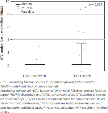
Fibroblast growth factor receptor 1, but not FGFR2–4, discordance, was significantly associated with CTC positivity. Circulating tumour cells were detected in 7/11 cases (63.6%) with FGFR1 discordance and in 1/11 cases (9.1%) with concordant FGFR1 status (p = 0.024) (Table 3). In the combined FGFR analysis restricted to cases with complete paired FGFR1–4 status data, CTCs were detected in 8/17 cases (47.1%) with at least one FGFR status conversion, whereas no CTCs were detected in fully FGFR- concordant cases (0/4); however, this association was not statistically significant (p = 0.131).
Discussion
The present study enabled assessment of phenotypic changes between primary breast tumours and matched LNM, with particular focus on FGFR1–4 expression as a non-classical dimension of inter-site tumour heterogeneity. Immunohistochemistry-based BC intrinsic subtype switch was infrequent, whereas FGFR discordance was common, suggesting that FGFR1–4 expression may capture phenotype changes not reflected by standard receptor reassessment. When these tissue-based findings were related to CTCs, neither CTC presence nor CTC abundance was associated with IHC-defined subtype switch. However, CTC presence was associated with FGFR1 discordance, and CTC burden correlated inversely with FGFR1 expression in LNM, suggesting a link between FGFR1-related heterogeneity and early blood-based dissemination.
The fibroblast growth factor receptor findings represent the main novel aspect of the study. Emerging evidence indicates that, beyond the well-established role of FGFRs in BC progression, tumour-microenvironment crosstalk and resistance to therapy, the FGF/FGFR axis may also contribute to tumour plasticity and dissemination [1, 12–15]. In paired primary tumour-LN analyses in BC, FGFR heterogeneity between these sites has been so far addressed only in lobular breast carcinoma. Given that CTC enumeration is widely recognised as a hallmark of tumour metastatic potential [16, 17], the frequent FGFR1 discordance observed in our paired samples, together with the association between CTC positivity and FGFR1 discordance, may suggest that FGFR-mediated inter-site heterogeneity represents a biologically relevant component of locoregional progression not captured by routine biomarker assessment. The inverse correlation between CTC burden and FGFR1 expression in LNM, and the restriction of FGFR1 discordance to HER2-negative tumours, support this interpretation, although these findings should still be regarded as exploratory and require validation in larger paired cohorts.
One of the more unexpected findings was the lack of association between CTC positivity and LN status. At first glance this may seem counterintuitive, because nodal involvement is classically regarded as a marker of dissemination. However, the relationship between CTCs and nodal status in early BC is not straightforward [5, 7, 18–20]. Hall et al. [21] or Serrano et al. [22] showed in nonmetastatic BC that preoperative CTC detection was not associated with LN status, tumour size, or grade, yet still showed adverse prognostic value. It shows that blood-based dissemination and locoregional spread to axillary LN are related, but not interchangeable, processes.
Another important finding was the low frequency of IHC-based BC subtype switch between PT and matched LNM. In our cohort, BC subtype switch was observed in only 3/23 paired cases, although isolated marker changes, particularly in Ki-67, were more frequent. This is lower than the discordance rates reported for individual routine biomarkers in many paired primary tumour-axillary metastasis series, but is more in line with recent observations that actual BC subtype conversion is less common than single-marker change [23–25]. The low rate of BC subtype conversion appears biologically plausible in a treatment- naïve cohort restricted to stage I–IIIA breast cancers.
This study has several strengths despite its still small number of included cases. It combines paired tissue analysis with blood-based CTC assessment, includes only treatment-naïve cases, and evaluates all four FGFR receptors rather than one selected marker. The limitations are also clear. The cohort was relatively small, the paired subgroup was limited, and the analyses were exploratory. The study was restricted to loco-regional progression and did not include distant metastases. Survival analysis was underpowered because of the very low number of events. Finally, FGFR assessment was based on immunohistochemistry, which is clinically accessible, but does not directly resolve the genomic or transcriptomic basis of the observed discordance.
Overall, the data suggest that in early BC, CTCs do not simply mirror LN status or routine IHC-based BC intrinsic subtype switch. Instead, they appear to identify tumours with FGFR-related inter-site heterogeneity, particularly involving FGFR1.
Conclusions
Fibroblast growth factor receptor discordance between primary breast tumours and matched LNM was frequent, whereas routine IHC-based BC subtype switch was uncommon. Circulating tumour cell positivity was associated with FGFR1 discordance, but not with LN status or BC subtype switch. These findings support combined tissue-based FGFR profiling and liquid biopsy as complementary approaches to capture biological heterogeneity during BC progression. Although exploratory, the association between CTCs and FGFR1 discordance suggests that FGFR1-related plasticity may contribute to early blood-based dissemination, particularly in HER2-negative breast cancer.










