
The prevalence of aneurysmal coronary artery disease in patients undergoing coronary angiography varies from 0.2% to 10%, with such a wide range primarily reflecting the different angiographic criteria used to define coronary artery aneurysm (CAA) [1]. CAA may infrequently grow large enough to be called “giant”, defined as ≥ 8 mm in diameter [2]. Several clinical and treatment questions on this matter remain unanswered together with the current natural history of this condition.
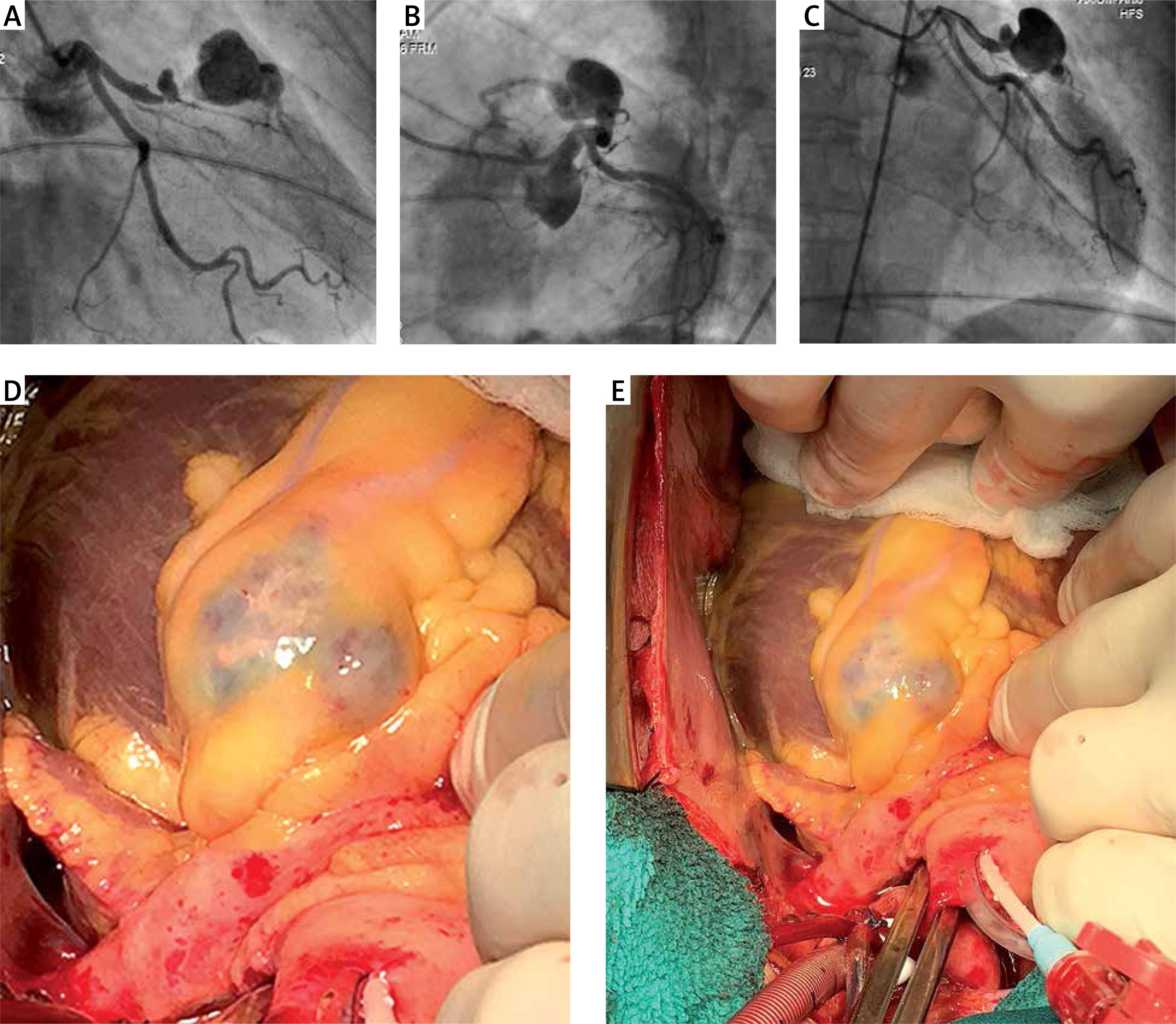
We present a case of a 63-year-old female patient with a history of coronary artery disease (CAD). The patient was admitted to the cardiac surgery clinic for elective coronary artery bypass grafting. Three years prior to admission, the patient sustained anterior wall ST-elevation myocardial infarction that was treated with fibrinolytic therapy in a regional centre. At that time, the patient was not submitted to coronary angiography. Following the last admission due to progressive chest pain, the patient underwent coronarography which demonstrated a sub-occlusive lesion on the left anterior descending (LAD) artery with a huge mid-level LAD aneurysm (Figure 1 A). Echocardiography revealed moderately reduced left ventricular ejection fraction of 45% and otherwise normal findings. The patient was admitted for surgery. The LAD aneurysm was easily identified, measuring 35 mm in diameter (Figure 1 B). Total exclusion of the aneurysm was performed with clipping the artery proximally and distally in relation to the LAD aneurysm. On-pump left internal mammary artery to LAD anastomosis was subsequently performed. The patient was discharged in a good condition on the 7th postoperative day.
Figure 1
A–C – Coronarography demonstrating large LAD mid-level aneurysm with proximal LAD sub-occlusive lesion. D, E – intraoperative finding of giant LAD aneurysm
The pathogenesis of CAA is not clearly understood, but there is compelling evidence of associations between certain risk factors and CAAs: genetic susceptibility, CAD, certain vasculitis and connective tissue diseases, and local wall injury following intracoronary manipulation [3]. CAD was most likely the underlying cause of CAA development in our patient.
Most CAAs are incidentally detected during coronary angiography or computed tomography. The majority of cases remain clinically silent, although a variety of clinical scenarios may develop: 1 – effort angina and acute coronary syndrome due to concomitant coronary artery disease, 2 – distal embolization, 3 – compression of local structures in cases of giant CAAs, 4 – aneurysm rupture, 5 – stress-induced ischemia due to microvascular dysfunction [3].
The treatment of CAAs remains speculative in the absence of clinical trials. Medical therapy generally consists of attempts to prevent thromboembolic complications and mainly relies on using antiplatelet medications and/or anticoagulants. For patients with coexisting obstructive lesions and symptoms or signs of significant ischaemia, percutaneous and/or surgical coronary revascularisation can safely and effectively restore normal myocardial perfusion. Percutaneous coronary intervention of an aneurysmal vessel has been consistently associated with lower procedural success and a higher rate of complications, especially in the setting of acute coronary syndrome. Surgical tactics include aneurysm ligation with distal bypass grafting, isolated coronary artery bypass grafting, aneurysm plication and saphenous vein patch repair of the aneurysm [4]. Surgical revascularization is the preferred approach for addressing giant coronary aneurysms [5]. Lastly, it is important to acknowledge that the treatment for each patient is individual and based on the location of the aneurysm and clinical context.


