Atherosclerotic carotid artery stenosis remains a significant cause of ischaemic stroke, accounting for approximately 20–25% of all cerebrovascular events [1]. Medical treatment constitutes important first-line management – but evidence is clear today that even maximised pharmacotherapy, paired with coached lifestyle improvement, is not sufficiently effective in prevention of carotid-related strokes [2–4]. Carotid-related strokes are often large and disabling, carrying profound consequences to stroke-affected individuals and their families [1, 3, 4].
The decision to pursue revascularisation – using the endovascular route (carotid artery stenting, CAS) or open surgery (carotid endarterectomy, CEA) – requires careful patient evaluation, weighing statistical risk and benefits and the patient individual factors that may modify the statistical risk and benefits [1, 5]. The patient, placed in the very centre of the decision-making process regarding their care, needs to be fully informed about the treatment options and the patient preferences need to be respected [1]. The hospital and physician expertise in delivering competent revascularisation needs to be considered when discussing with the patient the locally available treatment options or external referrals [1]. The Neuro-Vascular Team (“Carotid Team”) concept, mirroring the multi-disciplinary Heart Team, embraces diverse specialties, tailors personalised, stratified approaches to individual patient needs, and integrates innovative imaging and risk-assessment biomarkers [1]. Involving the patient and, with consent, their carers or family in the decision-making process may also help with long-term adherence to medical therapy that plays an important part in the long-term efficacy of the intervention [1].
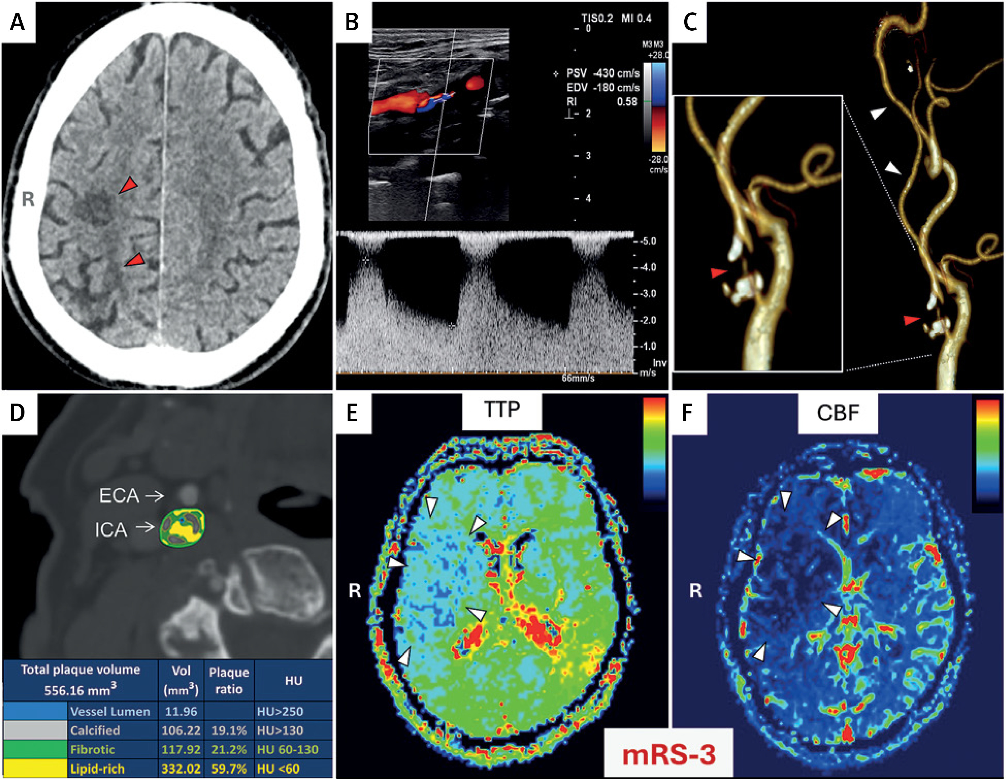
Herein, we illustrate image-guided decision-making in contemporary carotid revascularisation by sharing the diagnostic and management process in a 71-year-old woman with right internal carotid artery stenosis evaluated for carotid revascularisation on top of maximised medical treatment versus maximised medical treatment alone. Eleven months previously, the patient had sustained an ischaemic right (dominant) haemispheric stroke, with residual left-sided pyramidal hemiparesis. Modified Rankin Scale (mRS) score was 3 upon referral to our attention for consideration of potential revascularisation. Medical history included arterial hypertension, heart failure with preserved ejection fraction, hypercholesterolemia, obesity, and psoriasis. According to the current definition [1, 2], this previously symptomatic patient, at the point of referral to our institution, should be labelled “asymptomatic” because of a period of > 6 months from the point of ipsilateral retinal or cerebral ischaemic symptoms attributable to the carotid artery stenosis [1, 2, 5]. However, different cutoffs (such as > 1 month, > 3 months, > 12 months) or no time-based cutoff (i.e., patient with a history of carotid stenosis symptoms considered “symptomatic”) have also been used [6]. Routine non-contrast computed tomography of the brain demonstrated a hypodense region in the right (dominant) hemisphere, consistent with a peracted cerebral infarct (Figure 1 A). A relatively extensive area of porencephaly and gliosis was identified, consistent with sequelae of prior ischaemic injury in the right fronto-parietal border zone and right insular region. Routine duplex ultrasound performed in our institution demonstrated a morphologically mixed, predominantly soft, critical stenosis of the right internal carotid artery with very high flow velocities (peak systolic 430 cm/s; peak diastolic 180 cm/s; Figure 1 B). Computed tomography angiography showed a collapsed, nearly-occluded right internal carotid artery with extraluminal calcific content in the proximal and distal portion of the stenotic segment (Figure 1 C).
Figure 1
Pre-procedural, routine (top) – and additional (bottom) – imaging to guide clinical decision-making. A – Routine non-contrast computed tomography of the brain demonstrating a hypodense region in the right (dominant) hemisphere (R), consistent with a peracted cerebral infarct (red arrowheads). B – Routine duplex ultrasonography of the right internal carotid artery showing a critical stenosis with very high flow velocities (peak systolic 430 cm/s; peak diastolic 180 cm/s; near-occlusion with high-velocity flow). C – Three-dimensional computed tomography reconstruction of the right carotid arteries, demonstrating a collapsed (white arrowheads), nearly-occluded right internal carotid artery with extraluminal calcific content in the proximal and distal portion of the stenotic segment. The inset on the right is a detailed image of the stenotic segment with a critical lumen reduction and a focal lack of lumen continuity as per computed tomography resolution (near-occlusion; red arrowhead). D – Colour-coded computer angiography imaging of the stenotic lesion, demonstrating a large-volume (550.16 mm3), predominantly non-calcified (80.9%) atherosclerotic plaque with a dominating lipid-rich content (59.7%). Disseminated conglomerates of calcium constitute 19.1% of plaque volume. ECA – external carotid artery, ICA – internal carotid artery. Hounsfield units (HU)–based colour code of the plaque key components is provided at the bottom of the image. This plaque morphology is consistent with (1) and increased risk of (in this patient – recurrent) stroke, and (2) an increased risk of cerebral embolism with classic endovascular management. Contemporary level 1 (randomised controlled clinical trial) evidence indicates, for this patient, at least 50% reduction in ipsilateral stroke risk over 4 years (with average procedural risk of ~1.3% accounted for) with endovascular revascularisation (carotid artery stenting) added to maximized medical management; surgical management with carotid endarterectomy provides no significant clinical benefit [2]. Subcritical internal carotid artery stenosis resulting in vessel collapse as a result of substantially reduced flow (white arrowheads in C), coexisting with the ipsilateral cerebral infarcted tissue (red arrowheads in A) constitute important risk factors for cerebral bleed in the early period after carotid revascularisation that is fatal in ~20–40% cases [11]; the risk of haemorrhage is estimated at 0.47% with CAS and up to 1.3% with CEA [11–13]. There is no evidence for any increased cerebral haemorrhage risk with single or double antiplatelet treatment in CAS [15]. E, F – Magnetic resonance cerebral perfusion imaging colour-coded maps of time-to-peak perfusion (TTP, E) and cerebral blood flow (CBF, F). White arrowheads indicate a large area of reduced cerebral perfusion in the right hemisphere (R), extending beyond the infarcted region (cf., A) and involving the frontal, parietal, and temporal lobes. The patient lost functional independence as a result of the prior stroke (modified Rankin Scale, mRS, 3), that had been feasible for emergency revascularisation therapy but the patient had not been referred for the stroke interventional management [3, 4, 29])
The two primary concerns in this patient are (1) risk of a further stroke (that would likely lead to further neurologic deterioration) that had to be balanced against (2) the risks of intervention [1, 5]. The patient, unreferred for emergency endovascular treatment at the time of acute stroke (which should have been the primary management strategy) [3] had been subsequently turned down for revascularization, due to a perceived high risk of intervention, by two centres offering both CEA and CAS but the patient and family were severely concerned about the further stroke risk. In absence of intervention, the annual ipsilateral ischaemic stroke risk is estimated at least 2% [1], translating into > 30% risk of ipsilateral ischaemic stroke during the remaining average statistical lifespan of 15 years in this patient (stat.gov.pl). The functional benefit of potentially restoring normal perfusion to the dominant hemisphere has not been quantified precisely (and the revascularisation procedure may add to the ischaemic cerebral injury [1, 6, 7]) but it is likely [1, 8].
The patient met inclusion criteria of the recent CREST-2 study [2, 9] that provided appropriately powered, level-1 evidence to guide management [10]. In patients with “asymptomatic” carotid stenoses of 50–99% (including those with a history of previous stroke or TIA attributable to target lesion > 6 months before randomisation), CAS, but not CEA, was found to be superior to medical management and lifestyle modification alone [10]. Over 4 years of follow-up, CAS reduced the risk of ipsilateral ischaemic stroke by > 50% taking into account the risk of intervention [2, 9]. However, in this patient (near-occlusion, collapsed artery, ipsilateral infarcted cerebral tissue, Figures 1 A and C), the risk of cerebral haemorrhage as a complication of hyperperfusion syndrome with carotid revascularisation required careful consideration. Intracerebral haemorrhage in relation to carotid revascularisation carries a 37% to 80% mortality rate and a 20% to 37% risk of poor recovery in survivors [11]. Overview of 86 cases of intracerebral haemorrhage post carotid revascularisation indicated that critical (> 90%) ipsilateral ICA stenosis and previous ipsilateral cerebral infarction (ischaemic stroke) are central to the risk of intracerebral bleed (with ~70% mortality) while periprocedural antithrombotics were not not a risk factor [12]. Studies indicated the incidence of intracerebral haemorrhage associated with hyperperfusion syndrome following CAS of 0.67% whereas post-CEA cerebral bleed occurred in up to 1.2% [11, 13]. Recent meta-analytic evidence confirmed a higher risk for cerebral hyperperfusion syndrome with CEA compared to CAS (pooled odds ratio = 1.432 [95% confidence interval 1.078–1.901]; p = 0.015) [13]. Impaired cerebral perfusion volume ratio and large artery stroke history are significantly associated with a higher risk of cerebral hyperperfusion (p = 0.012 and p = 0.028, respectively) [14]. Notably, in patients undergoing CAS dual antiplatelet therapy is associated with a reduced risk of ischaemic and haemorrhagic complications compared with monotherapy without an increase in bleeding complications [15].
Following discussion with the patient and her daughters, analysis of the plaque morphology and further cerebral imaging were performed to obtain further information to guide decision-making, with the idea that lack of any significant hypoperfusion of the ipsilateral hemisphere would indicate not proceeding to elective revascularisation. Medical management was reviewed; anti-hypertensive therapy was modified to include ramipril for its clinically-relevant anti-atherosclerotic effect, and the statin dose was increased to achieve guideline-indicated goal of the low-density cholesterol level [1].
A recent meta-analysis of 64 prospective studies in 20,751 participants with asymptomatic carotid stenosis demonstrated a pooled prevalence of high-risk plaques of 26.5% [16]. Lipid-rich core constituted, along with neovascularisation and echolucency, one of the top three high-risk plaque features [16]. The incidence of ipsilateral ischaemic cerebrovascular events in severe stenoses was 3.7 events per 100 person-years [16]. Plaque morphology played an even greater role than stenosis severity, with the incidence of ipsilateral ischaemic cerebrovascular events higher in patients with high-risk plaques (4.3 events per 100 person-years) than in those without (1.2 events per 100 person-years); odds ratio of 3.0 (95% CI: 2.1–4.3) [16]. Overall evidence indicates an important role of plaque morphology in the primary and secondary risk of carotid-related stroke [1, 17]. Hounsfield Unit–determined fibrotic, lipid-rich, and calcific content of the stenotic lesion was also determined (Figure 1 D). This showed a mixed, predominantly lipid-rich content (59.7% of the plaque volume) with diffuse calcium conglomerates accounting for nearly 20% of the plaque volume, and a very severe lumen compromise (Figure 1 D). Typical cutoffs are < 60 HU for lipidic tissue, 60–130 HU for fibrous tissue, and > 130 HU for calcium tissue [18]. Recent data suggest that intra-plaque haemorrhage (a high-risk feature typically evaluated on plaque imaging using magnetic resonance [1, 17, 19]) can be further distinguished on computed tomography within the lipidic tissue by using the threshold of < 25 HU [18]. According to a proposed plaque classification, the plaque (Figure 1 D) exhibited Type 5 calcium configuration (mixed bulky calcifications); a phenotype with a ~2-fold greater prevalence in symptomatic lesions than absence of calcifications (Type 1 plaque) [20].
Cerebral perfusion is emerging as an important parameter of carotid revascularisation, in particular in secondary stroke prevention and it is presently an area of intensive research [8, 14, 21]. Magnetic resonance cerebral perfusion imaging indicated a large area of reduced cerebral perfusion in the right hemisphere, extending beyond the infarcted region and involving the frontal, parietal, and temporal lobes (colour-coded maps of time-to-peak perfusion and cerebral blood flow are presented in Figures 1 E, F), shifting – together with the high-risk plaque morphology – the pre-procedural risk/benefit ratio towards the intervention [5].
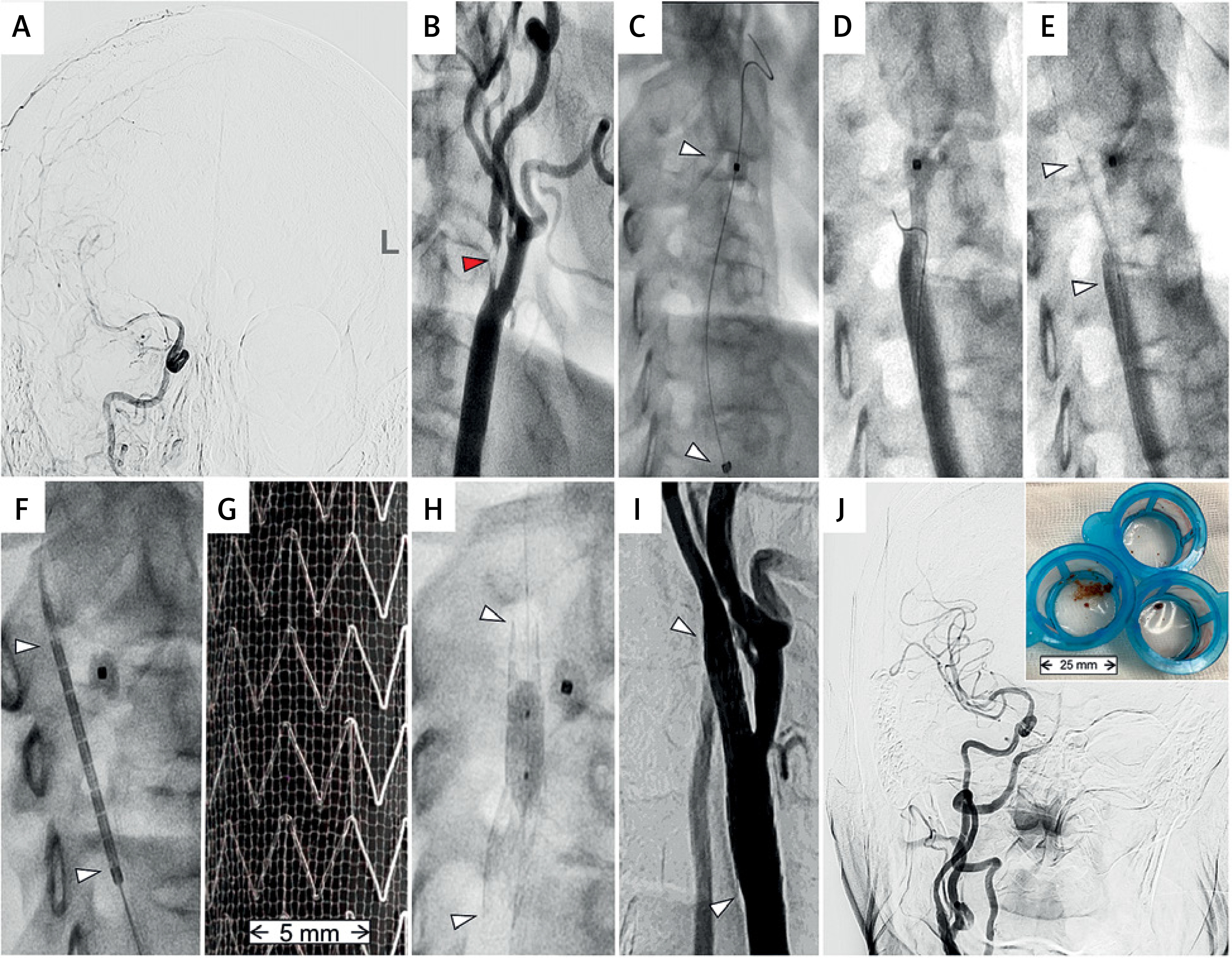
Uneventful CAS with an optimal effect of complete endovascular reconstruction was performed using transfemoral access, proximal cerebral protection [22, 23] with a dual-ballon system [24] and a second-generation MicroNet-covered anti-embolic stent [7, 25–30] (Figure 2).
Figure 2
Procedural images of proximal-protected endovascular reconstruction of carotid bifurcation, using a MicroNet-covered anti-embolic stent. A – Baseline cerebral angiography demonstrating a markedly reduced supply from the right internal carotid artery to the right hemispheric vessels. B – Critical, thrombus-containing near-occlusion (red arrowhead; part of the flow channel is totally invisible) of the right internal carotid artery with antegrade filling of the distal (collapsed) segment. C – Mo.Ma dual-balloon proximal cerebral protection device (catheter) in its operational position, inserted into the target carotid bifurcation over a 0.018” wire. Note the already inflated occlusive balloon in the external carotid artery (white arrowhead, top) and the common carotid artery balloon marker (white arrowhead, bottom). D – Crossing the thrombus-containing subocclusion with a coronary wire (WhisperMS 0.014”) under transient flow reversal (“back” pressure 52/41 mm Hg). Note the contrast slow washout from the internal to the common carotid artery (any debris, via an open stopcock of the catheter, are being evacuated from the patient vasculature). E – Initial predilatation with a 2.0 × 20 mm coronary balloon (white arrowheads) was followed by routine lesion preparation with a 4.0 × 20 mm balloon (not shown). F – Positioning of a MicroNet-covered self-expanding neuroprotective stent (CGuard 10 × 30 mm) using the geographic landmarks of the ECA balloon and the vertebral column. G – Magnified photograph of the MicroNet-covered second-generation dual-layer carotid stent. The atherothrombotic plaque material-sequestrating, neuroprotective MicroNet can be seen outside the open-cell laser-cut nitinol struts [25]. This combination of the presently most open-cell (nitinol frame) and presently most closed-cell (MicroNet layer) carotid stent design demonstrated best-ever clinical outcomes across the United States Food and Drug Administration rigorously controlled carotid trials [5, 45]. H – Post-dilatation optimisation of the neuroprotective stent: sequential post-dilatation with a 4.5 × 15 mm non-compliant balloon (not shown) was followed by a focal post-dilatation a 5.0 × 8 mm non-compliant balloon at 20 atm (white arrowheads indicate stent edges). Finally, the proximal cerebral protection balloons are deflated under active aspiration and antegrade flow is restored. Note that, in most cases, intraprocedural occlusion of the external carotid artery is required to achieve an effective proximal protection (otherwise the internal carotid artery can be supplied from the external carotid artery that conis well-collateralised, resulting in antegrade flow to the brain and thus absence of cerebral protection [24]. I – Final angiographic result of the carotid bifurcation reconstruction with the MicroNet-covered stent. The stent (white arrows denote edges) SmartFit adjustment to the internal and common carotid artery diameters [25, 26] enables achieving a ‘complete endovascular reconstruction’. Note that the internal carotid artery distal to the stent is still partly collapsed; with flow-mediated dilatation the vessel diameter will be gradually further restored. J – Final cerebral angiogram demonstrating a post-procedural near-normalisation of cerebral supply from the stent-reconstructed carotid artery. The Mo.Ma proximal cerebral protection device system baskets (top-right inset) contain the organised thrombi and plaque material evacuated during this revascularization procedure. Note that – in case of absence of cerebral protection – this material would have significantly embolised the right hemispheric cerebral arteries. Evidence shows an additive cerebral neuroprotective effect when the intra-procedural cerebral protection by transient flow reversal is paired with the permanent neuroprotective effect of the MicroNet (permanent lesion insulation) [7, 30]. The arterial access site (right common femoral artery) was sealed with AngioSeal 8F vascular plug, and the patient was discharged home on the following day on double anti-platelet therapy for 3 months (followed by single anti-platelet therapy) and guideline-directed anti-hypertensive and maximised anti-hyperlipidemic therapy
Conventional carotid artery stenting using first-generation (single-layer) stents demonstrates, according to a recent analysis from US Vascular Quality Initiative, a statistically significant relationship between the lesion calcium volume percent and percent residual stenosis (odds ratio 1.324; 95% confidence interval, 1.005–1.746; p = 0.046), with plaques with calcium volume of > 18.7% (19.1% in our patient, Figure 1 D) showing a 2-fold increase in the risk of increased residual stenosis (odds ratio = 2.532; 95% confidence interval, 1.049–6.115; p = 0.039) [31]. Kaplan-Meier analysis in that study demonstrated a statistically significant increase in the rate of in-stent restenosis during a 36-month follow-up with a cutoff of > 8.2% of lesion calcium volume (p = 0.0069) [31], pointing to the need to optimise the carotid stent residual stenosis/residual stenosis after carotid stenting. Virtual histology carotid plaque imaging [32] revealed increased dense calcium volume in patients with ipsilateral cerebral embolism detected with diffusion-weighted magnetic resonance imaging that has a growing role in evaluating the safety of carotid revascularisation (33.2 vs. 24.5 mm3 vs. 11.4 vs. 6.1 mm3; p = 0.08) [7, 28, 32–34].
In a recent study of risk factors for cerebral embolism with conventional carotid percutaneous revascularisation, including 107 patients undergoing distal filter-protected single-layer closed-cell stent CAS, lesion calcification was found to be an independent risk factor for perioperative cerebral embolisation (OR = 5.68; 95% CI: 1.12–28.79; p = 0.04) [35]. This highlights the need to optimise cerebral protection in endovascular management of calcific lesions. Today, evidence is increasing that proximal cerebral protection and the anti-embolic MicroNet-covered stent (Figure 2) act synergistically to maximise cerebral protection in CAS [30] because the anti-embolic MicroNet stent layer exerts its permanent protective action only during and after stent implantation, whereas the embolic risk occurs already with the lesion wiring, predilatation, and stent insertion [22, 23, 36–39].
A significant proportion of patients presenting for consideration of carotid revascularisation have coronary artery disease [1], with myocardial infarction as a complication of carotid revascularisation and a significant proportion of patients dying of coronary disease in follow-up [40]. Because we routinely perform coronary angiography (except when done within prior 6–12 months or clinically contraindicated) in patients undergoing CAS to identify any coexisting severe coronary artery stenosis that may indicate changing the order of intervention (e.g., critical left main coronary artery disease, major proximal flow-limiting stenosis) or – when carotid intervention should not be delayed – a simultaneous percutaneous coronary intervention and CAS [41], we chose, following discussion with the patient, the transfemoral (less-invasive) rather transcarotid access for carotid revascularisation employing flow reversal cerebral protection and anti-embolic stent [42].
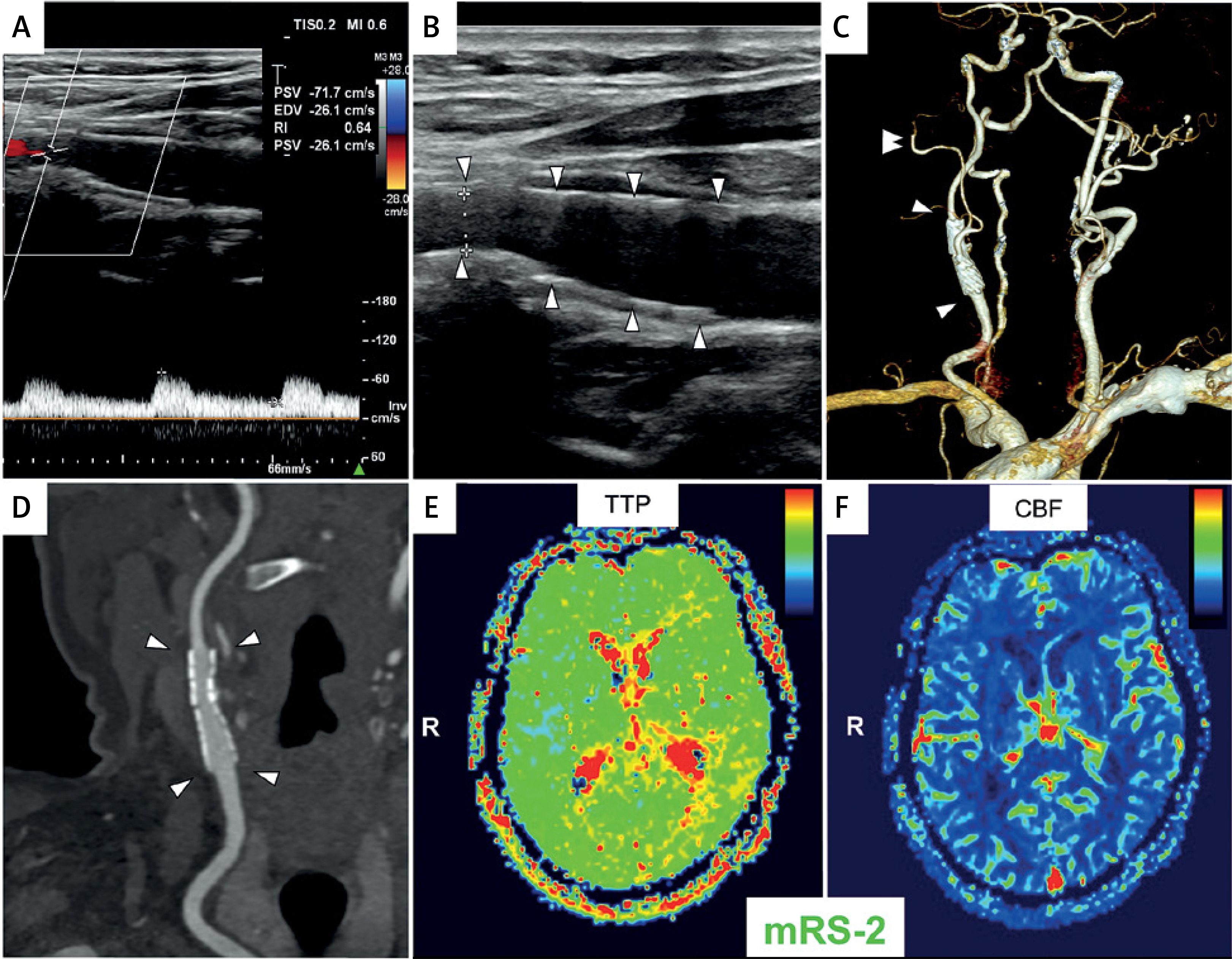
Post-procedural follow-up has been uneventful except for a mild headache lasting 2–3 h after the procedure and responsive to paracetamol. There was rigorous control of post-procedural arterial blood pressure to ensure its systolic values below 100-110 mm Hg. Discharge duplex ultrasonography confirmed optimal stent expansion and apposition in absence of any in-stent material, and normal in-stent velocities that remained normal at 12 months (Figure 3). Computed tomography angiography demonstrated the effect of a complete anatomic reconstruction of the target carotid artery bifurcation normal anatomy (Figures 3 C, D). Magnetic resonance cerebral perfusion imaging at 12 months showed a fully restored normal (symmetrical) perfusion of the right and left cerebral hemisphere, including the previously underperfused areas in the right frontal, parietal, and temporal lobes. The patient neurologic deficit severity decreased to the modified Rankin Scale (mRS) of 2 – functional independence. The patient is part of an all-comer, unselected consecutive-patient study of endovascular revascularisation in symptomatic and asymptomatic atherosclerotic carotid stenosis in primary and secondary stroke prevention using MicroNet-covered anti-embolic stents (PARADIGM-EXTEND; NCT04271033).
Figure 3
Post-procedural and follow-up imaging. A and B are routine discharge duplex ultrasonography images of the stent that illustrate its optimal expansion and apposition (minimal stent diameter of 4.58 mm in the proximal right internal carotid artery) in absence of any in-stent material, and normal in-stent velocities (peak systolic 71.7 cm/s; peak diastolic 26.1 cm/s). Note that the in-stent velocities remained normal at 12 months (peak systolic 72.4 cm/s; peak diastolic 31.2 cm/s) in continued absence of any in-stent material (effective lumen reconstruction). C – Three-dimensional imaging (computed tomography angiography, volume render technique) of the aortic arch arteries demonstrating the effect of a complete reconstruction of the target carotid artery bifurcation normal anatomy with the MicroNet-covered stent (white arrows denote stent edges; double arrowhead shows a fully patent right external carotid artery) on follow-up at 3 months. D – Computed tomography angiography planar image (curved planar reformation) demonstrating an optimal lumen gain in the carotid bifurcation fully revascularised using the endovascular approach. White arrowheads denote edges of the CGuard stent implant in this patient. Note a fully restored arterial lumen patency in absence of any instent material. The residual plaque material (cf., Figures 1 C, D and Figure 2 J inset) is not visible; it is fully (and permanently) isolated from the reconstructed lumen by the (now healed) MicroNet-covered stent implant [36]. E, F – Magnetic resonance cerebral perfusion imaging colour-coded maps of time-to-peak perfusion (TTP, E) and cerebral blood flow (CBF, F) at 12 months post-procedure, demonstrating a fully restored normal (symmetrical) perfusion of the right and left cerebral hemisphere, including the previously underperfused areas in the right frontal, parietal, and temporal lobes (cf., Figures 1 E, F). The patient’s neurologic deficit severity decreased to the modified Rankin Scale (mRS) of 2; i.e. functional independence.
Minimally invasive treatment, in correctly identified and risk-stratified patients, carrying a long-term efficacy and stroke prevention durability, is one of primary goals of today’s carotid revascularisation performed on top of maximised medical management to reduce stroke, including recurrent stroke, risk [43–45]. In this short communication we have demonstrated the role of contemporary clinical evidence and cerebrovascular imaging in guiding clinical and procedural decision-making in atherosclerotic carotid artery stenosis. The diagnostic steps and decision-making process, along with selection and justification of the revascularisation strategy were discussed at the carotid session of 2025 Paris Vascular Insights Course.