A 74-year-old man with multiple comorbidities, including severe calcific aortic stenosis, chronic kidney disease, chronic obstructive pulmonary disease, and permanent atrial fibrillation, was admitted with non-ST-elevation myocardial infarction, NYHA class III heart failure symptoms, and recurrent syncope. On admission, the patient was hemodynamically stable despite advanced heart failure, with systolic blood pressure ranging from 95 to 116 mm Hg and without the requirement for vasopressor or inotropic support.
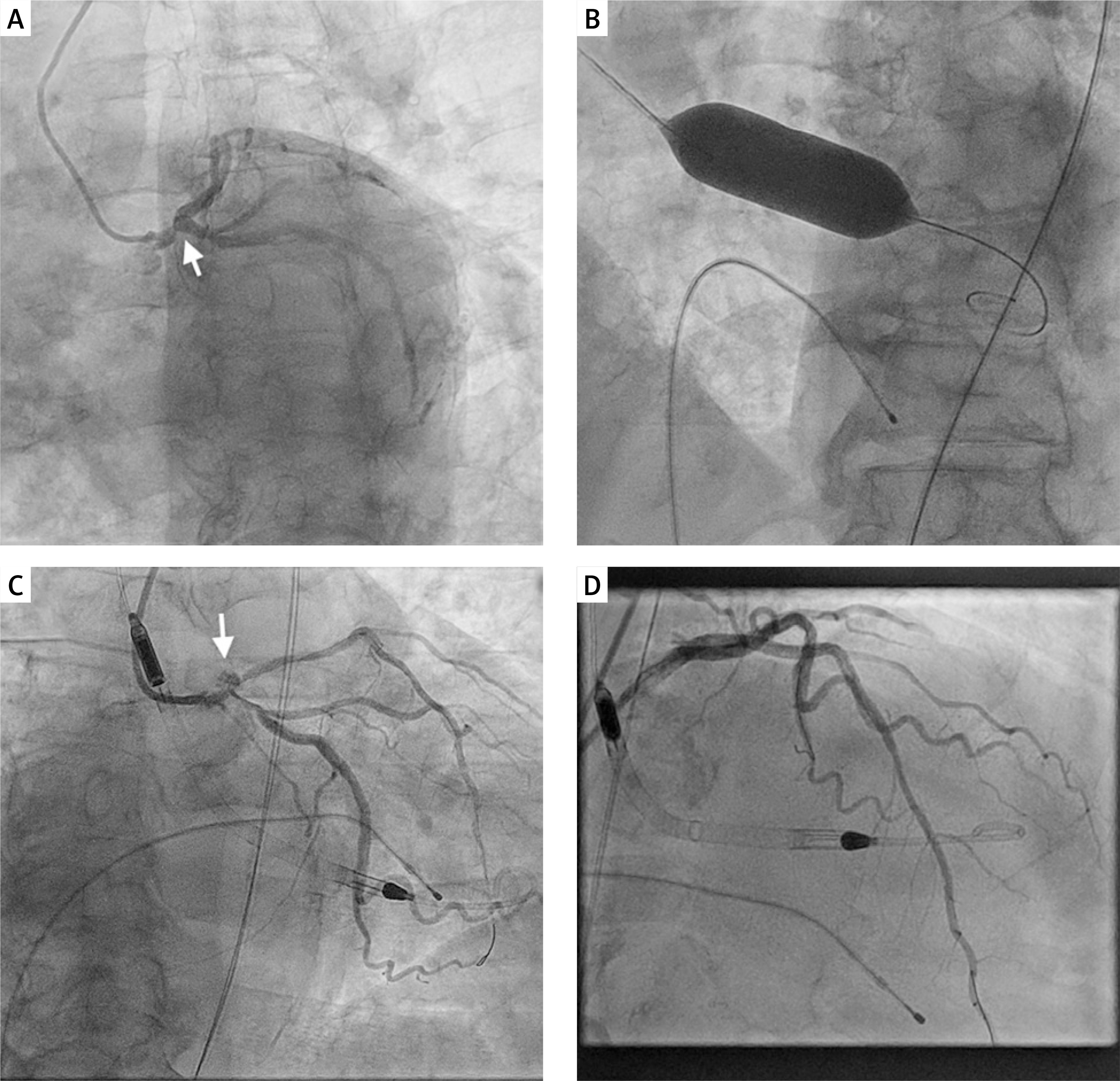
Transthoracic echocardiography demonstrated severe aortic stenosis (Vmax 4.23 m/s, mean gradient 50 mm Hg, aortic valve area 0.5 cm²) and severely impaired left ventricular systolic function (LVEF 25%). Urgent coronary angiography revealed heavily calcified multivessel disease, including approximately 80% stenosis of the distal left main (LM) bifurcation with a calcified nodule, diffuse disease of the left anterior descending artery (LAD), and significant (~80%) mid-right coronary artery (RCA) stenosis (Figure 1 A) (SYNTAX score 34) [1].
Figure 1
Impella-supported bailout strategy in cardiogenic shock with severe aortic stenosis and complex coronary artery disease. A – Coronary angiography demonstrating heavily calcified distal left main bifurcation stenosis involving the LAD and LCx (arrow). B – Balloon aortic valvuloplasty performed via left femoral access using a 20 mm Valver balloon. C – Coronary perforation (arrow) at the left main bifurcation eventually leading to cardiac tamponade requiring urgent pericardiocentesis. D – Complex left main PCI with multiple stents implanted using a culotte technique under Impella support
Given the prohibitive surgical risk (EuroSCORE II 21.4%, STS 19.2%), severe aortic stenosis, advanced left ventricular dysfunction, and complex calcified left main disease, the Heart Team elected to pursue a staged percutaneous strategy. Balloon aortic valvuloplasty was performed as a bridge-to-stabilization approach to improve forward flow and facilitate Impella-supported high-risk percutaneous coronary intervention (PCI), while definitive transcatheter aortic valve implantation (TAVI) was deferred until clinical stabilization [2].
A stepwise approach was adopted. Balloon aortic valvuloplasty (BAV) via left femoral access (14 Fr) was performed using a 20 mm Valver balloon (Balton, Poland), resulting in a reduction of the mean transaortic gradient from 50 mm Hg to 25 mm Hg and transient hemodynamic improvement (Figure 1 B). Mechanical circulatory support with an Impella CP SmartAssist device (Abiomed) was then established via the same access [3].
High-risk PCI of the LM bifurcation (LM/LAD/LCx) was performed via right radial access using a 7 Fr EBU 3.5 guiding catheter. IVUS was used both for lesion characterization and procedural optimization. It demonstrated extensive calcification involving the LM, LAD, and proximal LCx, while post-PCI imaging confirmed satisfactory stent expansion and apposition. Lesion preparation included two runs of orbital atherectomy over a ViperWire, complicated by transient crown entrapment and a small coronary perforation in the distal LM bifurcation region (Figure 1 C). Repeated angiographic reassessment demonstrated only limited contrast staining without persistent contrast extravasation, while serial intraprocedural transthoracic echocardiography showed no significant pericardial effusion. Given the bifurcation location of the perforation, implantation of a covered stent was considered but deferred because of the risk of side-branch compromise and an unfavorable final bifurcation result.
Additional plaque modification with intravascular lithotripsy and cutting balloons was unsuccessful due to repeated balloon rupture. Adequate preparation was achieved using 3.5 and 4.0 mm non-compliant balloons.
Three Promus Elite drug-eluting stents (2.75 × 20 mm, 3.0 × 32 mm, 3.5 × 28 mm) were implanted in the LAD, followed by culotte stenting with Xience PROs stents (3.5 × 33 mm LM–LCx and 4.0 × 23 mm LM–LAD). Final kissing balloon inflation (3.5 mm) and proximal optimization (4.5 mm) were performed (Figure 1 D).
PCI of the RCA was performed via right femoral access (7 Fr). After orbital atherectomy over a ViperWire, the lesion was predilated (3.5 × 15 mm NC balloon). Two Xience PROs stents (3.5 × 38 mm and 3.5 × 28 mm) were implanted, followed by postdilatation (4.0 mm NC balloon). IVUS confirmed adequate expansion and TIMI 3 flow. Repeated intraprocedural transthoracic echocardiography showed no significant pericardial effusion, and repeated angiographic reassessment demonstrated no persistent contrast extravasation. Procedural anticoagulation included unfractionated heparin, targeting ACT values > 250 s during device insertion and the procedure, followed by maintenance ACT values of approximately 160–180 s, and dual antiplatelet therapy was initiated. Following a period of hemodynamic stabilization, the Impella device was removed in the catheterization laboratory. At procedure completion, vascular closure was achieved using a MANTA closure device at the Impella access site and an AngioSeal device at the right femoral access site.
Complete revascularization during the index procedure was chosen to avoid a second high-risk intervention requiring repeat mechanical circulatory support and vascular access.
Shortly after the procedure, the patient developed hemodynamic deterioration with cardiogenic shock, hypotension despite vasopressor support, and escalating catecholamine requirements. Emergency echocardiography revealed cardiac tamponade, and pericardiocentesis evacuated 360 ml of blood with good clinical effect. Protamine administration was considered but ultimately not required because hemodynamic recovery was achieved following pericardiocentesis and there was no evidence of ongoing coronary extravasation.
Gradual clinical recovery was achieved with stabilization of hemodynamics and discontinuation of vasopressor support. The patient was discharged in stable condition. Two months later, he underwent successful transcatheter aortic valve implantation (26 mm Edwards valve). At follow-up, left ventricular ejection fraction improved to 39%, and at 6 months he remained clinically stable.
Management of patients with severe aortic stenosis, severely impaired left ventricular systolic function, and complex calcified left main coronary artery disease remains particularly challenging. Surgical revascularization and valve replacement were considered prohibitive in the present case because of advanced comorbidity burden and very high operative risk [4]. Consequently, a fully percutaneous strategy was selected by the Heart Team as the only realistic therapeutic option.
BAV may serve as a bridge-to-decision, bridge-to-intervention, or bridge-to-TAVI strategy in selected high-risk patients with severe aortic stenosis. In the present case, BAV was performed primarily to improve hemodynamic tolerance of subsequent complex PCI and facilitate the safe use of Impella support. This approach allowed complete coronary revascularization before definitive valve treatment.
Treatment of heavily calcified left main bifurcation lesions frequently requires advanced calcium-modification techniques. Intravascular imaging played a critical role in procedural planning and optimization, demonstrating extensive calcification and confirming adequate final stent expansion. This case also highlights the importance of immediate preparedness for perforation-related complications during complex calcium-modification procedures, particularly in large proximal coronary segments.
Finally, this report illustrates that successful clinical recovery remains possible despite life-threatening complications, including coronary perforation, delayed cardiac tamponade, and cardiogenic shock. Such procedures should be performed only in experienced centers with immediate access to mechanical circulatory support, intraprocedural echocardiography, pericardiocentesis, and multidisciplinary Heart Team decision-making. As a single case report, our findings cannot be generalized but may provide practical insights into the management of similarly complex patients.