Summary
In 417 patients undergoing iliac artery intervention, higher pre-procedural inflammatory burden index was independently associated with contrast-associated acute kidney injury and complex TASC II C–D lesions. It showed better discrimination than other inflammatory indices and may support pre-procedural risk assessment. External validation is required.
Introduction
Peripheral artery disease is a common manifestation of systemic atherosclerosis and is associated with impaired functional capacity, cardiovascular morbidity, and mortality [1]. Endovascular therapy has become a widely used treatment strategy for iliac artery disease because of its high technical success rates and less invasive nature [2]. However, these procedures require the intra-arterial administration of iodinated contrast media and may therefore be complicated by contrast-associated acute kidney injury (CA-AKI). Although its reported incidence varies according to patient characteristics, procedural setting, and diagnostic criteria, CA-AKI remains a clinically relevant complication associated with adverse renal and clinical outcomes [3, 4].
The development of CA-AKI is multifactorial and involves renal vasoconstriction, medullary hypoxia, oxidative stress, inflammation, and direct tubular toxicity [5, 6]. Conventional risk assessment mainly relies on factors such as advanced age, pre-existing renal dysfunction, diabetes mellitus, anemia, hemodynamic instability, and contrast volume. However, these variables may not fully reflect individual biological susceptibility to contrast-mediated renal injury [5, 6]. Systemic inflammation may contribute to renal microvascular dysfunction and amplify oxidative and ischemic injury after contrast exposure. Accordingly, routinely available inflammatory markers, including C-reactive protein (CRP), the neutrophil-to-lymphocyte ratio (NLR), the systemic immune-inflammation index (SII), and the aggregate index of systemic inflammation (AISI), have been investigated as potential predictors of CA-AKI [7–11].
The inflammatory burden index (IBI), calculated as CRP multiplied by the NLR, integrates the biochemical component of systemic inflammatory activity with leukocyte-derived inflammatory activity. This combined structure may provide a more comprehensive estimate of systemic inflammatory burden than isolated biochemical or hematologic markers [12]. Elevated IBI has been associated with cardiovascular disease, adverse clinical outcomes, and mortality in different populations [13–16]. To our knowledge, evidence regarding the association between IBI and CA-AKI has thus far been limited to the coronary setting, where IBI was independently associated with CA-AKI after percutaneous coronary intervention (PCI) in patients with ST-segment elevation myocardial infarction (STEMI) [17]. Its role in predicting CA-AKI after peripheral endovascular interventions, particularly iliac artery interventions, remains poorly defined, and its discriminative performance relative to other commonly used inflammatory indices is unclear.
Aim
The primary aim of this study was to evaluate the association between pre-procedural IBI and CA-AKI in patients undergoing endovascular intervention for iliac artery disease. We also compared the discriminative performance of IBI with CRP, NLR, SII, and AISI and examined the dose–response relationship between IBI and CA-AKI. As a secondary exploratory objective, we investigated whether IBI was independently associated with complex TASC II C–D iliac lesion morphology.
Material and methods
Study design and patient population
This retrospective observational study was conducted at a tertiary cardiovascular center and included consecutive patients who underwent endovascular intervention for iliac artery disease between January 2020 and December 2025. Clinical, demographic, laboratory, angiographic, procedural, and follow-up data were obtained from the institutional electronic medical records and angiography database.
Patients were eligible for inclusion if they were ≥ 18 years of age, underwent endovascular treatment for symptomatic or clinically significant iliac artery stenosis or occlusion, had available pre-procedural complete blood count and biochemical parameters required for calculation of inflammatory indices, and had baseline and post-procedural serum creatinine measurements allowing assessment of contrast-associated acute kidney injury (CA-AKI). Patients with unilateral or bilateral iliac artery lesions were included, and lesion morphology was evaluated angiographically according to the TransAtlantic Inter-Society Consensus II (TASC II) classification.
Patients were excluded if they had missing baseline or post-procedural creatinine values, missing laboratory parameters required for calculation of the inflammatory indices, insufficient angiographic data for assessment of lesion characteristics or TASC II classification, end-stage renal disease requiring chronic dialysis, an estimated glomerular filtration rate (eGFR) < 30 ml/min/1.73 m², active infection or acute coronary syndrome at the time of the procedure, active malignancy, chronic inflammatory, autoimmune, or hematologic disease, or Rutherford category 6 disease with extensive tissue loss. Patients receiving systemic anti-inflammatory or immunosuppressive therapy and those undergoing emergency procedures under hemodynamically unstable conditions that could independently affect inflammatory markers or renal function were also excluded. Chronic kidney disease was defined as a documented history of CKD or an eGFR < 60 ml/min/1.73 m² persisting for at least 3 months.
After application of the inclusion and exclusion criteria, a total of 417 patients constituted the final study cohort. The study protocol was approved by the institutional ethics committee. Given the retrospective design, the requirement for written informed consent was waived by the ethics committee. The study was conducted in accordance with the principles of the Declaration of Helsinki.
Laboratory measurements and inflammatory indices
Baseline venous blood samples were obtained before the endovascular procedure and before contrast exposure. Complete blood count parameters, including neutrophil, lymphocyte, monocyte, and platelet counts, as well as hemoglobin and white blood cell count, were recorded. Biochemical parameters, including serum creatinine, blood urea nitrogen, eGFR, albumin, CRP, lipid parameters, liver enzymes, electrolytes, and uric acid, were obtained from the same pre-procedural laboratory evaluation whenever available. Serum CRP was measured using a conventional CRP assay and was reported in mg/l. High-sensitivity CRP (hs-CRP) was not used. Post-procedural serum creatinine values were recorded for the diagnosis of CA-AKI.
The IBI was calculated using the following formula: IBI = CRP × neutrophil count/lymphocyte count.
Because NLR is dimensionless, IBI values retain the numerical scale determined by CRP expressed in mg/l. Accordingly, the reported IBI cutoff of 40.6 is applicable when CRP is expressed in mg/l.
Other inflammatory indices were calculated as follows: NLR = neutrophil count/lymphocyte count, SII = platelet count × neutrophil count/lymphocyte count, AISI = neutrophil count × monocyte count × platelet count/lymphocyte count.
All inflammatory indices were calculated from pre-procedural laboratory parameters. Because these indices reflect overlapping components of systemic inflammation, they were evaluated both descriptively and comparatively for CA-AKI prediction. Owing to their right-skewed distributions, IBI, SII, and AISI were natural-log transformed before regression analyses.
Patients were dichotomized into IBI-low and IBI-high groups according to the receiver operating characteristic (ROC) curve–derived cutoff value for CA-AKI prediction, selected using the Youden index. Based on this analysis, an IBI cutoff value of 40.6 was used to define the IBI-low and IBI-high groups.
Endovascular procedure and angiographic assessment
All endovascular procedures were performed by experienced interventional cardiologists according to standard clinical practice. A nonionic, low-osmolar contrast agent (iohexol, Omnipaque 350 mg/ml) was used in all procedures. In patients who underwent contrast-enhanced CT during the diagnostic work-up, imaging was performed at least 2 weeks before the therapeutic procedure. When lesion assessment was performed by conventional angiography, diagnostic angiography and endovascular treatment were completed during the same session, and the total contrast volume administered throughout the session was recorded as procedural contrast exposure. As part of the institutional protocol for the prevention of CA-AKI, intravenous hydration with 0.9% sodium chloride was initiated 12 h before the procedure at a rate of 1 ml/kg/h and continued for 12 h after the intervention. In patients with a left ventricular ejection fraction (LVEF) < 40% or clinically significant heart failure, the infusion rate was reduced to 0.5 ml/kg/h to minimize the risk of volume overload. The procedural strategy, including balloon angioplasty, stent implantation, stent type, stent diameter, stent length, and use of additional techniques such as kissing stent implantation, was determined by the operator according to lesion morphology, anatomical suitability, and clinical indication.
Iliac angiographic images were reviewed to assess lesion characteristics, including lesion localization, stenosis severity, lesion length, presence of total occlusion, bilateral disease, lesion ulceration, dissection, thrombus, and procedural features. TASC II lesion classification was assessed by two observers experienced in peripheral endovascular interventions, based on review of the procedural angiographic images. Iliac lesions were classified according to the TASC II classification, with TASC II A and B lesions considered non-complex and TASC II C and D lesions defined as complex lesion morphology. All analyses were performed at the patient level, with each patient counted only once. In patients with bilateral iliac disease, TASC II classification was assigned according to the more advanced lesion, with the higher TASC II category used for patient-level classification.
Technical success was defined as successful recanalization and treatment of the target lesion with restoration of adequate antegrade flow and without immediate need for surgical conversion. Procedural complications, acute reintervention, acute surgery, and in-hospital mortality were recorded from hospital records.
Study outcomes
The primary outcome of the study was CA-AKI after iliac endovascular intervention. CA-AKI was defined as an increase in serum creatinine of ≥ 0.5 mg/dl or ≥ 25% from baseline, based on the peak value within 72 h after contrast exposure, in the absence of an alternative evident cause of acute kidney injury [18]. For patients discharged before 72 h, serum creatinine measurements obtained at the scheduled third-day outpatient follow-up were also included. This definition was selected to capture clinically relevant early renal function deterioration after contrast administration.
The secondary exploratory outcome was complex iliac lesion morphology, defined as TASC II C-D lesions.
Statistical analysis
Continuous variables were expressed as mean ± standard deviation or median (25th–75th percentile), as appropriate, and categorical variables as n (%). Between-group comparisons were performed using appropriate parametric or nonparametric tests for continuous variables and the χ2 or Fisher’s exact test for categorical variables. For patients with bilateral treated segments, continuous lesion- and procedure-related measurements recorded separately for both sides were averaged to obtain a single patient-level value.
The discriminative performance of IBI for CA-AKI was evaluated using ROC curve analysis and compared with CRP, NLR, SII, and AISI using DeLong test. The optimal IBI cutoff was determined using the Youden index. Multivariable logistic regression analyses were performed to evaluate the associations of IBI with CA-AKI and, in an exploratory analysis, with TASC II C–D lesion complexity. Covariate selection was based on clinical relevance, previous evidence, and univariable findings. IBI, NLR, SII, and AISI were natural-log transformed because of their right-skewed distributions, and odds ratios for IBI were additionally expressed per doubling of IBI.
Missing covariate data used in multivariable analyses were handled using multiple imputation by chained equations, with estimates pooled across 20 imputed datasets using Rubin’s rules. The extent and handling of missing data are summarized in Supplementary Table SI. Model discrimination and calibration were assessed using standard performance measures. The association between IBI and CA-AKI was further examined using restricted cubic spline analysis, and internal validation was performed using bootstrap resampling. Detailed statistical procedures, including model specification, multiple imputation, spline modeling, and bootstrap validation, are provided in the Supplementary Methods.
All statistical tests were two-tailed, and p < 0.05 was considered statistically significant. Analyses were performed using R version 4.5.3 (R Foundation for Statistical Computing, Vienna, Austria).
Results
Baseline characteristics and group comparison
A total of 417 patients who underwent iliac endovascular intervention were included. The mean age was 63.0 ±9.2 years, and 371 (89.0%) were male. Patients were stratified into IBI-low (n = 292) and IBI-high (n = 125) groups using the prespecified cutoff of 40.6. Baseline demographic, comorbidity, and procedural characteristics are summarized in Table I. Compared with the IBI-low group, patients in the IBI-high group were older (65.5 ±9.0 vs. 62.0 ±9.1 years, p < 0.001) and more frequently had chronic kidney disease (18.4% vs. 10.3%, p = 0.022). The two groups were comparable with respect to sex, smoking status, diabetes, hypertension, hyperlipidemia, and coronary artery disease (all p > 0.05).
Table I
Baseline demographic, comorbidity, and procedural characteristics according to IBI score groups
[i] Data are presented as n (%), mean ± standard deviation, or median (25th–75th percentile) as appropriate. Patients were stratified into IBI-low and IBI-high groups according to a receiver operating characteristic–derived cutoff for contrast-associated acute kidney injury prediction (see Methods). Categorical variables were compared using the χ2 or Fisher’s exact test; continuous variables using the Student t-test, Welch’s t-test, or Mann-Whitney U test according to distribution and variance homogeneity. Bold p-values indicate statistical significance (p < 0.05). CABG – coronary artery bypass grafting, IBI – inflammatory burden index, PCI – percutaneous coronary intervention, TASC – TransAtlantic Inter-Society Consensus.
Lesion complexity differed markedly between groups. TASC II C-D lesions were substantially more common in the IBI-high group, whereas TASC II A-B lesions predominated in the IBI-low group (p < 0.001). The IBI-high group had a higher rate of total occlusion (61.6% vs 38.4%, p < 0.001), longer lesions, greater contrast volume (median 120 vs. 60 ml, p < 0.001), and longer total stent length (all p < 0.001). CA-AKI occurred in 70 (16.8%) patients overall and was far more frequent in the IBI-high group (44.8% vs. 4.8%, p < 0.001).
Baseline and post-procedural laboratory parameters and systemic inflammatory indices are presented in Table II. In addition to higher CRP and lower albumin, the IBI-high group had lower baseline hemoglobin and eGFR and higher creatinine (all p < 0.05). Composite inflammatory indices, including IBI, NLR, SII, and AISI, were significantly higher (all p < 0.001).
Table II
Baseline, post-procedural laboratory parameters, and systemic inflammatory indices according to IBI score groups
[i] Data are presented as median (25th–75th percentile) or mean ± standard deviation as appropriate. Groups were compared using the Student t-test, Welch’s t-test, or Mann-Whitney U test according to distribution and variance homogeneity. Bold p-values indicate statistical significance (p < 0.05). AISI – aggregate index of systemic inflammation, ALT – alanine aminotransferase, AST – aspartate aminotransferase, BUN – blood urea nitrogen, CRP – C-reactive protein, eGFR – estimated glomerular filtration rate, HDL – high-density lipoprotein, IBI – inflammatory burden index, LDL – low-density lipoprotein, MCV – mean corpuscular volume, MPV – mean platelet volume, NLR – neutrophil-to-lymphocyte ratio, RDW – red cell distribution width, SII – systemic immune-inflammation index, WBC – white blood cell.
Discriminative performance of IBI and comparison with other inflammatory markers
ROC curve analysis for the prediction of CA-AKI is shown in Figure 1. IBI demonstrated the highest discriminative ability among the evaluated inflammatory markers, with an area under the curve (AUC) of 0.857 (95% CI: 0.810–0.904). This was higher than CRP (AUC = 0.776; 95% CI: 0.721–0.831), NLR (AUC = 0.704; 95% CI: 0.642–0.766), SII (AUC = 0.677; 95% CI: 0.609–0.744), and AISI (AUC = 0.670; 95% CI: 0.601–0.739).
Figure 1
Receiver operating characteristic curves for the prediction of contrast-associated acute kidney injury by IBI and other inflammatory markers. IBI had the highest area under the curve among the evaluated markers
AISI – aggregate index of systemic inflammation, AUC – area under the curve, CRP – C-reactive protein, IBI – inflammatory burden index, NLR – neutrophil-to-lymphocyte ratio, SII – systemic immune-inflammation index.
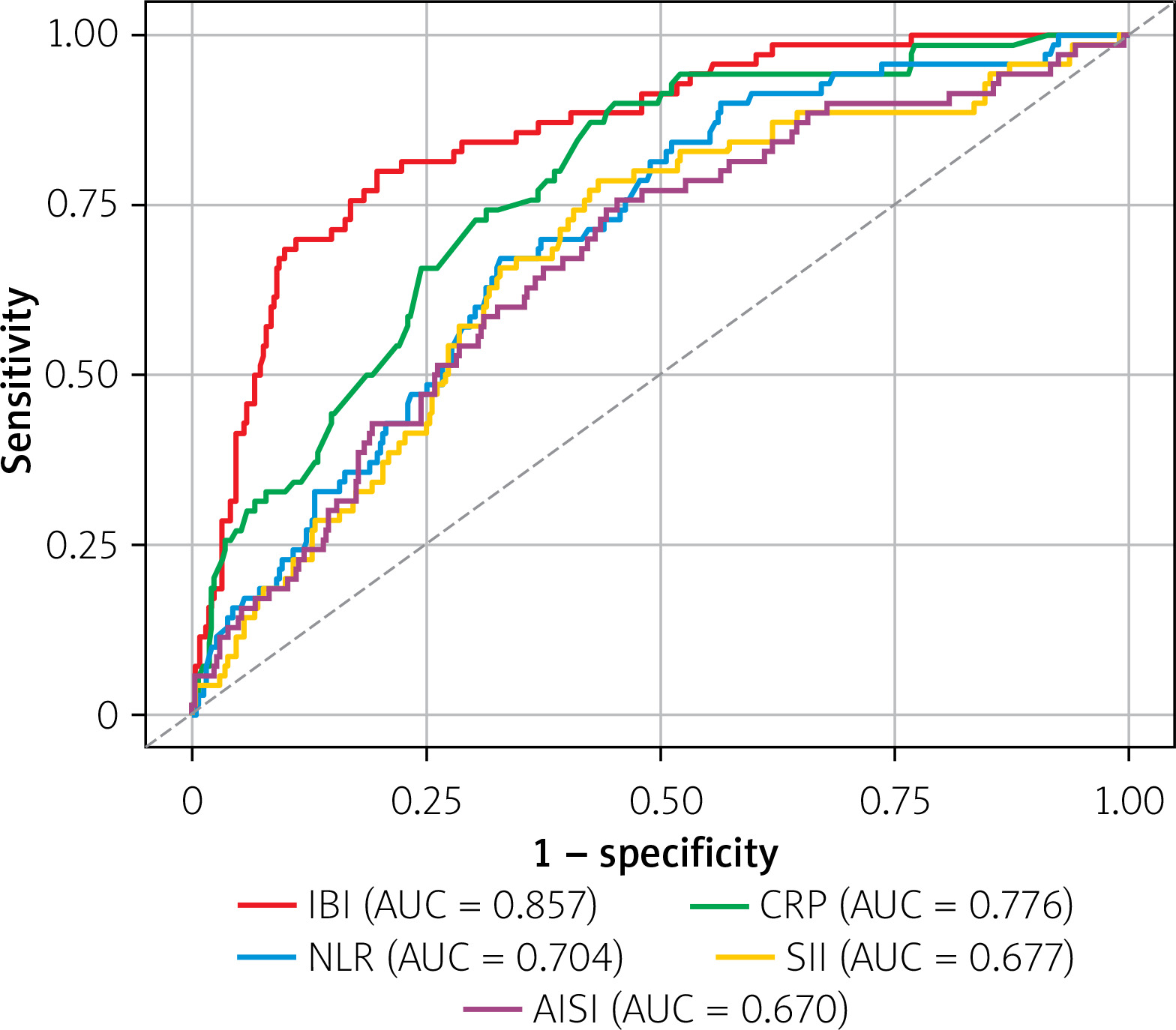
Pairwise DeLong tests showed that the AUC of IBI was significantly higher than that of each comparator: CRP (ΔAUC = 0.081, p < 0.001), NLR (ΔAUC = 0.153, p < 0.001), SII (ΔAUC = 0.180, p < 0.001), and AISI (ΔAUC = 0.187, p < 0.001).
At the cutoff of 40.6, IBI provided a balanced apparent performance, with a sensitivity of 80.0% and a specificity of 80.2%. By contrast, CRP showed higher sensitivity (90.0%) but lower specificity (54.9%), indicating that CRP behaved as a more sensitive but less specific marker. NLR showed a sensitivity and specificity of 67.0% and 67.0%, respectively; SII showed 78.6% and 56.7%; and AISI showed 75.7% and 54.7%.
Independent association between IBI and CA-AKI
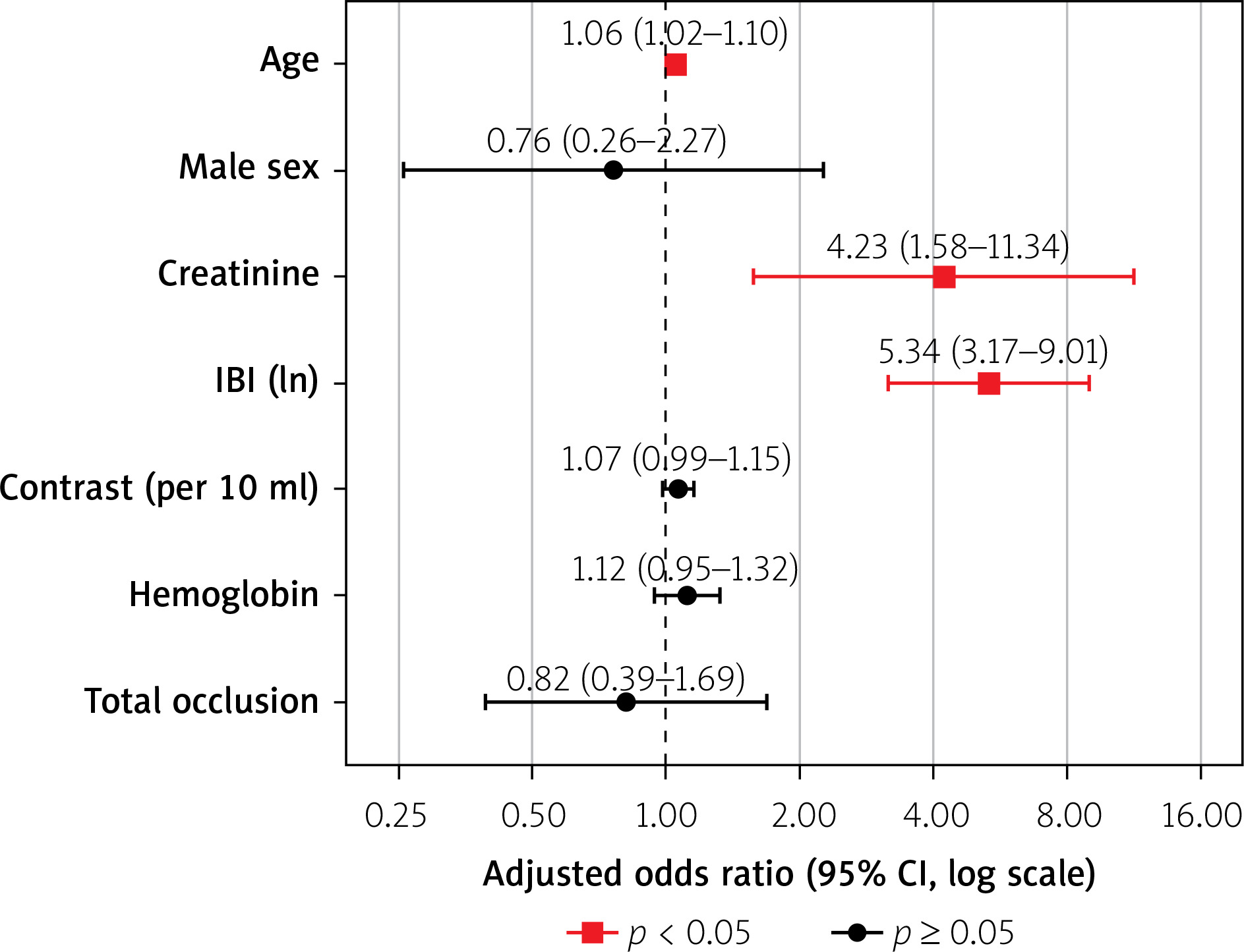
In multivariable analysis, ln-transformed IBI remained significantly associated with CA-AKI (adjusted OR = 5.34; 95% CI: 3.17–9.01; p < 0.001), corresponding to an adjusted OR of 3.19 (95% CI: 2.22–4.59) per doubling of IBI (Table III, Figure 2). Age (adjusted OR = 1.06; 95% CI: 1.02–1.10; p = 0.005) and serum creatinine (adjusted OR = 4.23; 95% CI: 1.58–11.34; p = 0.004) were also independently associated with CA-AKI. Contrast volume was not an independent predictor in the adjusted model (adjusted OR = 1.07 per 10 ml; 95% CI: 0.99–1.15; p = 0.110), nor were male sex, hemoglobin, or total occlusion. The model showed good discrimination (AUC = 0.873) and adequate calibration (Hosmer-Lemeshow p = 0.200), with variance inflation factors between 1.06 and 1.40 indicating no relevant multicollinearity.
Table III
Univariate and multivariable logistic regression analysis factors of contrast-associated acute kidney injury
[i] Odds ratios for continuous variables (age, creatinine, IBI, contrast volume, hemoglobin) reflect the change in odds per one-unit increase; odds ratios for categorical variables (male sex, total occlusion) reflect the comparison versus the reference category (female sex and absence of total occlusion, respectively). IBI was natural-log (ln) transformed prior to modeling owing to its right-skewed distribution; contrast volume is reported per 10 ml increase for clinical interpretability. Variance inflation factors for all predictors ranged from 1.06 to 1.40, indicating no relevant multicollinearity. Bold p-values indicate statistical significance (p < 0.05). CI – confidence interval, IBI – inflammatory burden index, ln – natural logarithm, OR – odds ratio.
Figure 2
Forest plot of the multivariable logistic regression model for contrast-associated acute kidney injury. Adjusted odds ratios are shown with their 95% confidence intervals on a logarithmic scale; the dashed vertical line denotes the null value (OR = 1). Red squares indicate variables with statistical significance (p < 0.05), and black circles indicate variables without statistical significance. Inflammatory burden index (IBI) was natural-log transformed, and contrast volume is expressed per 10 ml increment
CI – confidence interval, OR – odds ratio.
In sensitivity analyses, the association between IBI and CA-AKI remained significant after additional adjustment for TASC II C–D lesion status (adjusted OR = 6.16; 95% CI: 3.52–10.77; p < 0.001) and lesion length (adjusted OR = 5.17; 95% CI: 3.08–8.69; p < 0.001). The association also remained robust when both measures of lesion complexity were included simultaneously (adjusted OR = 6.11; 95% CI: 3.47–10.74; p < 0.001; Supplementary Table SII).
In incremental predictive analyses, addition of IBI to the base clinical model increased the AUC from 0.801 (95% CI: 0.743–0.858) to 0.873 (95% CI: 0.824–0.918), corresponding to a ΔAUC of 0.070 (DeLong p < 0.001). Among the evaluated inflammatory markers, IBI provided the largest improvement in discrimination and model fit (Supplementary Table SIII).
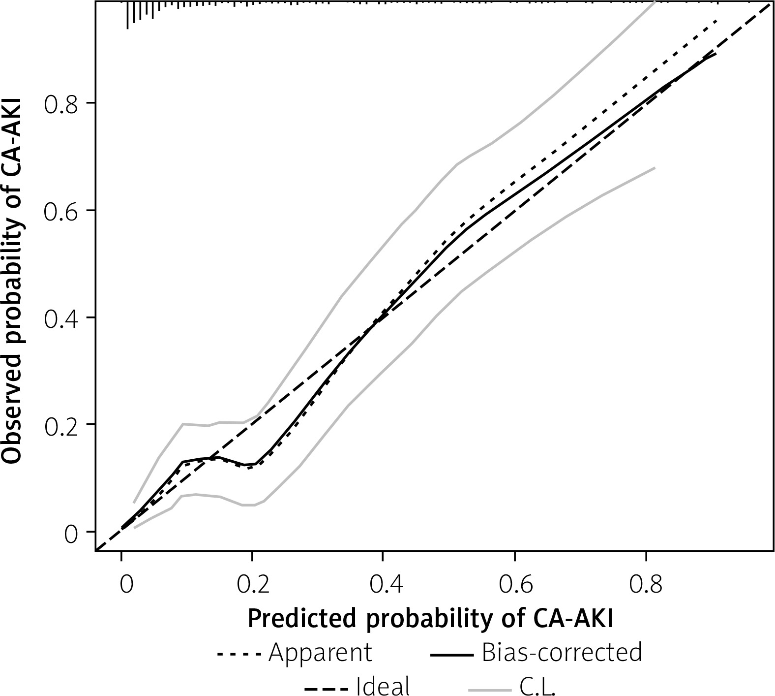
Restricted cubic spline analysis showed a significant nonlinear association between IBI and CA-AKI (p for nonlinearity < 0.001; Figure 3). Relative to the median reference value, the adjusted odds ratio remained below unity at lower IBI values, increased progressively across the intermediate range, and tended to level off at higher values. Calibration of the multivariable CA-AKI model is shown in Figure 4. Detailed results of the internal validation are provided in Supplementary Table SIV.
Figure 3
Restricted cubic spline model showing the association between IBI and the adjusted risk of contrast-associated acute kidney injury. The solid line represents the adjusted odds ratio, and the shaded area represents the 95% confidence interval; the dashed horizontal line marks the reference value (OR = 1), set at the median IBI (26.5). The relationship was non-linear (p for non-linearity < 0.001), with the odds ratio lying below the reference at lower IBI values, increasing across the intermediate range, and tending to level off at higher values
CA-AKI – contrast-associated acute kidney injury, IBI – inflammatory burden index, OR – odds ratio.
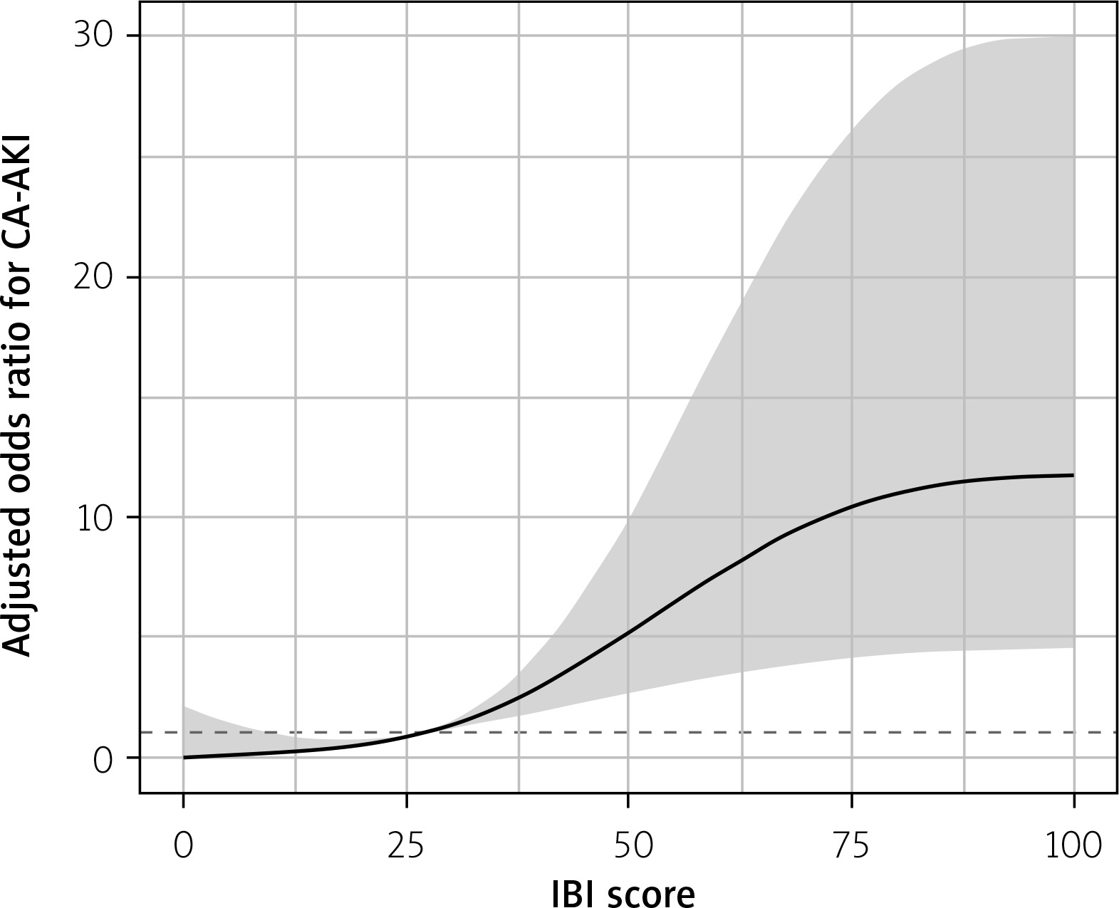
Figure 4
Calibration plot for the multivariable CA-AKI prediction model. The dotted line represents apparent calibration, the solid line represents the bootstrap bias-corrected estimate, and the dashed diagonal line represents ideal calibration; light grey lines denote the confidence limits. The bias-corrected curve generally followed the ideal line across most of the predicted probability range, suggesting reasonable agreement between predicted and observed probabilities
CA-AKI – contrast-associated acute kidney injury, C.L. – confidence limits.
IBI and lesion complexity
The association between IBI and TASC II C–D lesion complexity was further evaluated in a dedicated multivariable logistic regression model incorporating prespecified clinically relevant covariates (Table IV). After adjustment for demographic, comorbid, and laboratory covariates, ln-transformed IBI remained independently associated with complex TASC II C-D lesion morphology (adjusted OR = 4.68; 95% CI: 3.30–6.64; p < 0.001; adjusted OR = 2.91 [95% CI: 2.29–3.71] per doubling of IBI), whereas most conventional risk factors were not independently associated.
Table IV
Univariate and multivariable logistic regression analysis of factors associated with TASC II C-D (complex) lesions
[i] Odds ratios for continuous variables reflect the change in odds per one-unit increase; odds ratios for categorical variables reflect the comparison versus the reference category. IBI was natural-log (ln) transformed prior to modeling owing to its right-skewed distribution. Raw components and other composite inflammatory indices sharing IBI’s building blocks (CRP, neutrophil and lymphocyte counts, NLR, SII, AISI, albumin-to-lymphocyte ratio) were excluded owing to collinearity with IBI, and variables defining the TASC II classification itself (lesion length, total occlusion, bilateral involvement, stenosis, contrast volume) were excluded a priori to avoid circularity. Variance inflation factors ranged from 1.02 to 1.42, indicating no relevant multicollinearity. Bold p-values indicate statistical significance (p < 0.05). CI – confidence interval, IBI – inflammatory burden index, ln – natural logarithm, OR – odds ratio, TASC – TransAtlantic Inter-Society Consensus.
Discussion
In this retrospective cohort of 417 patients undergoing endovascular intervention for iliac artery disease, higher pre-procedural IBI was significantly associated with CA-AKI. IBI demonstrated better discriminative performance than CRP, NLR, SII, and AISI, with an AUC of 0.857 and a balanced sensitivity and specificity of approximately 80% at the optimal cutoff value of 40.6. This association remained significant after adjustment for demographic, renal, hematologic, and procedural variables, with each doubling of IBI associated with an approximately threefold increase in the odds of CA-AKI (adjusted OR = 3.19; 95% CI: 2.22–4.59). Restricted cubic spline analysis further demonstrated a nonlinear association in which CA-AKI risk increased across the intermediate range of IBI and tended to level off at higher values. The multivariable model showed good discrimination, satisfactory calibration, and limited optimism after bootstrap validation. In addition, higher IBI was independently associated with complex TASC II C–D iliac lesion morphology.
Reported rates of CA-AKI after peripheral endovascular interventions vary considerably according to the characteristics of the study population, vascular territory, contrast exposure, and diagnostic criteria. Previous studies involving lower-extremity peripheral interventions have reported CA-AKI or acute kidney injury rates ranging from approximately 3% to 13%, with higher rates observed in patients with chronic kidney disease or other high-risk features [19–21]. In the present cohort, CA-AKI occurred in 16.8% of patients, representing a somewhat higher incidence than that reported in several previous peripheral vascular cohorts. This difference may partly reflect the substantial proportion of complex TASC II C–D lesions in our population, which entailed greater procedural complexity and higher contrast exposure. Advanced age, impaired baseline renal function, anemia, and contrast volume are among the most consistently reported risk factors [5, 6, 22]. In line with previous evidence, older age and higher baseline serum creatinine were independently associated with CA-AKI in our study. Lower hemoglobin levels and greater contrast volume were also associated with CA-AKI in univariate analyses, although these associations were attenuated after multivariable adjustment.
The clinical relevance of CA-AKI extends beyond a transient increase in serum creatinine. Following peripheral vascular interventions, renal injury has been associated with increased in-hospital morbidity and mortality, prolonged hospitalization, and a greater risk of persistent deterioration in renal function, cardiovascular events, and long-term mortality [6, 19, 20]. These adverse consequences emphasize the importance of identifying susceptible patients before contrast exposure. Although the pathophysiology of CA-AKI is multifactorial and not completely understood, renal vasoconstriction and medullary hypoxia, direct tubular toxicity, oxidative stress, endothelial dysfunction, and inflammatory activation are considered central interacting mechanisms [23, 24].
Systemic inflammation has an established role in the development and progression of atherosclerosis and is associated with adverse outcomes across a broad spectrum of cardiovascular diseases [25]. In the setting of contrast exposure, an increased inflammatory burden may further impair endothelial and renal microvascular function, amplify oxidative stress, and increase susceptibility to ischemic and tubular injury [23]. Accordingly, several routinely available inflammatory markers, including CRP, NLR, SII, and AISI, have been investigated and shown to have potential value in identifying patients at increased risk of CA-AKI [7–11]. In the present study, we evaluated IBI, a composite index calculated by multiplying CRP by NLR. By integrating the biochemical component of systemic inflammatory activity reflected by CRP with the balance between neutrophil-mediated inflammation and lymphocyte-related immune regulation represented by NLR, IBI may provide a more comprehensive estimate of systemic inflammatory burden than either component considered separately.
The higher discriminative ability of IBI compared with the other inflammatory markers was one of the principal findings of this study. In ROC analysis, IBI achieved the highest area under the curve (0.857), and pairwise DeLong comparisons confirmed that its discriminative performance was significantly superior to that of CRP, NLR, SII, and AISI. At the optimal cutoff value of 40.6, IBI provided a sensitivity of 80.0% and a specificity of 80.2%, whereas CRP showed higher sensitivity but substantially lower specificity, and the cell count–based indices demonstrated more modest overall discrimination. Importantly, adding IBI to the conventional clinical model was associated with a significant improvement in discrimination, suggesting potential incremental predictive value beyond established clinical covariates. This performance may be related to the composite structure of IBI. CRP reflects the biochemical component of systemic inflammatory activity but provides limited information regarding the cellular immune response, whereas NLR and other count-based indices primarily reflect cellular inflammatory imbalance. By multiplying CRP by the neutrophil-to-lymphocyte ratio, IBI integrates these complementary dimensions into a single measure and places greater weight on patients in whom both components are concurrently elevated. Accordingly, IBI may capture the coexistence of biochemical and cellular inflammatory activation more comprehensively than either component or other hematologic inflammatory indices alone, potentially allowing more refined pre-procedural risk stratification. Nevertheless, the proposed cutoff and its clinical utility require confirmation in externally validated cohorts.
Our findings are concordant with the limited existing evidence on IBI and contrast-related renal injury. To date, this association has been described only in the coronary setting, where higher IBI was independently associated with contrast-associated acute kidney injury after percutaneous coronary intervention in patients with ST-segment elevation myocardial infarction, and a comparable nonlinear dose–response relationship was reported [17]. Similarly, in our restricted cubic spline analysis, the association was weak at lower IBI levels, became more pronounced across the intermediate range, and tended to level off at higher values. These consistent observations across different vascular territories, procedural settings, and patient populations support the potential value of IBI as a marker of increased risk of contrast-related renal injury. Importantly, the association between IBI and CA-AKI remained significant after additional adjustment for TASC II C–D status and lesion length, suggesting that anatomical lesion complexity alone may not fully account for the observed association. However, given the observational design, the relative contributions of biological susceptibility, lesion complexity, and procedural factors cannot be fully disentangled. The nonlinear pattern further indicates that the increase in risk may not be constant across the entire range of IBI values. To our knowledge, the present study is the first to evaluate IBI in patients undergoing iliac endovascular intervention and the first to directly compare its discriminative performance with that of several established inflammatory indices in this population, thereby extending the existing evidence to the peripheral vascular setting.
Beyond its association with CA-AKI, higher IBI was independently associated with complex TASC II C–D iliac lesion morphology. This finding suggests that elevated pre-procedural inflammatory burden may reflect not only susceptibility to contrast-related renal injury but also the extent and complexity of the underlying atherosclerotic disease. Previous studies have similarly linked inflammation- and nutrition-related indices with the severity and anatomical complexity of peripheral and aortoiliac arterial disease, supporting a broader relationship between systemic biological burden and advanced lesion morphology [26–29]. Because complex lesions are often accompanied by greater procedural demands and higher contrast exposure, systemic inflammation, anatomical complexity, and procedural risk are likely to represent overlapping rather than entirely independent domains. Accordingly, IBI may capture a broader high-risk phenotype characterized by both increased renal vulnerability and more advanced atherosclerotic burden. However, the observational design of this study does not allow the temporal or mechanistic relationships among these factors to be established, and these findings should therefore be regarded as hypothesis-generating.
From a clinical perspective, IBI may serve as a simple and readily obtainable adjunct to established pre-procedural risk assessment in patients undergoing iliac endovascular intervention. Because it is calculated from CRP and routinely available complete blood count parameters, it can be derived without specialized testing or substantial additional cost. When considered together with conventional risk factors such as age and baseline renal function, an elevated IBI may help identify patients who warrant more careful preventive management, including individualized hydration, minimization of contrast exposure, avoidance of additional nephrotoxic insults, and closer post-procedural monitoring of renal function. However, IBI should not be interpreted as a stand-alone decision tool, and the cutoff identified in the present cohort requires external validation. Whether incorporating IBI into existing risk-assessment strategies improves clinical decision-making or patient outcomes should be evaluated prospectively.
This study has several limitations. First, it was a single-center, retrospective, observational study, and despite multivariable adjustment and internal validation, residual confounding and selection bias cannot be excluded, and no causal relationship can be inferred. Second, CA-AKI was defined on the basis of the classic serum creatinine criteria rather than the broader KDIGO acute kidney injury definition; although this choice was intended to capture clinically relevant early renal deterioration and to maintain comparability with prior contrast-nephropathy studies, it may have affected the estimated incidence. Third, the inflammatory indices were measured at a single pre-procedural time point and therefore do not reflect the dynamic evolution of the inflammatory response. Fourth, the model was validated only internally through bootstrap resampling, and external validation in independent cohorts is required before clinical application. Moreover, the ROC-derived cutoff and its associated sensitivity and specificity were selected and evaluated in the same dataset and were not separately subjected to optimism correction; therefore, they should be regarded as apparent estimates. Fifth, the cohort was predominantly male and drawn from a single tertiary center, which may limit the generalizability of the findings. Finally, some laboratory-derived indices could not be evaluated because of missing data, and missing values in the retained variables were handled with multiple imputation, which, although statistically appropriate, carries its own assumptions.
Conclusions
Among patients undergoing endovascular intervention for iliac artery disease, higher pre-procedural IBI was independently associated with CA-AKI and showed better and more balanced discriminative performance than CRP, NLR, SII, and AISI. The association between IBI and CA-AKI was nonlinear, and higher IBI was also independently associated with complex TASC II C–D iliac lesion morphology. As a simple and readily available index, IBI may complement conventional pre-procedural risk assessment; however, prospective multicenter studies with external validation are required before routine clinical implementation.