Severe carotid artery calcification has traditionally been considered a contraindication to endovascular treatment, carrying a high risk of technical failure (suboptimal lumen gain, stent compression with high-grade residual stenosis, stent malapposition, stent fractures, increased rate of in-stent restenosis) and a greater risk of cerebral embolism [1–6]. Today, this paradigm is being challenged by the emergence of novel endovascular techniques that enable more effective (than plain balloon angioplasty) preparation of heavily calcified lesions to achieve a better lumen gain [7]. Accurate assessment of the severity and extent of the stenotic plaque calcification may aid selection of most appropriate plaque-modification strategy [8]. Intravascular lithotripsy (IVL) is a calcific plaque-modification technique that uses acoustic pressure waves to microfracture vascular calcium, converting non-compliant “stone”-like lesion content into more compliant lesion material. IVL already plays a growing role in managing highly calcified coronary [9, 10], iliofemoral [11] and below-the-knee [12] lesions. One, principal concern with the carotid use of IVL technology [13–15] is cerebral embolism that may carry far greater functional consequences than embolism to the coronary or limb territory [16]. Increasing severity of carotid plaque calcification appears proportional to the risk of ischemic cerebral events with conventional carotid artery stenting [6, 17].
Herein, we share our observations from the protocol development phase for PARADIGM-IVL trial with regard to determining the cerebral protection strategy for the study. IVL-assisted carotid artery stenting (CAS) procedure was performed in a 73-year-old patient with a computed tomography angiography-documented progression of angiographically-significant highly calcified stenosis of the left internal carotid artery (LICA) and a family history of stroke. The lesion fulfilled the highly calcified carotid stenosis criteria including calcific segment length to lesion length ratio ≥ 2/3, calcification circularity index ≥ 3, calcification peak thickness ≥ 3.0 mm, and calcification severity grade ≥ 3 [8]. The patient had clinical characteristics of increased risk for carotid endarterectomy, including heart failure with left ventricular ejection fraction of 20–25% and severe chronic obstructive pulmonary disease [18, 19].
Catheter angiography (Figure 1 A) confirmed high-grade carotid calcification severity (Grade 4/4) [8]. Intravascular ultrasound, an imaging technique unaffected by blooming artifacts and of a significantly greater resolution than computed tomography angiography, confirmed circumferential calcification (calcification circularity index of 4/4) [8] and visualized a “projecting (protruding) calcific nodules” lesion phenotype (Figure 1 D) consistent with an increased risk of spontaneous carotid-related stroke [20–23]. Distal filter was used for cerebral protection [13] (Figures 1 C–H). Intravascular lithotripsy (Shockwave C2+ rapid exchange system; 4.0 × 12 mm balloon [10, 13]; Figure 1 E) was performed, delivering a total of 120 impulses of calcium-cracking energy. A second-generation MicroNET-covered anti-embolic stent [24, 25] (CGuard 9 × 40 mm) was implanted (Figures 1 F, G) and was post-dilatation optimized using 5.5 mm and 6.0 mm diameter non-compliant balloons [8, 24] (Figure 1 H). Excellent angiographic result was confirmed by intravascular ultrasound demonstrating optimal stent expansion and apposition (Figure 1 E), consistent with achieving a complete endovascular reconstruction [24].
Figure 1
Imaging of distal (filter)-protected left internal carotid artery stenting in a patient with progressive highly calcified stenosis, high-risk clinical characteristics for carotid endarterectomy, and a family history of stroke. A – Baseline angiography in two contralateral projections; note critical stenosis (black arrow) of the left internal carotid artery (LICA) and heavy calcifications (red arrows); LECA – left external carotid artery. B – Attempt to pass (dotted arrow) an intravascular ultrasound (IVUS) probe (crossing profile ~1.20mm) through the lesion was unsuccessful due to intraluminal blockage by calcifications conglomerate. Subsequent filter delivery (nominal diameter 6 mm; crossing profile of ~1.05mm) was successful (C, green arrow denotes an open filter). C – Filter-protected (green arrow) initial predilatation of the lesion with a small coronary balloon (2.0 × 15 mm; white arrows denote balloon markers). D – IVUS evaluation of predilated lesion, demonstrating circular calcifications (acoustic shadowing; red arrowheads) and intraluminally projecting (protruding) calcific nodules (orange arrowheads, note zoomed image in top-right corner). E – Intravascular lithotripsy (IVL) Shockwave energy delivery using C2+ dual-emitter rapid exchange balloon catheter (4.0 × 12 mm) under distal cerebral protection (green arrow denotes a properly apposed filter). A zoomed image is shown on the right; white arrows denote balloon markers, blue arrows – lithotripsy energy emitters. 120 impulses (12 series of 10 impulses each) of calcium-cracking energy were delivered. Because maximum energy is delivered at the IVL emitter level [10], the IVL balloon was deflated and repositioned between the individual series. After the sixth series of IVL energy delivery, the patient reported neurologic symptoms of a markedly reduced sensation and aggravating weakness of the right limbs (exacerbating hemiparesis); this was initially attributed to cerebral flow reduction with the IVL balloon inflations. F – MicroNet-covered anti-embolic second-generation stent positioning (white arrowheads; CGuard 9 × 40 mm) [24, 57, 58, 60]. G – Photograph of the CGuard MicroNet-covered anti-embolic stent. H – Sequential post-dilation of the stent with a 5.5 × 20 mm and then 6.0 × 20 mm non-compliant balloon (stent edges are marked with white arrowheads). I – Final angiographic result (left) and IVUS images (right). Note optimal expansion (lack of residual stenosis) and apposition of the stent and the external carotid artery (ECA) patency in absence of any procedure-related stenosis – ‘a full endovascular reconstruction’ [24]. J – Cerebral angiogram in full lateral projection; note embolization of distal cerebral arterial branches supplying the parietal region (red arrowheads indicate missing vessel continuity). Insets are photographs of the filter used in this procedure; note embolic material in the filter basket as well as some material protruding (red asterixes/asterisks/arrowheads) through the filter pores of 70 to 200 μm. K – Venous phase of J; note visualization (red arrows) of the distal segments of some embolized vessels – now filling through retrograde collateral circulation
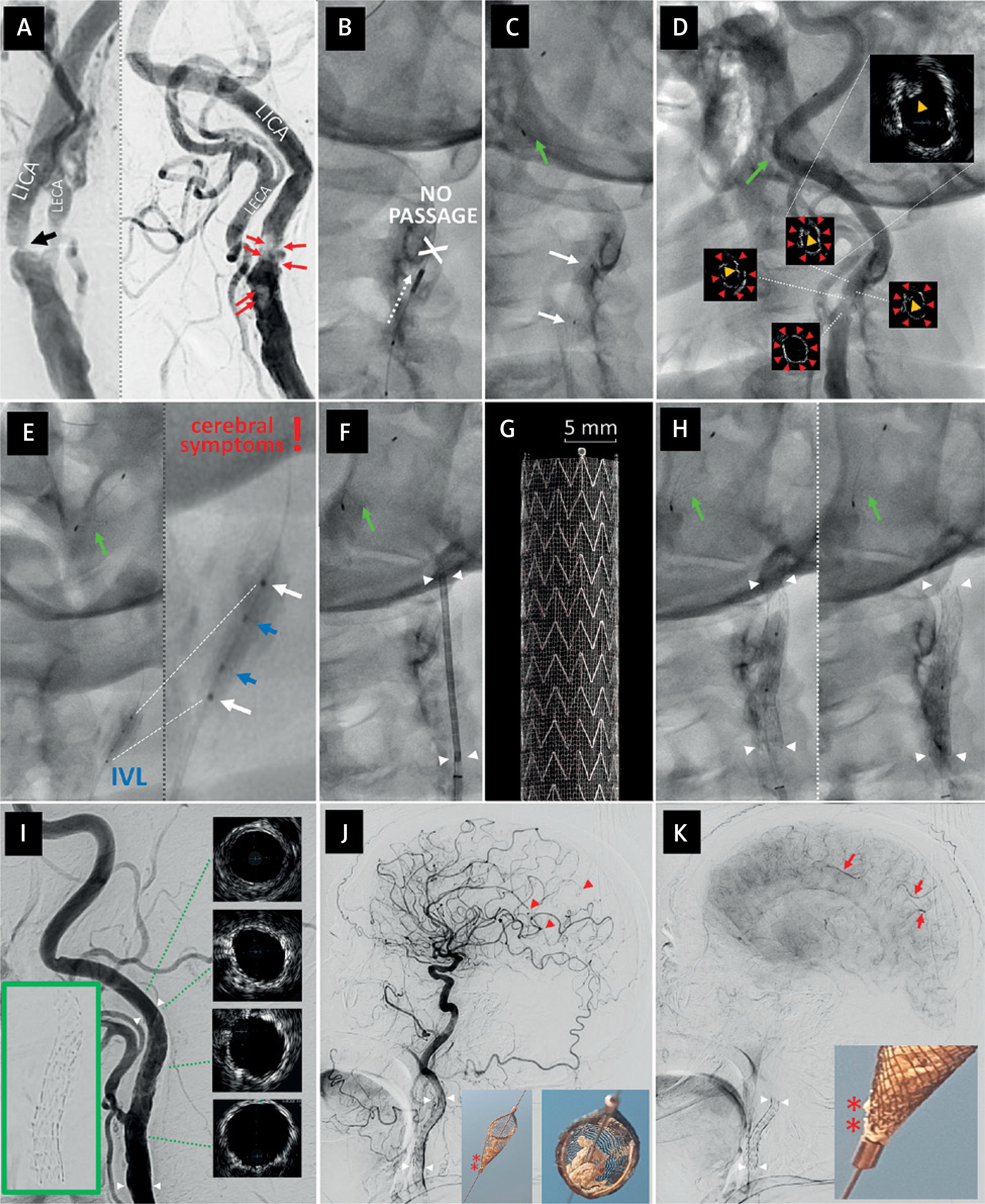
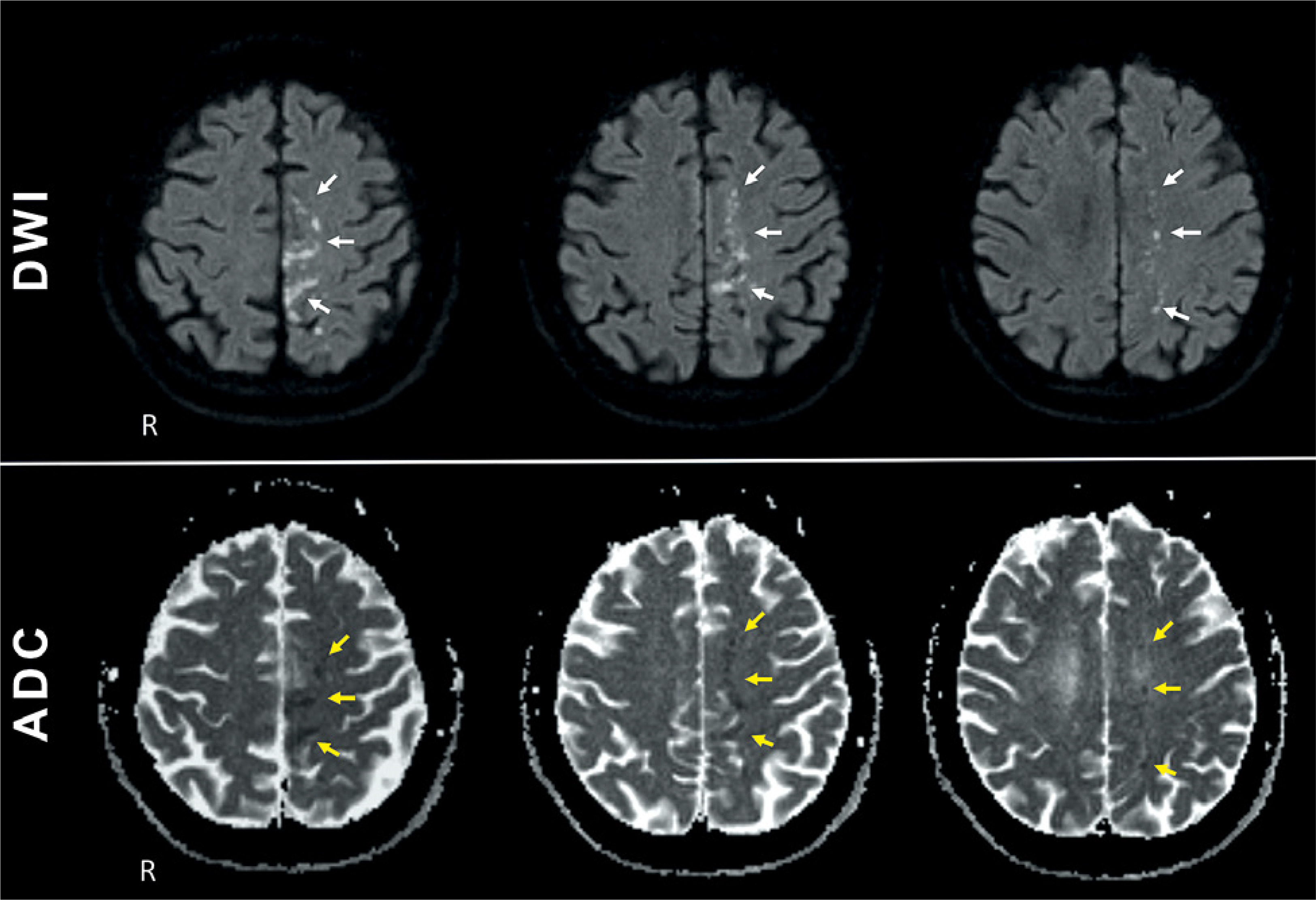
During lithotripsy, the patient developed symptoms of right-sided hemiparesis. The symptoms, initially believed to be associated with a transient reduction of the left-hemispheric cerebral perfusion with the IVL balloon inflations and calcium-cracking energy deliveries, persisted after retrieval of the distal protection filter. Completion cerebral angiogram demonstrated, in the arterial and parenchymal phase, embolization of distal cerebral arterial branches supplying the parietal region (Figure 1 J); in the venous phase there was retrograde filling of some embolized vessels (Figure 1 K). There was substantial embolic material in the filter basket, protruding through the filter pores (Figures 1 J and K insets). Post-procedural diffusion-weighted magnetic resonance imaging (DWI-MRI) revealed numerous small ischemic lesions (Figure 2), likely corresponding to particles smaller than the filter pore (cf., Figure 1 K inset). The patient underwent intensive neurological rehabilitation, and at 90 days was functionally independent (mRS 2). This substantial amount of embolic material in the filer basket protruding through the filer pores and embolizing to the brain (Figures 1, 2) shows that distal cerebral protection may not suffice in IVL-assisted endovascular management of highly calcified carotid stenoses, highlighting limitations of distal embolic protection filters that include the need to cross the lesion before cerebral protection can be exerted, risk of the basket malapposition and cerebral embolism with particles smaller in size than the filter pores (Figures 1 J, K) [26, 27]. Our present finding is consistent with several other incidental observations and moderately sized filter-protected series of IVL CAS [15, 28, 29]. Notably, also proximal-only cerebral protection may not be sufficient [30, 31] in the present-day IVL-assisted CAS that typically requires longer than usual procedure durations due to the time-requiring multiple charging of current-generation IVL emitters and cumbersome exchanges of over-the-wire peripheral systems that seem more powerful in carotid calcium cracking than the coronary rapid exchange systems [7, 30, 31]. Indeed, in a recent study of 58 patients undergoing IVL-assisted CAS under proximal cerebral protection, the average flow reversal duration was very long, averaging 25 min [30].
Figure 2
Cerebral post-procedural diffusion-weighted magnetic resonance imaging. Top panel shows diffusion-weighted images (DWI; three consecutive horizontal layers), illustrating temporo-parietal multiple scattered foci of diffusion restriction. Corresponding low signal intensities on apparent diffusion coefficient (ADC) maps are provided in the bottom panel, indicating acute ischemic cerebral injury corresponding to angiographic evidence of embolism in Figures 1 J, K
For these reasons, the PARADIGM-IVL Study protocol (A novel PARADIGM in management of highly calcified carotid stenoses: IntraVascular Lithotripsy and MicroNet-covered anti-embolic stents; ISRCTN94131858) recommends use of dual cerebral protection (a mandatory proximal system with addition of a distal filter if anatomically feasible). Other key findings from the PARADIGM-IVL protocol development phase [7] included (1) initial (i.e., prior to IVL) lesion predilatation to facilitate balloon insertion to minimize the risk of IVL balloon rupture on intraluminally protruding calcium, (2) non-compliant balloon optimization of lesion preparation after IVL (to maximize the lumen gain) and (3) post-dilatation stent optimization with 5.0–6.0 mm non-compliant balloon(s) followed (in case of large vessel diameters) by 6.5–8.0 semi-compliant balloon(s) use. Furthermore, we had found that performing IVL after (rather than prior to) stent implantation appeared less effective than maximal preparation of highly calcified lesions prior to stenting [7]; this, however, requires further systematic evaluation.
Cerebral injury on diffusion-weighted magnetic resonance imaging (DW-MRI) is an important outcome measure for procedure-related stroke risk [16, 32, 33]. The present findings (Figure 2), consistent with other reports [28, 34], extend our previous observations that embolic protection limited to a distal filter may be suboptimal in a subset of carotid lesions [27, 35]. Interestingly, Tulip et al. [36] reported that cerebral embolization, detected by transcranial doppler and DW-MRI, may occur with a similar frequency after CAS for asymptomatic and symptomatic carotid stenosis. With their finding of postprocedural ischemic brain injury in approximately half of asymptomatic patients, the authors concluded that the safety of CAS under filter embolic protection for asymptomatic carotid stenosis should be considered uncertain [36]. Evaluation of the relationship between atherosclerotic plaque composition assessed by virtual histology intravascular ultrasound [37] and DW-MRI cerebral embolization in filter-protected CAS revealed a larger median dense calcium volume in patients with ipsilateral cerebral embolism (33.2 ±24.5 mm3 vs. 11.4 ±6.1 mm3) [38]. This suggested that dense calcifications, rather than the proportion of necrotic core (traditionally considered the principal carotid plaque risk component [37]) may be associated with an increased risk of subclinical cerebral embolization after CAS when a filter device is used for embolic protection [38].
Projecting calcified nodules, an important mechanistic trigger of the carotid plaque symptomatic thrombus formation [21, 23], are present in ≈8% of carotid bifurcation atherosclerotic stenoses [39, 40] and they pose an important challenge to conventional endovascular revascularization [41, 42]. In a study of risk factors for cerebral embolism in patients undergoing distal filter-protected (Emboshield NAV6) conventional single-layer closed-cell stent (Xact) CAS, lesion calcification was found to be an independent risk factor for perioperative cerebral embolization (OR = 5.68; 95% CI: 1.12–28.79; p = 0.04) [40], pointing to the need to improve endovascular calcium handling techniques and optimize cerebral protection in calcific lesion CAS. This notion has been echoed by several other groups [1, 3–6, 43, 44].
There is a significant body of evidence that proximal cerebral protection can minimize cerebral embolism in CAS compared to filter protection [26, 27, 44–47]. Advantages of proximal protection are not limited to first-generation (single-layer) stents because cerebral embolism involves the CAS stages prior to stent implantation [26, 27, 48], and thus – in case of neuroprotective stent use – prior to the cerebral protection exerted by the stent anti-embolic layer [25]. Evidence is accumulating that second-generation (anti-embolic, dual-layer) stents can improve clinical outcomes when compared to single-layer stents and to carotid endarterectomy [49–52]. These stents may play a particularly important role in endovascular management of high-risk lesions, such as those thrombus-containing or highly calcified [8, 52–55]. Besides the level-1 advantage of MicroNET-mediated cerebral protection in highly emboligenic lesions [25] and some sealing characteristics [56], the stent frame unique properties of a high radial force and SmartFit self-apposition [24, 57] may be particularly relevant to endovascular management of highly calcified carotid stenoses. Recently, the MicroNet-covered stent [57–59] has delivered best ever clinical outcomes amongst rigorously controlled United States Food and Drug Administration carotid stent studies [60, 61].
Dual cerebral protection in CAS (i.e., transient flow cessation/reversal combined with a distal filer) can increase the completeness of embolic material evacuation [62, 63]. Recently, Vuruskan et al. [64] documented a significant reduction in the number of cerebral ischemic lesions on DW-MRI – from an average of 1.80 in the distal (filter) group, through 0.90 with proximal protection (dual balloon; Mo.Ma), to 0.55 in the double-protection group (p < 0.001), indicating a role for dual cerebral protection to minimize the risk of cerebral embolization in CAS. Dr. Juan Parodi, inventor of the endovascular flow reversal system for cerebral protection in CAS, postulated that the combination of 2 existing cerebral protection techniques (proximal flow reversal and distal filter) is likely to achieve what neither of them can do independently, and thus the “seat belt and air bag” approach may represent the ideal means of cerebral protection in endovascular management of carotid bifurcation stenoses in patients with a non-functioning circle of Willis [65]. We suggest extending this indication to lesions with a significant embolic risk when there may be a need for intermittent flow reversal due to a longer-than-in-average-CAS time requirement [8, 30] due to the device exchanges and current device limitations – such as the need for repeated charging of shockwave emitters in IVL [9, 10].
In conclusion, in complex lesions with high embolic potential, such as highly-calcified carotid stenoses, neither proximal nor distal protection alone can ensure cerebral safety of the endovascular revascularization procedure [28, 31, 34]. Using both together may address vulnerabilities of each of the two cerebral protection approaches – a strategy recommended in the study of a novel PARADIGM in management of highly calcified carotid stenoses: IntraVascular Lithotripsy and MicroNet-covered anti-embolic stents (PARADIGM-IVL) [66].