Ischaemia with non-obstructive coronary arteries (INOCA) is identified in approximately 40–50% of patients undergoing invasive coronary angiography for evaluation of stable angina [1–3]. Previously, such patients were often discharged without a clear diagnosis, resulting in numerous adverse patient outcomes [1, 4, 5]. However, INOCA is no longer viewed as a benign condition but as a heterogeneous disorder with distinct functional endotypes: coronary microcirculatory dysfunction (CMD) and vasospastic angina (VSA) [1, 5]. While standard coronary angiography is effective in identifying macrovascular anatomical obstructions, its assessment of the coronary circulation functional capacity is limited [1].
Thus, normal angiographic findings in a symptomatic patient should prompt further assessment, rather than concluding the diagnostic process, as in the following case [1].
A 60-year-old man with a history of long-standing type 1 diabetes mellitus, arterial hypertension, class I obesity (BMI 34.1 kg/m²), chronic obstructive pulmonary disease, and a 40-pack-year smoking history was referred for evaluation of a 7-month history of exertional retrosternal burning discomfort.
At the outpatient department visit the patient reported progressive deterioration in exercise tolerance accompanied by poorly controlled arterial hypertension. Transthoracic echocardiography revealed mildly reduced left ventricular systolic function, with a left ventricular ejection fraction (LVEF) of 45%, and regional wall motion abnormalities. Subsequent exercise stress testing demonstrated inducible myocardial ischaemia, prompting invasive coronary assessment.
During hospitalisation, invasive coronary angiography revealed 30% stenosis in the proximal and middle segments of the left anterior descending artery (LAD). Due to the absence of angiographically significant epicardial stenosis and persistent evidence of ischaemia, a comprehensive invasive diagnostic protocol (IDP) was undertaken.
Physiological assessment was performed in the LAD. Fractional flow reserve (FFR) and resting full-cycle ratio (RFR) values were 0.81 and 0.91, respectively. Obtained measurements have not met the conventional threshold for a significant epicardial stenosis. Maximal hyperaemia to evaluate FFR was obtained by continuous intravenous adenosine infusion at 140 μg/kg/min. Coronary microvascular function was further evaluated invasively, demonstrating a coronary flow reserve (CFR) of 5.2 and an index of microcirculatory resistance (IMR) of 15. Overall, invasive microvascular indices were within the normal range in the LAD.
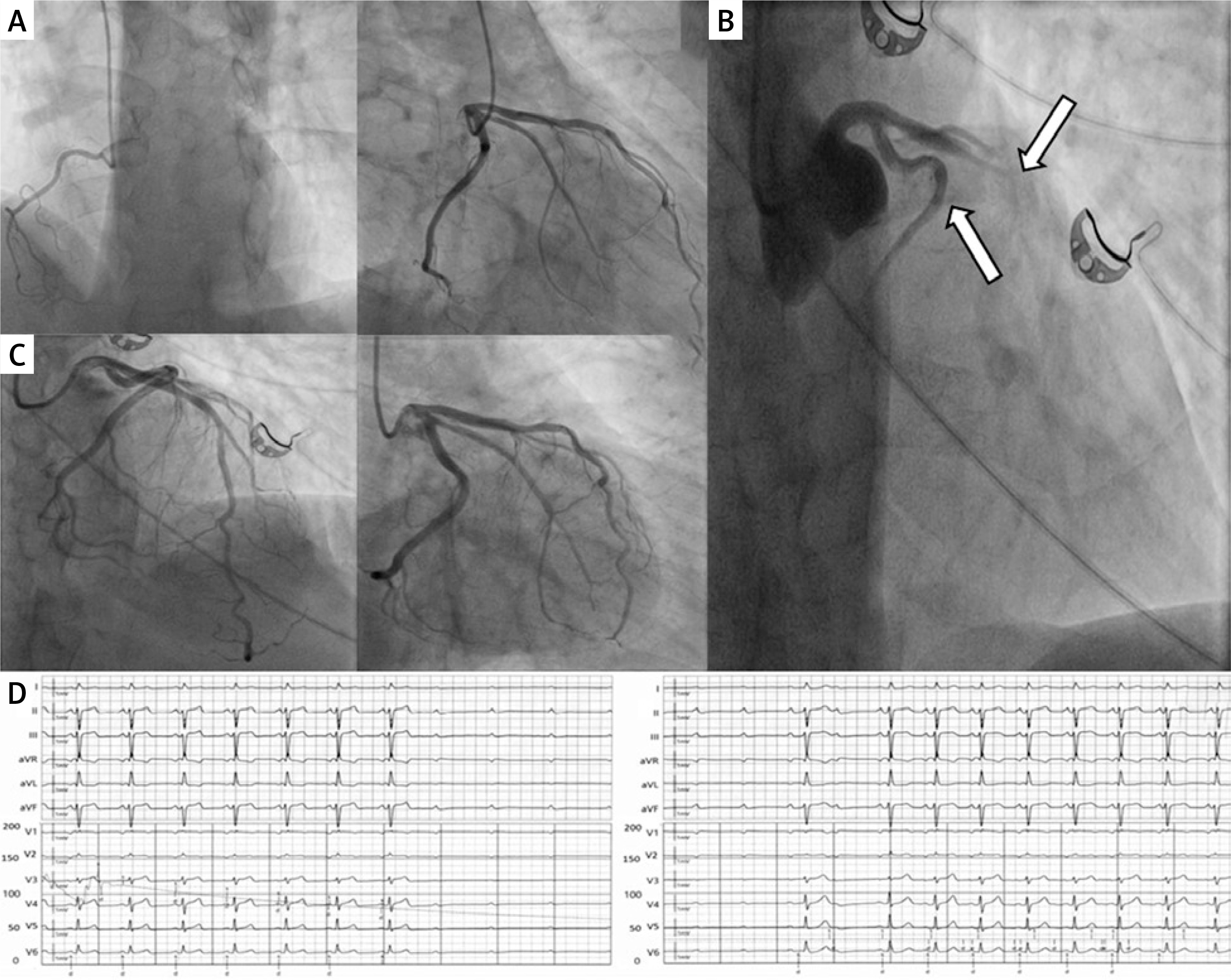
Subsequently, intracoronary acetylcholine (ACh) provocation testing was performed to assess vasomotor reactivity. Continuous infusion of ACh (20 μg) into the LAD via the guiding catheter induced diffuse, occlusive (100%) vasospasm of the proximal LAD and circumflex artery, accompanied by reproduction of the patient’s symptoms, ST-segment elevation on continuous electrocardiographic monitoring, and a transient 5-second episode of asystole. Prompt intracoronary nitroglycerin administration resulted in complete angiographic and clinical resolution. Examination was terminated immediately after symptoms occured together with coronary spasm and ischaemic ECG changes, thereby avoiding further dose escalation. These findings were diagnostic for epicardial vasospastic angina.
Targeted antianginal therapy with isosorbide mononitrate (60 mg once daily), molsidomine (2 mg once daily), trimetazidine (35 mg twice daily), short-acting sublingual nitroglycerin spray as needed, and the non-dihydropyridine calcium channel blocker (diltiazem at a dose of 60 mg once daily), along with optimisation of guideline-directed medical therapy for left ventricular systolic dysfunction and aggressive cardiovascular risk factor modification (including once daily: acetylsalicylic acid 75 mg, empagliflozin 10 mg, eplerenone 25 mg, ramipril 2.5 mg, and a combination of atorvastatin 40 mg and ezetimibe 10 mg), resulted in complete symptom resolution and improvement in LVEF to 60% at 6-month follow-up (Figure 1).
Figure 1
Invasive coronary angiography and acetylcholine provocation test. A – Baseline angiography of the right (RCA) and left (LCA) coronary arteries showing non-obstructive coronary artery disease. B – Massive, diffuse epicardial vasospasm (100% occlusion) of the proximal left anterior descending and circumflex arteries induced by a 20 μg acetylcholine continuous infusion (arrow). C – Angiographic resolution of the vasospasm: from partial response after the initial dose of intracoronary nitroglycerin to complete restoration of baseline vessel calibre following subsequent administration. D – ECG during the acetylcholine provocation test showed ST-segment elevation and a transient 5-second episode of asystole
Current guidelines, supported by clinical trials, emphasise the use of an IDP combining physiological assessment with pharmacological stress tests [1, 4]. This comprehensive approach enables identification of the underlying mechanisms and implementation of pharmacotherapy directed at a specific endotype of functional disorder found during IDP. Ultimately, the paradigm shift in the diagnostic approach to INOCA may result in complete symptom resolution in a patient cohort previously subject to/experiencing persistent symptoms, frequent hospitalisations, and a higher risk of major adverse cardiovascular events [1, 2, 4, 5].