
Acute coronary syndrome (ACS) is classically attributed to plaque rupture. However, plaque erosion is now recognized as an equally important mechanism, accounting for up to 40% of ACS cases. It is more common in women, younger patients, and smokers, and is associated with platelet-rich (“white”), often non-occlusive thrombi, more frequently leading to non-ST-elevation myocardial infarction (NSTEMI). High-resolution intravascular imaging, particularly optical coherence tomography (OCT), is considered the gold standard for differentiating these mechanisms. Type 2 diabetes mellitus (T2DM) remains a major cardiovascular risk factor, promoting endothelial dysfunction and increased platelet reactivity [1].
A 62-year-old woman with hypertension and newly diagnosed T2DM was admitted due to worsening exercise tolerance over 3 days and chest pain increasing in intensity over several hours. Electrocardiography showed ST-segment depression and T-wave inversion in leads III and aVF. Laboratory findings revealed a dynamic rise in troponin I (peak 139.68 ng/l), glycated hemoglobin of 8.3%, and non-high-density lipoprotein (HDL) cholesterol of 2.75 mmol/l. Urgent coronary angiography demonstrated significant stenosis of the right coronary artery with visible thrombotic material (Figure 1 A). Aspiration thrombectomy was performed, retrieving macroscopic thrombus, followed by implantation of a drug-eluting stent (Xience Pro 3.5 × 24 mm). Adjunctive therapy included intracoronary eptifibatide and intravenous infusion. Angiographic results were satisfactory, with restored distal flow (Figure 1 C) and no procedural complications. The patient was discharged on dual antiplatelet therapy with acetylsalicylic acid and ticagrelor.
Figure 1
Angiograms of the right coronary artery before and after percutaneous coronary intervention (PCI) and macroscopic appearance of the thrombus. A – Angiogram showing significant stenosis of the right coronary artery with visible thrombotic material (sites of stenosis are indicated by arrows). B – Macroscopically retrieved thrombus obtained by aspiration thrombectomy. C – Final angiographic result after thrombectomy and drug-eluting stent implantation, demonstrating restored coronary flow
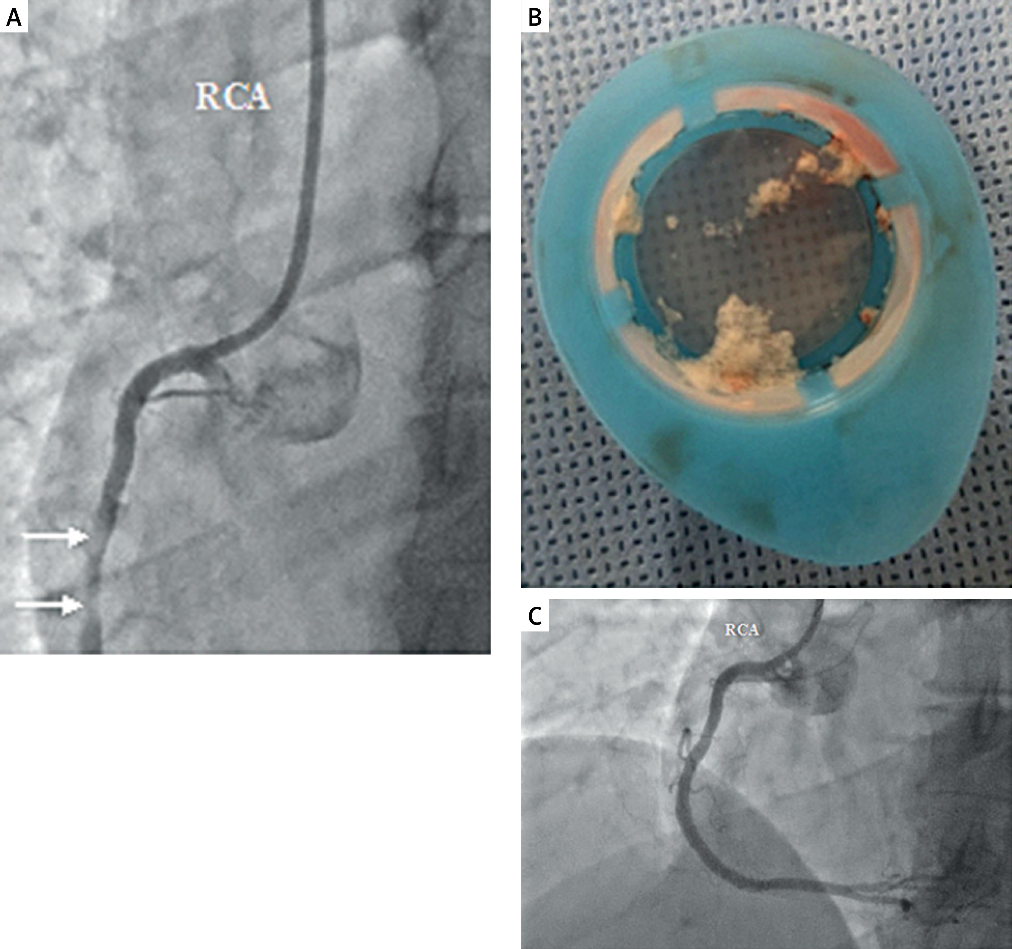
Macroscopically, the aspirated thrombus appeared pale and firm, suggesting a predominance of platelet components consistent with a “white thrombus” phenotype (Figure 1 B), typically associated with plaque erosion. However, this interpretation remains indirect, as no intravascular imaging or histopathological analysis was performed. Although routine use of aspiration thrombectomy is no longer recommended in current guidelines due to the lack of proven clinical benefit, selective use may be justified in cases with a high thrombus burden, as in this patient. From a technical standpoint, thrombectomy can improve coronary flow and lesion visualization, although its effectiveness depends on thrombus composition and operator experience [2, 3].
Plaque erosion has emerged as a major mechanism of ACS and is generally associated with NSTEMI, preserved coronary flow, and smaller infarct size compared with plaque rupture. It is less commonly observed in patients with T2DM, making this case potentially atypical. However, the patient’s diabetes was newly diagnosed, suggesting limited metabolic exposure. Given the heterogeneity of T2DM, vascular phenotype depends on disease duration and glycemic control, which may explain this presentation [4].
Recognition of plaque erosion has been facilitated by OCT, enabling in vivo differentiation from rupture and opening the possibility of tailored management strategies. In selected cases, a stentless approach with intensive antithrombotic therapy has been proposed. However, in the present case, stent implantation was performed due to significant residual stenosis and the absence of intravascular imaging [1].
Despite the lack of definitive thrombus characterization, this case highlights the importance of thrombus phenotype as a potential determinant of ACS pathophysiology and management. In NSTEMI, intravascular imaging should be considered essential for identifying the underlying mechanism and guiding tailored treatment.










