A 70-year-old man presented to the outpatient cardiology clinic with intermittent chest and back pain. His cardiovascular risk factors included hypertension, hyperlipidemia, and a history of smoking. Transthoracic echocardiography showed a left ventricular ejection fraction of 45%, posterior and inferior wall hypokinesia, mild mitral and tricuspid regurgitation, and no significant aortic stenosis or regurgitation; however, the sinus of Valsalva aneurysms were not clearly visualized, likely because of their complex root location and a limited acoustic window. The patient’s symptoms were considered multifactorial. Severe multivessel coronary artery disease, together with regional wall motion abnormalities, was thought to contribute to the chest pain, whereas the associated back pain raised suspicion for concomitant aortic root pathology.
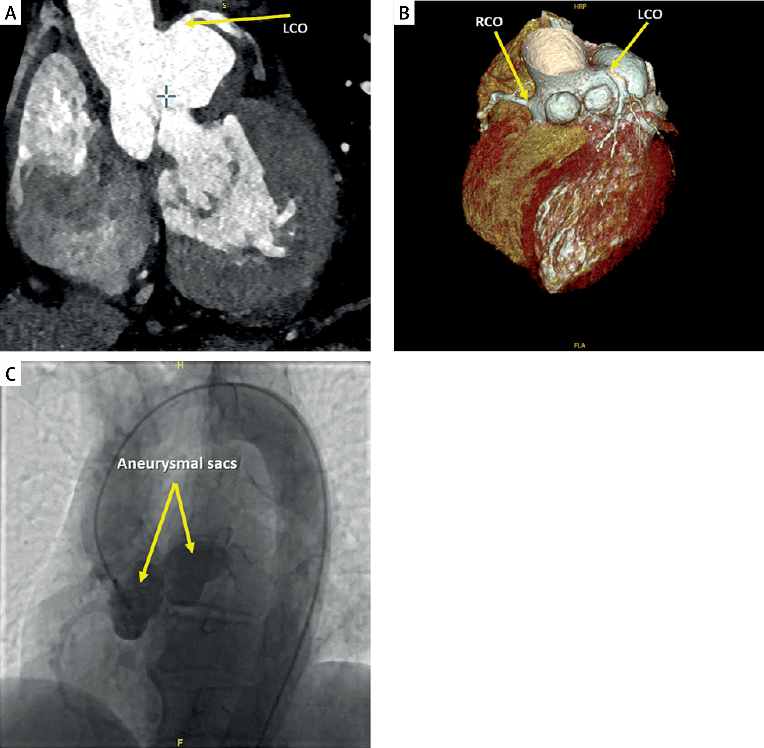
Because of the clinical suspicion of underlying coronary or aortic pathology, cardiac computed tomography was performed. Cardiac computed tomography (CT) angiography demonstrated a tricuspid aortic valve, a large left sinus of Valsalva aneurysm with a maximal diameter of 37 × 40 mm, and two additional aneurysms arising from the right sinus of Valsalva, measuring 15 × 15 mm and 11 × 20 mm. All aneurysm dimensions were obtained from multiplanar CT angiographic assessment, while three-dimensional reconstruction was used to better define the complex aortic root anatomy (Figure 1 A, B). Cardiac CT angiography provided high spatial resolution and enabled accurate assessment of the number, size, and anatomical relationship of the aneurysms to the coronary ostia.
Figure 1
Multimodality imaging of multiple sinus of Valsalva aneurysms. A – Cardiac CT angiography demonstrating a large aneurysm arising from the left sinus of Valsalva. The left coronary ostium (LCO) is indicated. B – Three-dimensional CT reconstruction showing complex aneurysmal involvement of the aortic root. The right coronary ostium (RCO) and left coronary ostium (LCO) are labeled. C – Aortography delineating the sinus of Valsalva aneurysms and their anatomical relationship with the coronary ostia. Yellow arrows indicate the aneurysmal sacs
Sinus of Valsalva aneurysm is a rare cardiac abnormality, with a reported prevalence of approximately 0.09% in the general population. Most aneurysms arise from the right coronary sinus, followed by the non-coronary sinus, whereas involvement of the left coronary sinus is uncommon [1]. Multiple sinus involvement is exceedingly rare, and intra-aneurysmal thrombus has been described only sporadically in isolated case reports [2, 3].
CT angiography suggested severe multivessel coronary artery disease, which was confirmed by invasive coronary angiography showing chronic total occlusions of the mid left anterior descending and circumflex arteries with distal collateral filling, and diffuse right coronary artery disease from the posterior descending artery segment onward. Aortography further delineated the sinus of Valsalva aneurysms and demonstrated their anatomical relationship to the coronary ostia (Figure 1 C). There was no clinical or laboratory evidence suggestive of connective tissue disease, infective endocarditis, prior chest trauma, previous cardiac surgery, vasculitis, or syphilitic aortitis. After multidisciplinary Heart Team evaluation, combined surgical treatment was planned. During surgery, organized thrombus was identified within the left sinus of Valsalva aneurysm sac and removed. Postoperative pathological examination confirmed thrombotic material within the aneurysm sac. No clinical neurological deficit or other evidence of systemic embolization was observed during the perioperative period. Although preoperative echocardiography showed no significant aortic regurgitation, the presence of multiple aneurysms involving both the left and right coronary sinuses, distortion of the aortic root anatomy, and intra-aneurysmal thrombus made isolated patch repair, root remodeling, or valve-sparing root replacement less favorable options. Given the concomitant severe multivessel coronary artery disease requiring surgical revascularization, Bentall surgery combined with coronary artery bypass grafting was preferred as a definitive surgical approach. Postoperative histopathological examination of the resected aneurysm wall demonstrated degenerative changes, consistent with a degenerative etiology.
The postoperative course was uneventful, with no major perioperative complications. Postoperative transthoracic echocardiography demonstrated a normally functioning aortic valve prosthesis without significant paravalvular leak. The patient was discharged in stable condition on postoperative day 8. At the 1-month follow-up, he remained clinically stable, with no recurrent chest or back pain and improved functional capacity.
This case highlights the value of advanced imaging techniques in identifying rare, complex sinus of Valsalva aneurysms and in informing surgical strategy.