Summary
This prospective multicenter randomized controlled trial compared a paclitaxel-coated balloon (DCB) with a rapamycin-eluting stent (DES) in 228 patients with de novo, non-complex coronary lesions. At 12-month follow-up, the DCB group showed comparable angiographic outcomes, major adverse cardiovascular event rates, and major bleeding events to the DES group, meeting non-inferiority criteria. Post-procedural angina frequency was lower in the DCB group, and both groups demonstrated significant improvements in cardiac function. The findings support DCB monotherapy as an effective alternative to DES for this lesion subset.
Introduction
Diabetes mellitus, hypertension, alcohol abuse, hyperuricemia, and psychological factors are closely associated with coronary artery disease (CAD) development [1]. These pathological states promote lipid deposition and atherosclerotic plaque formation within coronary arteries, leading to progressive luminal narrowing [2]. Percutaneous coronary intervention (PCI) remains a cornerstone treatment for CAD. Bare-metal stents (BMS) initially addressed limitations of balloon angioplasty, particularly acute closure, vascular recoil, and early restenosis [3]. Subsequent high in-stent restenosis (ISR) rates drove the development of drug-eluting stents (DES), which deliver antiproliferative agents to inhibit neointimal hyperplasia [4]. DES provide durable mechanical support against elastic recoil and acute occlusion while enabling sustained drug release to suppress intimal hyperplasia [5], proving particularly valuable for severe stenoses or unstable vessel structures [3]. Despite reducing restenosis and late lumen loss (LLL) in long-term follow-up [6], contemporary DES exhibit limitations in complex lesions such as small vessel disease (SVD). Stent thrombosis (especially late/very late stages) remains a critical concern, as it may potentially trigger acute myocardial infarction [7]. Chronic DES implantation may also induce inflammatory responses that impair vascular physiology [8], while metallic structures can cause abnormal remodeling in SVD with an increased LLL risk [9].
Drug-coated balloons (DCB) offer a metal-free alternative by directly delivering antiproliferative drugs to lesion sites [10]. Their rapid, high-concentration drug transfer effectively inhibits smooth muscle proliferation, minimizing restenosis [11]. Current guidelines endorse DCB for SVD, ISR, bifurcation lesions, and high-bleeding-risk patients [10], provided lesion preparation maintains < 30% residual stenosis without flow-limiting dissection or thrombus [12]. Shin et al. demonstrated that DCB-based strategies significantly reduce stent burden in multivessel PCI with lower major adverse cardiovascular event (MACE) rates than with DES alone [13].
Although newer-generation DES outperform BMS [14], robust evidence supporting their universal application remains limited. It was hypothesized that DCB would be non-inferior to DES with respect to LLL. Based on previous randomized controlled trial data comparing DES with bare-metal stents (BMS), the LLL benefit conferred by DES was approximately 0.4–0.5 mm. To preserve the majority of the DES effect and in line with the existing consensus on DCB for coronary small vessel disease, the non-inferiority margin (Δ) for LLL was set at 0.15 mm. Specifically, if the upper limit of the 95% confidence interval (CI) for the mean difference in LLL (DCB minus DES) was less than 0.15 mm, DCB would be considered non-inferior to DES.
Aim
This prospective randomized trial systematically compares DCB with rapamycin-eluting stents for de novo, non-complex small coronary vessel disease to elucidate their efficacy, safety, and clinical applicability.
Material and methods
Study population
A total of 228 patients with CAD referred for interventional therapy at Fuwai Yunnan Hospital, Chinese Academy of Medical Sciences between January 2022 and January 2024 were enrolled. Patients were allocated in a 1 : 1 ratio to either the DCB group (paclitaxel-coated balloon catheter) or the DES group (rapamycin-eluting metal stent) using a computer-generated random number table.
Inclusion criteria: (1) Patients with acute or chronic coronary syndrome meeting indications for stent intervention. (2) Patients with de novo, non-complex coronary lesions achieving successful pre-dilation. (3) Patients with ≥ 70% diameter stenosis confirmed by quantitative coronary angiography (QCA), with small vessel diameters measuring 2.25–2.75 mm. (4) Patients demonstrating good compliance and accepting clinical follow-up. (5) Patients aged ≥ 18 years. (6) Patients without contraindications to coronary revascularization, in whom intervention was indicated by angiography or functional testing. (7) Patients (and legal guardians) providing written informed consent. Exclusion criteria: (1) Patients who were pregnant or lactating. (2) Patients unable to receive antiplatelet/anticoagulant therapy. (3) Patients with allergies to heparin or contrast agents. (4) Patients with severe cardiomyopathy, valvular disease, cardiac insufficiency, cardiogenic shock, or heavily calcified lesions. (5) Patients refusing coronary intervention. (6) Patients with life expectancy < 1 year or anticipated follow-up difficulties. (7) Patients with severe hepatic/renal dysfunction.
Sample size calculation
Based on the sample size formula for non-inferiority trials, a one-sided α of 0.025, a power (1-β) of 0.80, an expected LLL difference of zero between the two groups, a non-inferiority margin (Δ) of 0.15 mm, and an assumed pooled standard deviation for LLL of 0.4 mm were used. The required sample size was calculated to be 90 patients per group. Considering an approximate 20% loss to follow-up and dropout rate for angiographic follow-up, a total of 228 patients were planned for enrollment.
Definitions
De novo lesion: A target vessel with no history of prior stent implantation.
Complex PCI was defined according to modified criteria from previously published standards, partially adopted in other clinical studies as meeting at least one of the following characteristics: (1) Treatment of ≥ 3 vessels. (2) Treatment of ≥ 3 lesions. (3) Total stent length > 60 mm. (4) Bifurcation stenting with 2 stents. (5) Use of any atherectomy device. (6) Left main coronary artery as target vessel. (7) Surgical bypass graft or chronic total occlusion as target lesion.
Successful lesion preparation: Balloon pre-dilation meeting all the following criteria: (1) TIMI grade 3 flow. (2) Absence of Type C-F dissection. (3) Residual stenosis < 30% after pre-dilation. (4) No severe coronary complications precluding PCI.
Diagnostic criteria
Mehran classification: Based on coronary angiography (CAG) findings: Type I: focal lesion (length ≤ 10 mm); Type II: diffuse lesion (length > 10 mm but confined within stent); Type III: proliferative lesion (length > 10 mm extending beyond stent edges); Type IV: total occlusion (TIMI flow grade 0).
Treatment methods
The DCB group underwent percutaneous intervention using DCB catheters. The drug coating consisted of paclitaxel (3 µg/mm²) combined with iopromide as the excipient (Shanghai Shenqi Medical Technology Co., Ltd., China). CAG was first performed to precisely identify the location and severity of the lesions. Lesion preparation was achieved through balloon pre-dilation to open the stenotic or occluded vessels. The balloon was then precisely delivered to the target lesion and inflated for 30–60 s to ensure adequate drug transfer to the vascular wall. Post-procedural antiplatelet therapy consisted of dual antiplatelet therapy (aspirin plus ticagrelor/clopidogrel) for 3 months, followed by single antiplatelet therapy (ticagrelor/clopidogrel) for 3 months, and finally aspirin monotherapy for secondary prevention over 18 months. Six patients who developed Type C dissection after balloon pre-dilation and required bailout stenting with DES were managed according to the intention-to-treat principle and remained in the DCB group for analysis.
The DES group received percutaneous intervention using rapamycin-eluting metal stents (Firebird 2, MicroPort Medical (Shanghai) Co., Ltd., China), which utilized a more inert SBS polymer as the drug-coating material for rapamycin. The pretreatment process was identical to that of the DCB group. The rapamycin-eluting stents were precisely deployed at the target lesions. Post-procedural antiplatelet therapy included dual antiplatelet therapy (aspirin plus ticagrelor/clopidogrel) for 3 months, followed by single antiplatelet therapy (ticagrelor/clopidogrel) for 9 months, and finally aspirin monotherapy for secondary prevention over 12 months.
Data collection
General data collection
Patient demographics including age, sex, smoking history, alcohol consumption, body mass index (BMI), and medical history (hypertension and diabetes mellitus) were recorded.
Laboratory measurements
Peripheral venous blood samples were collected from both groups within 24 h before treatment, immediately after the procedure, and at 6-month and 12-month follow-ups. The blood samples underwent anticoagulation treatment and were centrifuged at 3,000 rpm for 5 min to obtain serum. Serum levels of creatine kinase-MB (CK-MB), B-type natriuretic peptide (BNP), white blood cells (WBC), red blood cells (RBC), total cholesterol (TC), triglycerides (TG), high-density lipoprotein cholesterol, low-density lipoprotein cholesterol, fasting blood glucose (FBG), and thrombus precursor protein (TPP) were measured using the Beckman Coulter AU5800 automated biochemical analyzer (Beckman Coulter, USA).
Follow-up data
Primary outcomes
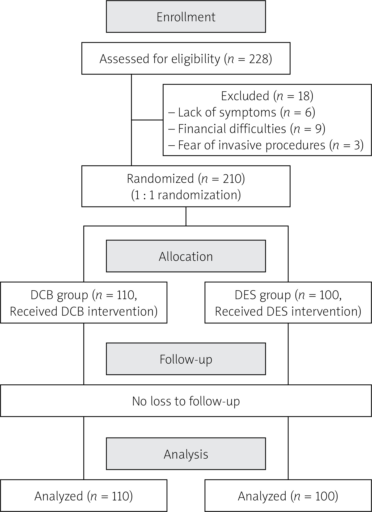
All patients were followed after discharge. Eighteen patients refused angiographic follow-up due to lack of symptoms, financial constraints, or fear of invasive procedures, resulting in a final cohort of 210 patients. Clinical follow-up, conducted via telephone or office visits, occurred every 3 months for 12 months, with the follow-up period concluding on January 30, 2025. Patients underwent angiographic follow-up at 6 months and clinical follow-up at 12 months post-procedure.
The primary endpoint was LLL at 6 and 12 months. LLL was defined as the difference between the post-procedure minimal lumen diameter (MLD) and the MLD at 6 and 12 months post-procedure. Based on the non-inferiority design, the non-inferiority margin (Δ) for LLL was set at 0.15 mm. If the upper limit of the 95% CI for the mean difference in LLL (DCB group minus DES group) was below 0.15 mm, DCB was considered non-inferior to DES. CAG was performed within 24 h before the procedure, immediately post-procedure, and at 6 and 12 months using standardized techniques. Nitroglycerin was administered routinely before angiography, and multiple projections were obtained during the procedure. Angiographic results were analyzed using QAngioXA software (version 7.2, Medis, Leiden, the Netherlands) by two experienced interventional cardiologists.
Secondary outcome measures
Angiographic secondary indicators: Minimal lumen diameter (MLD) and percentage stenosis of the target vessel lesions at different time points were measured and recorded in both groups using three-dimensional reconstruction quantitative analysis (QCA).
Clinical event indicators: During the 6- and 12-month follow-ups, major adverse cardiovascular events (MACE) were evaluated, defined as a composite of cardiac death, myocardial infarction, target vessel revascularization (TVR), and stent thrombosis. Specifically, MACE included cardiac death, non-fatal acute myocardial infarction, and target lesion revascularization (TLR). TLR was characterized by ISR within the previously treated lesion or the 5-mm segments proximal/distal to the stent edges, requiring repeat revascularization (PCI or coronary artery bypass grafting). All endpoint events and adverse events were independently adjudicated by the investigative team.
Safety indicators: Bleeding events were classified according to the BARC bleeding criteria: Type 0 (no bleeding), Type 1 (bleeding not requiring immediate intervention), Type 2 (any overt bleeding requiring intervention but not meeting criteria for Types 3–5, including those needing medical/non-surgical intervention, hospitalization, or evaluation), Type 3 (3a: overt bleeding with hemoglobin drop of 30–50 g/l or requiring transfusion; 3b: overt bleeding with hemoglobin drop ≥ 50 g/l, requiring surgical intervention or vasoactive drugs; 3c: intracranial or vision-impairing bleeding confirmed by autopsy/imaging/lumbar puncture), Type 4 (CABG-related bleeding), and Type 5 (fatal bleeding). Major bleeding events were defined as BARC Type ≥ 3.
Other indicators: Postoperative angina episodes were recorded in detail, including frequency (number of episodes per month) and duration (length of each episode) during the 12-month follow-up period. Cardiac function indicators, including left ventricular ejection fraction (LVEF) and left ventricular end-diastolic diameter (LVEDD), were measured via echocardiography preoperatively and at 6 and 12 months postoperatively.
Statistical analysis
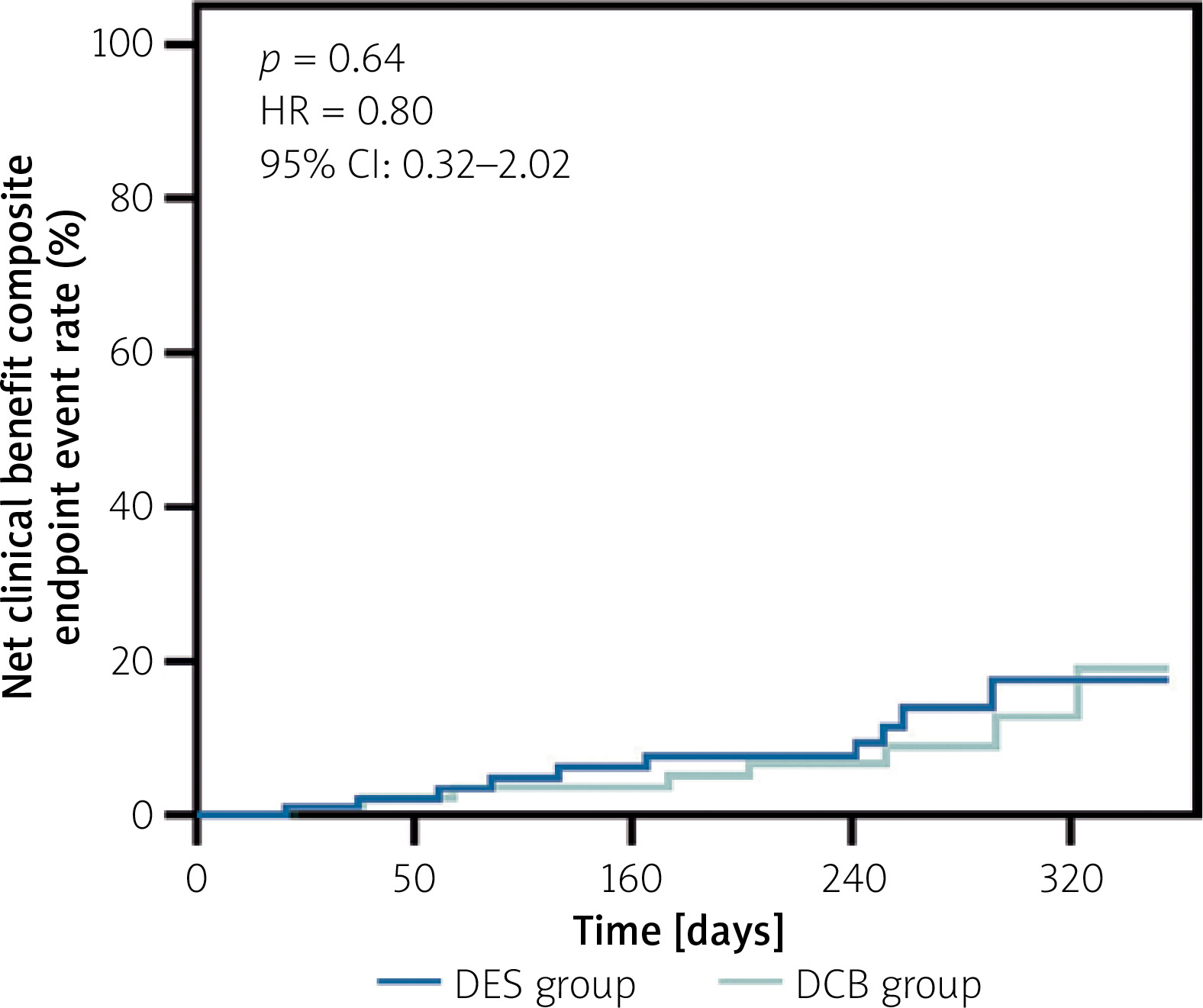
Statistical analysis was performed using SPSS Statistics version 26.0. The intention-to-treat (ITT) principle was followed, and the ITT analysis set was defined as all 228 randomized patients. Continuous variables were tested for normality. Normally distributed data were presented as mean ± standard deviation and compared using Student’s t-test. Non-normally distributed data were expressed as median (interquartile range) and analyzed using the Wilcoxon rank-sum test. Categorical variables were compared using the χ2 test. The primary endpoint was LLL at 6 and 12 months post-procedure. The mean difference between the two groups and its 95% CI (based on the t-distribution with Welch-Satterthwaite degrees of freedom) were calculated. A one-sided non-inferiority test was used with a significance level of α = 0.025. Non-inferiority was established if the upper limit of the 95% CI was below the non-inferiority margin of 0.15 mm. Kaplan-Meier survival analysis was used to compare net clinical benefit composite endpoint events between the DCB and DES groups. A p-value < 0.05 was considered statistically significant.
Results
Comparison of baseline characteristics between groups
During follow-up, 18 patients refused angiographic follow-up (14 in the DCB group and 4 in the DES group). A total of 210 patients ultimately completed angiographic follow-up, yielding an angiographic success rate of 92.11% (210/228). The CONSORT flow diagram is shown in Figure 1. The clinicopathological characteristics are detailed in Table I. The DCB group included 31 males and 79 females (age range: 52–69 years; mean: 60.74 ±8.25), while the DES group comprised 26 males and 74 females (age range: 54–69 years; mean: 61.67 ±7.41). No significant differences existed in age, sex distribution, smoking history, hypertension, diabetes mellitus, or BMI (all p > 0.05). Preoperative biochemical parameters (TG, TC, FBG, CK-MB, BNP) were comparable between groups (p > 0.05). Mehran classification showed significantly more Type I lesions in the DCB group than the DES group (p > 0.05), with fewer Type II–IV lesions. Regarding lesion characteristics, no statistically significant differences were observed between the two groups in lesion location, lesion length, reference vessel diameter, or the proportions of calcified lesions and bifurcation lesions.
Table I
Comparison of baseline characteristics between DES and DCB groups
[i] Statistical note: Normally distributed continuous variables are presented as mean ± SD (Student’s t-test); non-normally distributed data as median (IQR) (Wilcoxon rank-sum test); categorical variables as n (%) (χ² test). P < 0.05 was considered statistically significant. DES – drug-eluting stent, DCB – drug-coated balloon, BMI – body mass index, CK-MB – creatine kinase-MB, WBC – white blood cell count, RBC – red blood cell count, TC – total cholesterol, TG – triglyceride, HDL-C – high-density lipoprotein cholesterol, LDL-C – low-density lipoprotein cholesterol, FBG – fasting blood glucose, TPP – thrombus precursor protein, BNP – B-type natriuretic peptide.
Non-inferiority comparison of the primary endpoint between groups
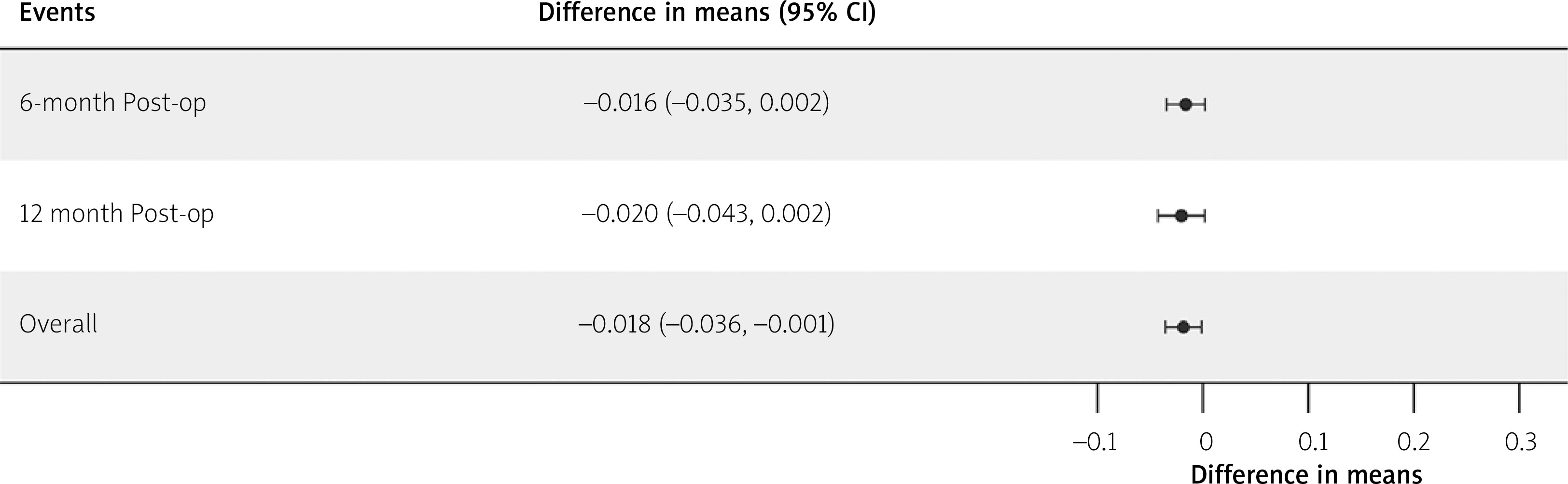
The non-inferiority analysis showed that at 6 months post-procedure, the 95% CI for the mean difference in LLL (DCB group minus DES group) was (–0.0346, 0.0020), with an upper limit (0.0020) far below the non-inferiority margin of 0.15 mm (p for non-inferiority < 0.001). At 12 months post-procedure, the 95% CI for the mean difference in LLL was (–0.0426, 0.0018), and the upper limit (0.0018) was also far below the non-inferiority margin of 0.15 mm (p for non-inferiority < 0.001). Furthermore, the 95% CIs for the mean difference in LLL at both 6 and 12 months contained 0, indicating that the difference in LLL between the two groups was not statistically significant, suggesting that the DCB group performed comparably to the DES group with respect to LLL.
Taking the average of LLL at 6 and 12 months for the overall analysis, the mean difference between the two groups was –0.0180 mm, with a 95% CI of (–0.0356, –0.0012). The upper limit of the 95% CI was far below the non-inferiority margin of 0.15 mm. In summary, when analyzed separately at 6 and 12 months, there was no statistically significant difference in LLL between the two groups; however, the overall mean analysis showed that LLL in the DCB group was significantly lower than that in the DES group (with the 95% CI not containing 0), further strongly supporting the non-inferiority of DCB (Table II, Figure 2).
Table II
Non-inferiority comparison of the primary endpoint (LLL at 6 and 12 months post-procedure) between groups
[i] Statistical note: Non-inferiority was assessed using a one-sided test with α = 0.025. The mean difference and 95% CI were calculated based on the Welch–Satterthwaite t-distribution. The non-inferiority margin (Δ) was set at 0.15 mm. Non-inferiority was concluded if the upper limit of the 95% CI for the mean difference was below 0.15 mm. P < 0.025 was considered statistically significant for the non-inferiority test. LLL – late lumen loss, SE – standard error, CI – confidence interval, DES – drug-eluting stent, DCB – drug-coated balloon.
Comparison of angiographic outcomes at different timepoints between groups
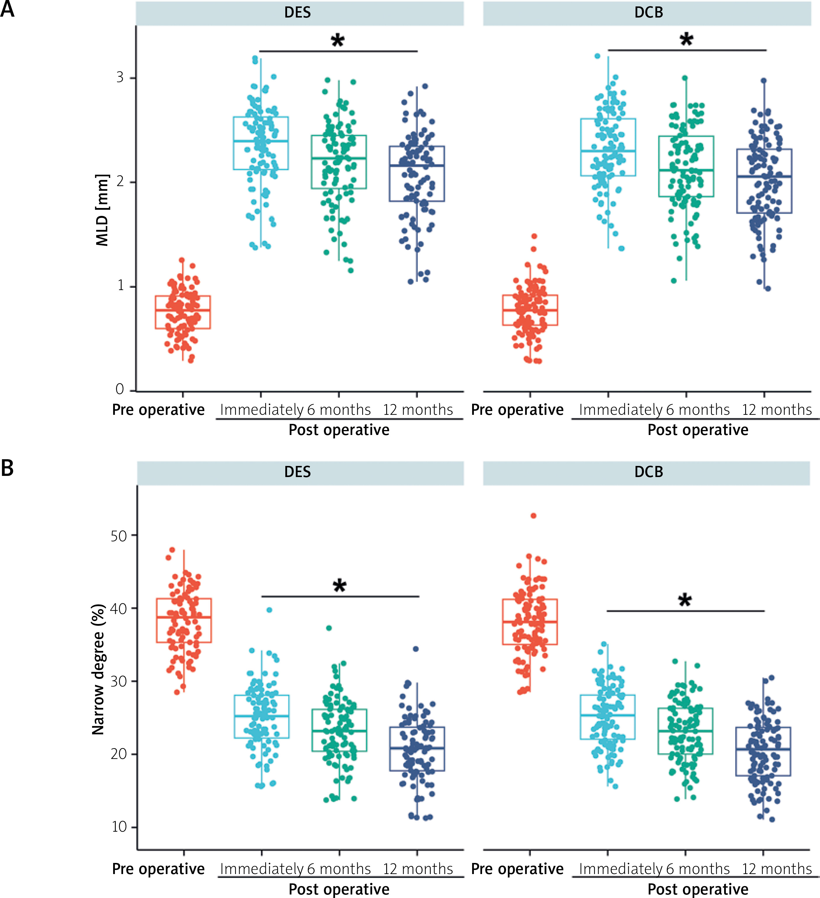
No intergroup differences were observed in MLD or percent stenosis at baseline or immediately post-procedure (all p > 0.05). At 6 and 12 months, both groups demonstrated significant improvements in MLD and percent stenosis versus baseline (all p < 0.05), with comparable efficacy between DCB and DES (all p > 0.05; Figure 3). These results indicate equivalent performance in lumen diameter improvement and stenosis reduction.
Comparison of MACE and postoperative angina between groups
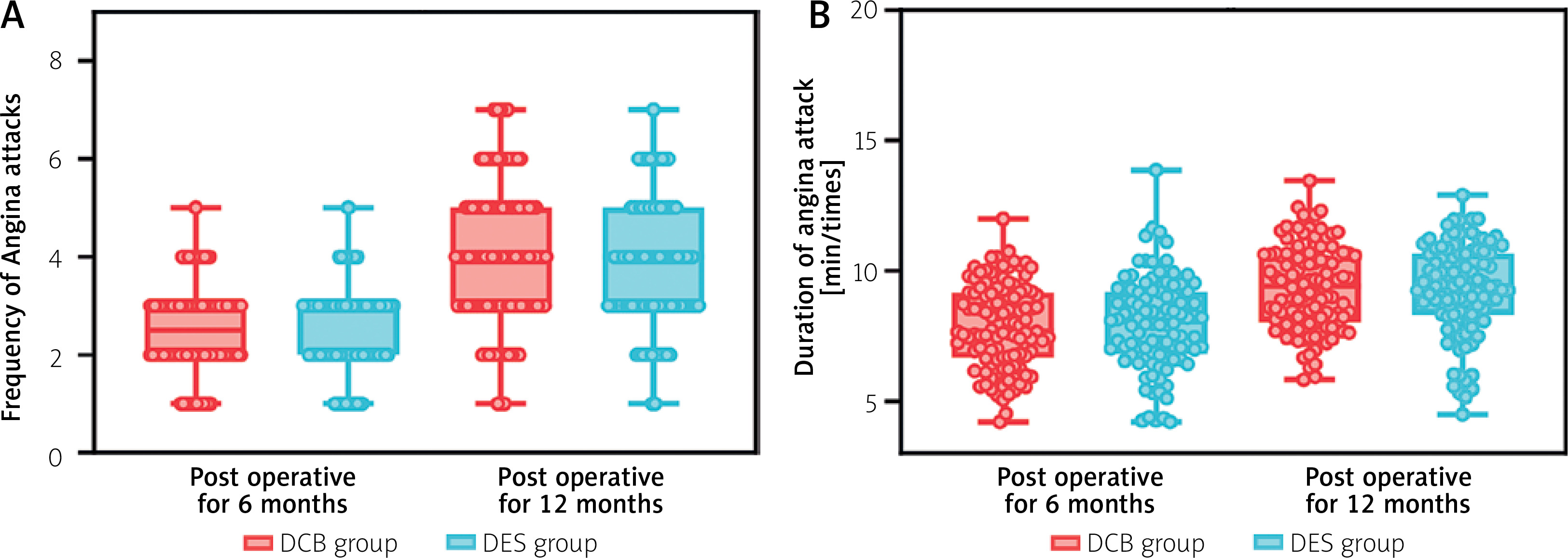
At 6-month follow-up, the MACE rate was 2.00% (2/100) in the DES group (1 stent thrombosis, 1 TVR) versus 4.55% (5/110) in the DCB group (1 cardiac death, 3 TVR, 1 stent thrombosis), with no significant difference (p > 0.05). By 12 months, MACE rates increased to 8.00% (8/100) and 9.09% (10/110) in the DES and DCB groups, respectively, remaining comparable (p > 0.05; Table III). The DCB group showed significantly lower post-procedural angina frequency than the DES group (p = 0.03), though angina duration was similar (p > 0.05). At 6 and 12 months, both angina frequency and duration showed no significant intergroup differences (p > 0.05; Figure 4).
Table III
Comparison of MACE between DCB and DES groups
Comparison of cardiac function and major bleeding events between groups
Both groups exhibited improved LVEF and reduced LVEDD after treatment versus baseline, with no significant intergroup differences (p > 0.05; Table IV). The DCB group showed a non-significant increase in BARC ≤ 2 bleeding events (p > 0.05), while BARC ≥ 3 events were comparable between groups (p > 0.05; Table V). Kaplan-Meier analysis demonstrated similar net clinical benefit (hazard ratio = 0.80, 95% CI: 0.32–2.02; log-rank p = 0.64; Figure 5).
Table IV
Comparison of cardiac function parameters between DCB and DES groups
[i] Statistical note: Normally distributed continuous variables are presented as mean ± SD (Student’s t-test). P < 0.05 indicates statistically significant differences versus preoperative values. DES – drug-eluting stent, DCB – drug-coated balloon, LVEF – left ventricular ejection fraction, LVEDD – left ventricular end-diastolic diameter.
Table V
Comparison of bleeding events between DCB and DES groups
Discussion
DES therapy has not been shown to reduce recurrent myocardial infarction or mortality. Endothelial dysfunction, neoatherosclerosis, and thrombotic complications following DES implantation significantly impair clinical outcomes and treatment efficacy [15]. While DCB has emerged as a promising strategy for ISR management, its adoption for coronary lesions remains limited due to insufficient clinical evidence. This study demonstrates comparable clinical outcomes between DCB and DES, providing new evidence to guide interventional strategies.
The evolution from BMS to DES as the primary PCI option has enhanced procedural safety and efficacy. However, persistent challenges including stent thrombosis, delayed healing, and hypersensitivity reactions have motivated the development of stent-free DCB approaches [16]. The lipophilic properties of paclitaxel enable effective vascular delivery, inhibiting smooth muscle proliferation through multiple molecular pathways [17]. DCB achieves direct drug transfer to vessel walls, providing potent suppression of neointimal hyperplasia [18]. The results of this study showed that LLL at 6 and 12 months post-procedure in the DCB group was non-inferior to that in the DES group (the upper limit of the 95% CI was well below the margin of 0.15 mm), and was significantly lower in the overall mean analysis. This is consistent with the study by Refaat et al., in which patients treated with DCB had significantly lower in-lesion LLL compared with DES [19], and also aligns with the findings of Shin et al. [20]. The stent-free DCB approach mediates anti-inflammatory and antiproliferative effects without permanent implants [10], while potentially mitigating the negative remodeling risks of conventional angioplasty. Late lumen enlargement occurs in 50.5–74% of DCB cases through plaque reduction and positive remodeling [21, 22]. The randomized controlled trial by Mahajan et al. compared the efficacy of paclitaxel drug-eluting balloon (DEB) versus second-generation DES for the treatment of bare metal stent ISR, with the primary endpoint of 6-month LLL. The results showed no significant difference in LLL between the two strategies, which is consistent with the findings of the present study [23]. However, it should be noted that the study population of Mahajan et al. consisted of ISR lesions, whereas the target lesions in the present study were de novo non-complex small-vessel lesions. Given the differences in pathophysiology and remodeling responses between ISR and de novo lesions, the conclusions of the present study should not be directly extrapolated to ISR or more complex lesion populations. Further investigation with larger, multi-center samples and extended follow-up is needed to clarify the precise role of DCB. In addition, a significant difference in adherence to follow-up angiography was observed between the two groups in this study (14 patients in the DCB group vs. 4 patients in the DES group did not undergo control angiography), which is related to the characteristics of the two treatment modalities. Patients in the DES group, having permanent metallic stents implanted, experienced greater psychological burden and required longer dual antiplatelet therapy; consequently, both physicians and patients were more vigilant about the risk of ISR, leading to higher adherence to angiographic follow-up. In contrast, patients in the DCB group had no foreign body implantation and experienced rapid symptom relief, often subjectively believing that they were “cured”. Unless angina recurred, they were frequently reluctant to undergo repeat invasive coronary angiography. Therefore, this asymmetric loss to follow-up carries a theoretical risk of survival bias. Although the robustness of the results is supported by the fact that the upper limit of the 95% CI for the mean difference in LLL (0.0020) was far below the non-inferiority margin (0.15 mm), future studies should explore more patient-friendly non-invasive imaging follow-up methods to reduce such bias [24, 25].
DES exhibit suboptimal drug distribution (< 20% vessel wall coverage), with heterogeneous diffusion patterns [26]. The high local drug concentrations required for efficacy may aggravate inflammation, delayed endothelialization, and thrombosis risk [27]. In contrast, DCB ensures uniform drug coverage across treated segments, preventing hemodynamic complications from uneven drug distribution [9]. This is supported by Gobić et al.’s reported 5.4% MACE rate with DES versus 0% with DCB [28], along with STEMI studies confirming DCB’s safety and efficacy at 1-year follow-up [29]. Although no statistically significant difference was observed in MACE incidence between the DCB and DES groups (9.09% vs. 8.00%) in this study, suggesting that the DCB strategy did not increase the risk of major adverse events, it must be emphasized that the study population mainly consisted of patients with de novo non-complex lesions and was not specifically designed as an acute myocardial infarction (AMI) cohort. Therefore, the data from this study are insufficient to draw definitive conclusions regarding the routine use of DCB in AMI management; the findings should be considered exploratory and require further validation in dedicated future studies.
The DCB group demonstrated lower post-procedural angina frequency compared to the DES group, likely due to more effective suppression of restenosis and subsequent reduction in myocardial ischemia [30]. However, angina duration showed no significant difference, potentially influenced by multiple factors including myocardial oxygen supply-demand balance and neural regulation. Both groups exhibited significant improvement in left ventricular function parameters (LVEF and LVEDD) from baseline without intergroup differences, suggesting comparable short-term cardiac recovery that may require extended follow-up to identify potential differential effects. The study revealed no statistically significant differences in MACE or BARC ≥ 3 bleeding events between groups (all p > 0.05), aligning with Scheller et al.’s findings of generally low major bleeding rates [31].
Although Mehran classification distribution differed significantly between groups, this did not translate to differences in clinical outcomes. This supports the clinical value of DCB for Type I lesions, offering advantages such as shorter dual antiplatelet therapy duration and avoidance of multiple stent layers, while achieving similar luminal gain to DES.
Several limitations warrant consideration: First, the primary endpoint of this study was LLL, an angiographic surrogate endpoint. Although it sensitively reflects local intimal hyperplasia, it cannot fully replace clinical hard endpoints such as cardiac death or myocardial infarction to adequately capture clinical benefit. Second, the follow-up duration was only 12 months, which is relatively short for evaluating late adverse events such as very late stent thrombosis with DES or long-term LLL with DCB, potentially underestimating long-term risks. This was a single-center prospective study with a limited sample size. Third, although the 6 patients who required bailout stenting due to dissection during DCB treatment were included in the analysis, 14 patients in the DCB group and only 4 in the DES group refused angiographic follow-up. In a non-inferiority trial, such unbalanced loss to follow-up could lead to convergence of the observed LLL values between the two groups, presenting a theoretical risk of false non-inferiority. However, the upper limit of the 95% CI for the mean difference in LLL observed in this study (0.0020) was far below the non-inferiority margin (0.15 mm), leaving a substantial margin. Even with the most conservative imputation methods for missing data or per-protocol sensitivity analysis excluding crossover cases, the non-inferiority conclusion remains robust. Therefore, large-scale, multi-center confirmatory trials with clinical endpoints and follow-up periods of 2–5 years are still needed to comprehensively evaluate the long-term safety and efficacy of DCB. Furthermore, the use of intravascular imaging (IVUS/OCT) was relatively low in this study; lesion assessment and dissection judgment relied mainly on coronary angiography, which may have missed some mild vessel wall injury or malapposition. Future studies guided by intracoronary imaging are needed to verify the long-term efficacy of DCB.
In conclusion, for de novo non-complex small-vessel lesions, DCB met the non-inferiority criterion compared with DES for LLL at 1-year follow-up, without significantly increasing MACE risk. It should be emphasized that the conclusions of this study apply only to coronary small-vessel lesions with diameters of 2.25–2.75 mm and should not be directly extrapolated to large-vessel or more complex lesion types. Given the limitations of the study, including the limited sample size and asymmetric loss to follow-up, the above findings should be considered exploratory and hypothesis-generating rather than definitive conclusions. Thus, the results provide preliminary evidence for DCB as an alternative treatment strategy for small-vessel disease. For diffuse or complex ISR lesions, further evidence is still needed to support the efficacy of DCB. Overall, the findings of this study await confirmation by future large-scale, multi-center confirmatory randomized controlled trials to verify long-term efficacy and safety under a more rigorous statistical framework.
Ethical approval
The study protocol was approved by the Ethics Committee of Fuwai Yunnan Hospital, Chinese Academy of Medical Sciences (approval number: IRB2022-BG-006), and the study was conducted in strict accordance with the ethical principles of the Declaration of Helsinki. Written informed consent was voluntarily provided by all participants and their legal guardians before enrollment.