
Introduction
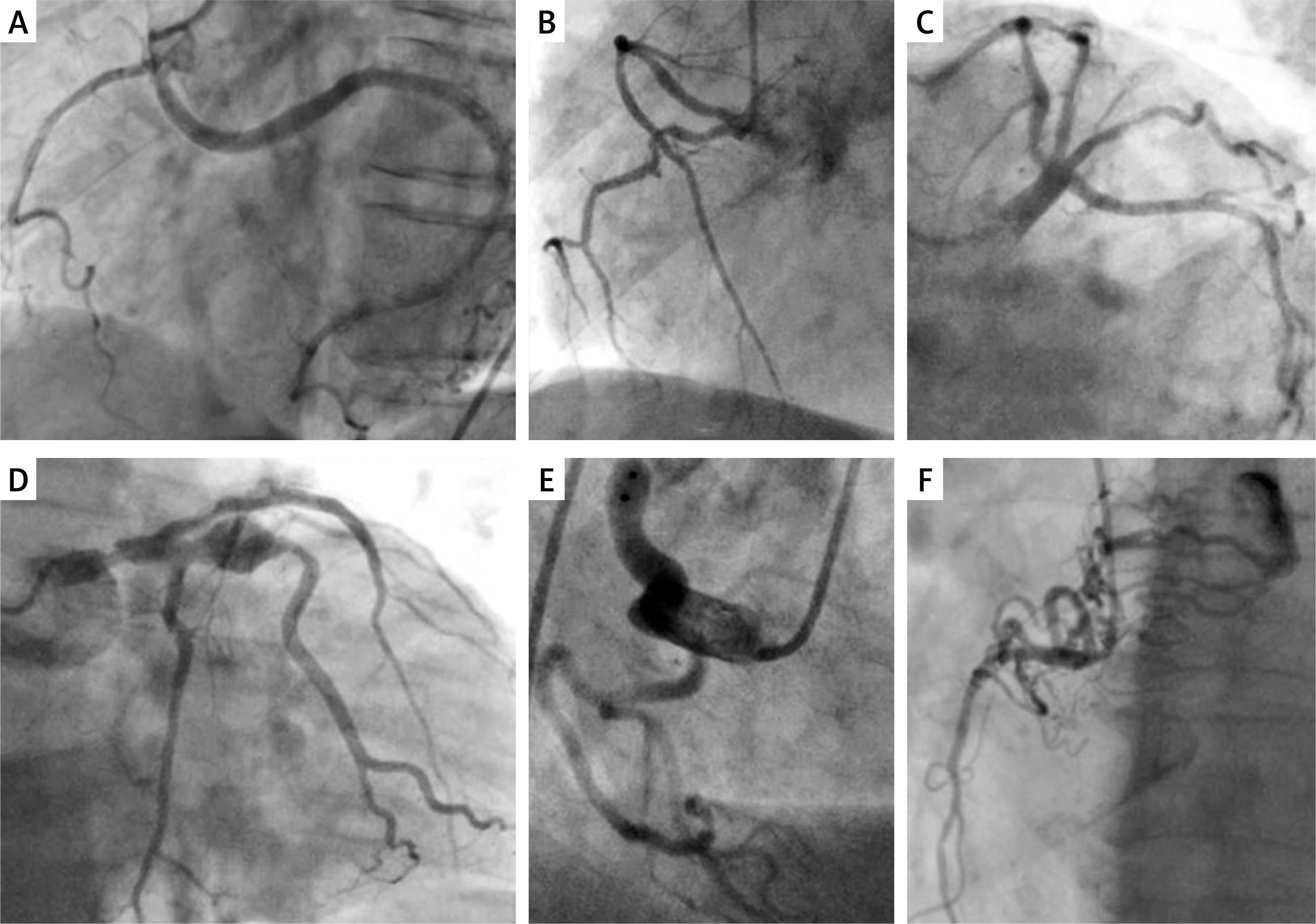
Anomalous coronary arteries (ACA) represent a heterogeneous group of congenital variants occurring in approximately 0.5–1.5% of the population and encompassing a wide anatomical spectrum (Figure 1). While many anomalies are incidental findings with limited clinical impact, selected patterns are associated with myocardial ischemia, arrhythmias, or sudden cardiac death. Despite their potential clinical relevance, communication and management of ACA remain inconsistent, largely due to the absence of standardized, clinically oriented reporting. Existing classification systems [1, 2], most notably Angelini’s framework, provide a scientifically robust anatomical description focused on coronary origin, course, and intrinsic morphology. However, their complexity and limited integration with risk stratification and procedural planning have contributed to underutilization in everyday clinical practice. To address these limitations, we propose the OCBMR mnemonic – Origin, Course, Branching, Morphology, Risk (“Observe Coronaries Before Making the Report”) – as a structured and practical framework that integrates detailed anatomical description with clinical and procedural relevance. OCBMR is intended as both an educational tool and a standardized reporting approach for coronary computed tomography angiography (CCTA) and invasive coronary angiography (ICA), while remaining fully compatible with established classification systems.
Figure 1
Invasive coronary angiography. A – Anomalous origin of the left circumflex coronary artery from the right coronary artery (OCBMR coding: Q2-C2-B1-M1-R0); B – Duplication of the right coronary artery (OCBMR coding: O1-C1-B2-M1-R0); C – Quadrifurcation of the left main coronary artery (OCBMR coding: O1-C1-B4-M1-R0); D – Aneurysm of the intermediate branch of the left coronary artery (OCBMR coding: O1-C1-B1-M4-R1); E – Large fistula from the right coronary artery to right atrium (OCBMR coding: O1-C1-B1-M5-R2); F – Fistula from the right coronary artery to the pulmonary trunk (OCBMR coding: O1-C1-B1-M5-R1)
OCBMR
The OCBMR Framework OCBMR is applied sequentially, progressing from Origin to Course, Branching, Morphology, and Risk (Table I).
Table I
The OCBMR framework of coronary artery anomalies
[i] ACA – anomalous coronary arteries, ALCAPA – anomalous left coronary artery from the pulmonary artery, ARCAPA – anomalous right coronary artery from the pulmonary artery, CCTA – coronary computed tomography angiography, ICA – invasive coronary angiography, IVUS – intravascular ultrasound, OCT – optical coherence tomography, OCBMR – Origin → Course → Branching → Morphology → Risk (Observe Coronaries Before Making the Report), PCI – percutaneous coronary intervention, SCD – sudden cardiac death.
Origin (O)
O1: Normal origin from the appropriate sinus
O2: Origin from the opposite sinus
O3: Single coronary artery
O4: Anomalous left coronary artery from the pulmonary artery (ALCAPA)
O5: Anomalous right coronary artery from the pulmonary artery (ARCAPA)
Precise definition of origin is critical for surgical and interventional planning [3–6], and CCTA is the preferred modality in stable patients [7, 8], although not always possible in urgent cases [9].
Course (C)
C1: Prepulmonic
C2: Retroaortic
C3: Interarterial (between the aorta and pulmonary artery)
C4: Proximal intramural course within the aortic wall
C3 is considered potentially malignant [10], with highest risk when combined with C4, slit-like ostium, or ischemia. Myocardial bridging is excluded from C4.
Branching (B)
B1: Standard bifurcation
B2: Duplication
B3: Complex branching (trifurcation)
B4: Complex branching (quadrifurcation)
B5: Complex branching (pentafurcation)
Although B3 is a common anatomical variant, it is included due to its significant impact on the complexity of percutaneous coronary intervention [11, 12].
Morphology (M)
M1: Normal ostium
M2: High take-off
M3: Slit-like, fish-mouth, or multiple ostia
M4: Aneurysmal dilation
M5: Coronary fistula
M1–M3 describe ostial morphology, while M4–M5 represent associated structural abnormalities relevant for intervention.
Risk (R)
R0: Benign anatomy without symptoms or ischemia
R1: Intermediate risk anatomy or equivocal findings
R2: High-risk anatomy and/or documented ischemia, arrhythmia, or cardiac events
Risk classification integrates anatomy, clinical presentation, and functional testing.
Clinical application
OCBMR is primarily derived from CCTA and ICA, supplemented by intracoronary imaging when necessary. A typical report follows the sequence O®C®B®M®R, ensuring completeness.
For example: O2-C3-C4-B1-M3-R2 describes an anomalous coronary artery from the opposite sinus with interarterial and intramural course, slit-like ostium, and high-risk classification (Figure 1).
Advantages
OCBMR standardizes reporting, enhances interdisciplinary communication, and directly links anatomy to procedural and clinical implications. Its mnemonic structure facilitates teaching and rapid adoption.
Limitations
OCBMR is not a validated prognostic score and does not include all anatomical modifiers, such as intramural length or acute take-off angle. Borderline cases may be subject to interpretive variability. Images from ICA may be inconclusive for exact OCBMR coding. In doubtful cases, evaluation with CCTA is necessary.
Conclusion
OCBMR provides a structured, clinically meaningful, and educational framework for reporting coronary artery anomalies. By integrating anatomical detail with clinical relevance, it complements existing classifications and supports consistent communication and patient-centered decision-making.










