
Introduction
Metastatic colorectal cancer (mCRC) represents a major global health burden and is one of the leading causes of cancer-related morbidity and mortality worldwide [1]. In 2022, colorectal cancer (CRC) was ranked as the third highest cause of death in the United States of America. In patients with mCRC the 5-year survival rate was approximately 15%, underscoring the ongoing urgent need to optimize screening and systemic therapy strategies [2]. Although CRC is a preventable malignancy due to excellent screening programs and the removal of pre-cancerous lesions, its incidence is anticipated to rise [3].
Despite the emergence of a new targeted therapy and the revolution in immunotherapy options, fluoropyrimidine-based multidrug regimens (MDR) continue to form the backbone of first-line treatment in mCRC. According to the National Comprehensive Cancer Network Clinical Practice Guidelines, regimens such as FOLFOX, FOLFIRI, and FOLFIRINOX remain essential treatment protocols and are considered the standard of care [4]. These protocols basically include two forms of 5-fluorouracil (5-FU): a short intravenous (IV) bolus, followed by a 46- to 48-hour continuous infusion, usually using a specialized pump [5].
For decades, fluorouracil has been considered the drug of choice in the treatment of mCRC. In the early 2000s, the standard of care evolved into MDR after it was discovered that adding oxaliplatin or irinotecan to the chemotherapy regimen can enhance efficacy, progression-free survival (PFS) and overall survival (OS) [6].
While the value of the 5-FU bolus was well established in the older single- drug regimens, its role in these newer multidrug combinations was never thoroughly tested and was largely just assumed, and there is no general consensus concerning the best schedule or the optimal dose for this treatment [7].
The 5-FU is not one drug; the efficacy and toxicity differ according to dosage, method and route of administration. The different modes of 5-FU delivery, either IV bolus or continuous infusion produce distinct pharmacodynamics and molecular effects. Bolus IV administration generates a rapid and high peak plasma concentration of 5-FU and its downstream metabolites, favouring the accumulation of fluorodeoxyuridine triphosphate (FdUTP) and fluorouridine triphosphate (FUTP) [8].
Following their misincorporation into DNA and RNA, respectively, these metabolites cause cytotoxicity by interfering with the synthesis and function of nucleic acids. On the other hand, continuous infusion sustains the formation of fluorodeoxyuridine monophosphate (FdUMP) by maintaining a prolonged systemic exposure to 5-FU [7]. With thymidylate synthase (TS) and reduced folates, this metabolite forms a stable ternary complex that results in long-lasting TS inhibition and the subsequent depletion of intracellular thymidine pools, especially during the S-phase of the cell cycle. Because of this prolonged TS suppression, continuous infusion regimens have historically shown better antitumor activity in CRC when compared to bolus administration [9].
The toxicity spectrum also diverges according to the administration schedule: bolus 5-FU is more frequently associated with myelosuppression, whereas continuous IV infusion is more commonly linked to gastrointestinal side effects, including diarrhoea, mucositis, nausea, vomiting, and hand-foot syndrome [8].
There are limited high-quality studies evaluating whether omission of the 5-FU bolus compromises treatment efficacy. Notably, capecitabine, an oral fluoropyrimidine that mimics the pharmacokinetic profile of continuous infusion of 5-FU and lacks any bolus component, has demonstrated comparable efficacy to IV 5-FU in multiple randomised trials, suggesting that the bolus may not be essential for therapeutic effect [10]. A recent questionnaire of gastrointestinal oncologists reported that 20–40% routinely omit the 5-FU bolus preemptively, particularly among senior clinicians and those specializing as experts in gastrointestinal malignancies [11].
Existing retrospective data provide conflicting findings, but most have been limited by small sample sizes and inadequate adjustment for baseline differences between treatment arms [12]. Moreover, the recent 5-FU drug shortages have shifted the focus to the priority of identifying drug components of standard regimens that may be safely reduced or omitted without compromising survival outcomes. Given the reported haematologic and specific toxicity associated with 5-FU bolus, predominately neutropenia, mucositis, and cytopenias, the priorities of focused studies to answer whether the bolus contributes meaningful clinical benefits, especially in the era of modern oncology practice, were urged.
To address this clinically relevant and increasingly common question, we conducted a retrospective cohort study to evaluate the impact of omitting the 5-FU bolus on survival outcomes and toxicity among patients receiving standard multidrug 5-FU-based regimens for mCRC.
Material and methods
Study design and patient population
This retrospective, multicentre cohort study was carried out in two university hospitals. It evaluated patients newly diagnosed with mCRC who received first-line mFOLFOX6-based chemotherapy. The study was conducted between June 2020 and June 2024 and adhered to the Strengthening the Reporting of Observational Studies in Epidemiology guidelines for observational research.
A total of 267 patients were included in the analysis: 126 received the standard mFOLFOX6 regimen (with 5-FU bolus), while 141 received a modified regimen without the 5-FU bolus (nbFOLFOX6). All regimens were administered with or without concurrent bevacizumab or an anti- epidermal growth factor receptor (EGFR) monoclonal antibody, as per standard clinical practice.
Patients were eligible if they met all of the following: age ≥ 18 years at the time of treatment initiation and histologically confirmed metastatic colorectal adenocarcinoma not amenable to either upfront surgery or conversion treatment followed by surgery. All the patients were newly diagnosed and did not receive any chemotherapy as an adjuvant treatment. Patients were excluded if any of the following applied: failure to attend scheduled follow-up visits or loss to follow-up immediately after treatment initiation, transfer of care to another institution before primary endpoint assessment or incomplete or missing medical records that prevented reliable evaluation of survival outcomes or toxicity profiles. Patients who had oligometastatic disease that could be surgically resected either upfront or after conversion chemotherapy were excluded as well to minimize confounding factors that may alter survival analysis. Patients who received a dose density less than 75% were excluded as well. To reduce selection bias and guarantee representativeness of actual clinical practice, consecutive eligible patients were included.
Sample size and sampling method
All consecutive patients who met eligibility criteria during the study period were included (convenience consecutive sampling). No prospective formal sample-size calculation was performed because the study is retrospective and exploratory; the final sample size reflects real-world caseload at the two centres in using this regimen per se in the metastatic setting.
Ethical consideration
The study protocol was reviewed and approved by the Institutional Review Board (IRB) of the sharing centres. In compliance with local regulations, the IRB waived the need for written informed consent because the study used de-identified retrospective clinical data. The study was not a prospective interventional trial and therefore was not registered in a clinical trials registry.
Data sources and collection procedures
Clinical and laboratory data were abstracted from electronic health records and paper charts using a standardized case report form. Extracted variables included demographics (age, sex), performance status (Eastern Cooperative Oncology Group – ECOG), tumour characteristics (primary tumour sidedness, sites of metastasis), molecular markers (RAS, BRAF, MSI status), comorbidities, prior systemic therapies, chemotherapy dosing and schedule, cycle intervals, baseline and on-treatment laboratory values (complete blood count, renal and hepatic panels), and documentation of adverse events and supportive care (including granulocyte colony-stimulating factor [G-CSF] use).
Laboratory assays (complete blood counts, renal and hepatic panels) were performed in each centre’s accredited clinical laboratory using standard automated analysers. Reference ranges and instrument models are recorded in the study database and can be provided on request. Molecular testing (RAS, BRAF, MSI) was performed according to local laboratory protocols (PCR-based platform); the testing method and date for each patient are recorded and were considered in sensitivity analyses.
Data abstraction was performed by trained clinical research staff at each site using a common data dictionary. A 10% random sample of records was re-abstracted by a second reviewer to assess inter-rater reliability; discrepancies were resolved by consensus. Abstractors were not blinded to the treatment group because treatment assignment is recorded in the chart; however, outcome measurement for survival and radiologic progression used objective documentation (death dates, radiology reports) to reduce measurement bias.
Metastatic CRC patients were stratified into two exposure groups based on chemotherapy administration records. The standard (FOLFOX6) arm group: patients received a 5-FU bolus of 400 mg/m2 on day 1 of cycle 1, along with leucovorin and the planned infusional 5-FU. However, the non-bolus (nbFOLFOX6) arm group: patients did not receive the 5-FU bolus on day 1 of cycle 1 but received the same planned infusional 5-FU. For both arms the planned continuous infusion dose of 5-FU was 2400 mg/m2 per cycle. Concomitant agents (leucovorin, bevacizumab, anti-EGFR) and any dose modifications, delays, or early discontinuations were recorded. Supportive measures like antiemetics, G-CSF administration, and transfusions, along with their timing in relation to chemotherapy cycles, were recorded.
Outcome measures and definitions
Primary outcomes
The primary outcomes of this retrospective study included the haematologic adverse events and the PFS. According to the Common Terminology Criteria for Adverse Events (CTCAE) version 5.0, the haematologic adverse events were defined as the incidence and severity of anemia, neutropenia, and thrombocytopenia occurring within the treatment cycles. Progression-free survival was calculated as the time from initiation of treatment to clinically or radiologically documented disease progression or death from any cause, whichever came first.
Secondary outcomes
Secondary outcomes of our study were overall survival, non-haematologic adverse effects (AEs), or G-CSF use. Overall survival was measured as the time from treatment initiation to death from any cause or last follow-up. Non-haematologic AEs included incidence and grade of diarrhoea, vomiting, neuropathy, and other treatment-related toxicities (CTCAE v5.0). Receiving of G-CSF by patients after chemotherapy and timing relative to neutropenic events was assessed as a secondary outcome.
Adverse events were captured from clinical notes, nursing documentation, and laboratory results. Neutropenia thresholds followed CTCAE definitions. Laboratory assays were performed in accredited hospital laboratories at each site using routine clinical methods; calibration and normal ranges were those reported by the local laboratory and are available on request.
The extent and pattern of missing data for key variables were assessed and reported. Primary analyses used complete-case methods; if missingness exceeded 10% for a key covariate, multiple imputation by chained equations was performed as a sensitivity analysis under the missing at random assumption. Sensitivity analyses also included models restricted to patients with complete molecular marker data.
Statistical analysis
Analyses were pre-specified in a statistical analysis plan. All tests were two-sided with α = 0.05. Data management and analyses were performed using IBM SPSS Statistics for Windows, version 27.0. Categorical variables are presented as counts and percentages; continuous variables as mean ± standard deviation or median (inter quartile range) depending on distribution. Normality was assessed with the Kolmogorov-Smirnov test. Independent Student t test was performed for normally distributed continuous variables; Mann-Whitney U test for non-normal continuous variables; χ2 test for categorical variables with Fisher’s exact test or Monte Carlo correction when cell counts were small. Survival analysis: Kaplan-Meier method was used to estimate PFS and OS; groups were compared with the log-rank test. Cox proportional hazards regression was used to estimate hazard ratios (HR) with 95% confidence intervals; proportional hazards assumptions were evaluated using Schoenfeld residuals.
Adjustment for confounding: propensity scores for receiving the 5-FU bolus were estimated by logistic regression including baseline covariates (age, sex, ECOG, primary tumour sidedness, sites of metastasis, RAS/BRAF/MSI status, prior chemotherapy, comorbidities). Multivariable logistic regression was used for binary toxicity outcomes and multivariable Cox models for survival outcomes adjusted for clinically relevant covariates. Post hoc subgroup analyses by molecular status and concomitant targeted therapy are reported with interaction tests. All effect estimates are presented with 95% confidence intervals.
Results
This retrospective study enrolled 267 patients with mCRC. The baseline demographic and tumour-related characteristics of the mCRC patients, as illustrated in Table 1, demonstrate excellent pre-treatment comparability between the standard FOLFOX (n = 126 patients) and nbFOLFOX (n = 141patients) groups, with statistically non-significant differences in all demographic or tumour- related characteristics (p > 0.05 for all), herewith enhancing the validity of subsequent toxicity and survival comparisons and minimizing any confounding bias. In both arms, the mean age was approximately ~53.7 years (p = 0.850), with a slight male predominance (60.3% vs. 50.4%, p = 0.102). Regarding the tumour site, left-sided mCRC predominated in both groups (62.7% in FOLFOX and 55.3% in nbFOLFOX), with a balanced distribution (p = 0.221). Notably, distribution of metastatic sites was comparable, as there were no significant differences between the two groups (p = 0.748), with liver metastasis in 46% and 44% and multi-site metastasis in 27.8% and 31.2% ensuring comparable metastatic burden.
Table 1
Comparison between FOLFOX and nbFOLFOX groups according to demographics data and tumour-related characteristics
The distribution of ECOG performance status, a strong prognostic factor in mCRC that influences both treatment tolerance and survival outcomes, was well-balanced between the two groups (p = 0.347), indicating no significant difference in baseline functional status. Notably, the majority of patients in both groups had good performance status, where PS0, PS1, and PS2 were 53.2%, 33.3%, and 13.5%, and 52.5%, 39.0%, and 8.5%, respectively (Table 1).
Overall as shown in Table 1, the molecular characteristics were well-balanced between the two groups, with no statistically significant differences observed (p > 0.05 for all). The Kirsten rat sarcoma viral oncogene homolog (KRAS) mutant was observed in 56.3% vs. 58.9% (p = 0.678), the B-Raf proto-oncogene, serine/threonine kinase (BRAF) mutant showed low prevalence, in 6.3% vs. 6.4% (p = 0.991), and only 4.0% vs. 3.5% (p = 1.000) of patients had high microsatellite instability (MSI). Thus, this comparable molecular profile indicates generalizability to actual mCRC populations and enhances the study’s internal validity.
Table 1 demonstrates a balanced use of targeted agents between the FOLFOX and nbFOLFOX groups (p = 0.357). The most frequently used agent was the monoclonal antibody bevacizumab (57.9% in FOLFOX vs. 61.0% in nbFOLFOX). The overall proportions of the EGFR inhibitor agents (cetuximab 19.0% vs. 22.7%; panitumumab 23.0% vs. 16.3%) were comparable. Nevertheless, G-CSF use was the only baseline difference between the FOLFOX and nbFOLFOX groups that was statistically significant (45.2% vs. 30.5%; p = 0.013) which reflects the higher need for G-CSF with the use of IV bolus.
In the FOLFOX arm, 13 of 126 patients (10.3%) required a 20% dose reduction, resulting in an average dose density of 84% within this subgroup and an overall dose density of 98.54% for the entire arm. In the nbFOLFOX arm, 12 of 141 patients (8.5%) underwent dose reductions of the same magnitude, yielding an average dose density of 84% among these patients and an overall dose density of 98.6% for the group. Dose-density values were therefore highly comparable between the two treatment arms, and all patients met the predefined threshold of ≥ 75% dose density.
Table 2 demonstrates a clinically significant reduction in neutropenia with omitting the 5-FU bolus, while anemia and thrombocytopenia rates remained comparable between the FOLFOX and nbFOLFOX groups (p = 0.019) in CBC analysis. A notable finding is absence of neutropenia in 43.3% in nbFOLFOX compared to 28.6% in the FOLFOX group, along with, high-grade neutropenia detected in 25.4% of the FOLFOX group compared to only 14.9% in the nbFOLFOX group. With comparable distributions of low- and high-grade events, anemia and thrombocytopenia did not differ significantly (p = 0.822 and p = 0.817, respectively).
Table 2
Comparison between FOLFOX and nbFOLFOX groups according to toxicity profile
In terms of gastrointestinal toxicity, there are no statistically significant differences between the FOLFOX and nbFOLFOX (n = 141) groups regarding diarrhoea or vomiting. Diarrhoea is found in comparable rates (none: 60.3% vs. 65.2%; low-grade: 34.1% vs. 29.8%; high-grade: 5.6% vs. 5.0%; p = 0.706), and vomiting showed a similar distribution. Neuropathy rates did not differ statistically significantly between the two groups (p = 0.148): no neuropathy: 93.7% in FOLFOX vs. 87.2% in nbFOLFOX, low-grade: 6.3% vs. 12.1%, and high-grade: 0% vs. 0.7% as displayed in Table 2.
Regarding disease progression and survival outcomes, nearly all patients in both arms experienced disease progression (100% vs. 97.9%) indicating an adequate follow-up duration of almost 21 months, and Table 2 shows no significant difference in disease control or progression rates between the standard and bolus-free groups (p = 0.250). Such behaviour is to be expected in mCRC, where progression is common and intended curative action is not typical despite treatment.
Tables 3, 4 and Figures 1 A, B present the results of Kaplan-Meier survival curve analysis for PFS and OS and their relation to the 5-FU bolus omission in studied patients. There was an observed statistically significant improvement in PFS when the 5-FU bolus was omitted. The mean PFS was 10.029 months in nbFOLFOX compared to 9.319 months in the FOLFOX group. Additionally, the HR was 1.532 (95% CI: 1.194–1.967), favouring nbFOLFOX. However, the absolute benefit is less than one month which is clinically irrelevant.
Table 3
Kaplan-Meier survival curve for progression-free survival
| Parameters | Mean [month] | Median [month] | Percentage at risk 1 year | Log rank | ||
|---|---|---|---|---|---|---|
| χ2 | p-value | |||||
| Arm | ||||||
| FOLFOX | 9.319 | 9.500 | 4.0 | 11 527* | 0.001* | |
| nbFOLFOX | 10.029 | 9.833 | 17.7 | |||
Table 4
Kaplan-Meier survival curve for overall survival
| Parameters | Mean [month] | Median [month] | Percentage at risk 1 year | Percentage at risk 2 year | Log rank | ||
|---|---|---|---|---|---|---|---|
| χ2 | p-value | ||||||
| Arm | |||||||
| FOLFOX | 20.713 | 20.633 | 96.8 | 15.1 | 2888 | 0.089 | |
| nbFOLFOX | 21.711 | 20.667 | 97.1 | 28.9 | |||
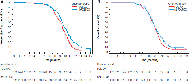
Interestingly, the Kaplan-Meier estimates for OS in the nbFOLFOX and standard FOLFOX groups showed similar OS benefit in both groups. The mean OS was 21.7 months in the nbFOLFOX group compared to 20.7 months in the FOLFOX group, and the median OS was 20.667 months compared to 20.633 months (log-rank χ2 = 2.888, p = 0.089). In comparison to 96.8% and 15.1% for the FOLFOX group, the 1-year and 2-year OS for the nbFOLFOX group were 97.1% and 28.9%. Meanwhile, the HR was 1.242 (95% CI: 0.966–1.597), suggesting that bolus administration may carry a higher risk.
Univariate and multivariate Cox proportional hazards models that identify factors linked to PFS in the entire cohort (264 progressed vs. 3 non-progressed events) are shown in Table 5. Several important predictors were highlighted by the univariate analysis: arm (nbFOLFOX vs. FOLFOX), the HR was 1.532 (95% CI: 1.194–1.967, p = 0.001), indicating that bolus omission resulted in superior PFS. Use of panitumumab is linked to an HR of 1.494 (p = 0.013). In addition to low-grade vomiting and low-grade neuropathy, good performance status demonstrated protective HR indicating that fragile patients are less likely to benefit from treatment. Multivariate analysis maintained independent significance for the arm with bolus inclusion, where the HR was 1.433 (95% CI: 1.111–1.848, p = 0.006), and for the use of panitumumab, HR was 1.408 (p = 0.044). Low-grade neuropathy of HR of 0.454 (p = 0.002) may suggest a protective effect, however, the incident events are too low to depend on (6–13%). Age, gender, side of the tumour, genetic profile (KRAS/BRAF/MSI) and haematologic/GI toxicities were not significant factors.
Table 5
Univariate and multivariate COX regression analysis for the parameters affecting progression-free survival in total sample
Cox proportional hazards models identifying factors associated with OS in the entire cohort (247 deaths vs. 20 survivors) are illustrated in Table 6, which highlights neuropathy and performance status as important prognostic variables while showing no significant effect of bolus omission on OS. In line with the borderline OS benefit seen in the Kaplan-Meier analysis, the treatment arm (nbFOLFOX vs. FOLFOX) displayed a non-significant trend towards improved OS with bolus omission (univariate HR 1.242, p = 0.091), which diminished in multivariate modelling. On univariate analysis, low-grade vomiting, low-grade neuropathy, and an ECOG performance status of 1 emerged as protective factors (p = 0.024, p < 0.001, and p < 0.001, respectively). In multivariate analysis, low-grade neuropathy and good performance status remained independent protective variables, although the very limited number of neuro-- pathy events warrants cautious interpretation.
Table 6
Univariate and multivariate COX regression analysis for the parameters affecting overall survival in total sample
Discussion
Numerous risk factors contribute to the development of CRC, broadly categorized into genetic and behavioural factors. Patients with diabetes mellitus, colonic polyps, inflammatory bowel disease, or a family history of CRC face significantly increased risk [13]. The stage at diagnosis has an impact on the survival rate of CRC, with later-stage diagnoses having a lower survival rate [3].
This retrospective study we conducted to evaluate the impact of omitting the 5-FU bolus on survival outcomes and toxicity among patients receiving standard multidrug 5-FU-based regimens for mCRC. A total 267 patients with mCRC were enrolled and were stratified into two exposure groups based on chemotherapy administration records. Our findings demonstrate that omission of the 5-FU bolus (nbFOLFOX, n = 141) compared to standard administration (FOLFOX, n = 126) resulted in a statistically significant but clinically marginal improvement in PFS (mean PFS 10.029 vs. 9.319 months, HR 1.532, 95% CI: 1.194–1.967, p = 0.001), with the absolute benefit being less than one month. Overall survival was comparable between groups (mean OS 21.7 vs. 20.7 months, median OS 20.667 vs. 20.633 months, HR 1.242, 95% CI: 0.966–1.597, p = 0.089). Multivariate analysis confirmed bolus inclusion as an independent predictor of inferior PFS (HR 1.433, p = 0.006) but showed no significant effect on OS. These results suggest that the 5-FU bolus does not contribute meaningful survival benefit in the context of modern multi-- drug 5-FU-based regimens for mCRC.
Our findings align with emerging evidence questioning the necessity of the 5-FU bolus in contemporary practice. A large multicentre study by Peng et al. [14] analysed 11,765 patients with advanced gastrointestinal cancers from the Flatiron Health database and found that omission of the 5-FU bolus from FOLFOX, FOLFIRI, and FOLFIRINOX regimens was not associated with decreased OS after adjusting for baseline clinical factors (HR 0.99, 95% CI: 0.91–1.07, p = 0.74). Similarly, in a propensity-score matched analysis of 6,126 patients with mCRC, the same group demonstrated no association between 5-FU bolus use and OS (HR 0.98, 95% CI: 0.91–1.06, p = 0.64) [15]. These results, derived from a substantially larger cohort than ours, provide robust support for our conclusion that the bolus component does not enhance survival outcomes.
The modest PFS benefit observed in our nbFOLFOX arm, while statistically significant, is of questionable clinical relevance given the less than one-month absolute difference. A retrospective study by Basilio et al. [16] at the Moffitt Cancer Center evaluated 133 patients receiving first-line mFOLFOX6 with or without 5-FU bolus and found no statistically significant differences in median PFS (8.12 vs. 6.64 months, p = 0.787) or OS (29.36 vs. 21.6 months, p = 0.395). Interestingly, their study reported a numerically higher median OS in the bolus arm, though this difference was not statistically significant and may have been confounded by baseline imbalances in metastatic burden between groups. In contrast, our study showed a trend toward improved OS with bolus omission, though this also did not reach statistical significance. These discrepant trends underscore the lack of consistent survival advantage attributable to the bolus component across different patient populations.
The pharmacologic rationale for omitting the bolus is supported by indirect evidence from capecitabine-based regimens. Capecitabine, an oral fluoropyrimidine that mimics continuous infusion of 5-FU without any bolus component, has demonstrated non-inferiority to FOLFOX regimens in multiple randomised trials. A meta-analysis by Guo et al. [17] including 4,363 patients from eight randomised controlled trials found no statistical differences in OS or objective response rate between XELOX (capecitabine plus oxaliplatin) and FOLFOX. The NO16966 trial, which randomised 2034 patients, showed median OS of 19.8 months with XELOX vs. 19.5 months with FOLFOX (HR 0.95, 97.5% CI: 0.85–1.06), confirming non-inferiority [18]. These findings strongly suggest that the bolus component is not essential for therapeutic efficacy when adequate infusional 5-FU is administered, as the mechanism of action predominantly relies on sustained TS inhibition achieved through continuous exposure.
Our study adds to a growing body of evidence regarding toxicity differences between bolus-containing and bolus- omitted regimens. Previous studies have consistently demonstrated increased haematologic toxicity with bolus administration. Areepium et al. [1] studied 110 patients receiving mFOLFOX6 at the King Chulalongkorn Memorial Hospital and found that patients receiving the 5-FU bolus had significantly lower absolute neutrophil counts compared to those without bolus (mean difference 43.13, 95% CI: 20.74–65.51, p = 0.0002). In a first-line treatment subgroup, this difference was even more pronounced (mean difference 46.01, 95% CI: 19.99–72.03, p = 0.0007). The large Flatiron Health database study confirmed these findings at scale, reporting that bolus omission was associated with significant reductions in neutropenia (10.7% vs. 22.7%, p < 0.01), thrombocytopenia (11.2% vs. 16.1%, p < 0.01), and use of granulocyte colony-stimulating factors (19.6% vs. 29.1%, p < 0.01) [14].
A Japanese study by Tezuka et al. [19] comparing modified FOLFOX7 plus bevacizumab (mF7 + BV, without bolus) found substantially less grade 3/4 neutropenia (7.8%) compared to historical controls receiving mFOLFOX6 plus bevacizumab (24%), with comparable efficacy (median PFS 11.8 months, response rate 50%). Similarly, Okita et al. [20] reported that the stop-and-go modified FOLFOX6 with bevacizumab (which includes bolus) was associated with grade 3/4 neutropenia in 40% of patients, substantially higher than the 7.8% observed in the bolus-omitted mFOLFOX7 regimen [19]. These data collectively support the conclusion that bolus omission reduces haematologic toxicity without compromising efficacy as observed in our study.
The differences in pharmacodynamics between bolus and continuous infusion of 5-FU account for their distinct effects. Bolus administration generates rapid, high peak plasma concentrations favouring the accumulation of FdUTP and FUTP, which are mis-incorporated into DNA and RNA respectively, causing cytotoxicity primarily through nucleic acid disruption [8]. In contrast, continuous infusion maintains prolonged systemic exposure that sustains formation of FdUMP, which forms a stable ternary complex with TS and reduced folates, resulting in durable enzyme inhibition and depletion of thymidine pools during the S-phase of the cell cycle [8]. Historical studies have demonstrated superior antitumor activity with continuous infusion compared to bolus administration in colorectal cancer, primarily due to prolonged suppression of TS [9]. In the context of modern MDR where continuous infusion is already employed alongside oxaliplatin or irinotecan, the additional bolus component appears to contribute primarily to toxicity rather than efficacy.
The clinical implications of our findings are substantial. A survey of gastrointestinal oncologists revealed that 20– 40% routinely omit the 5-FU bolus preemptively, with higher rates among senior clinicians and gastrointestinal malignancy specialists [11]. Our study indicates that experienced oncologists may have recognized the limited benefit of the bolus component in treatment regimens. This finding is particularly significant given recent 5-FU drug shortages, which require prioritizing essential treatment components. The data suggest that omitting the bolus may not negatively impact survival outcomes, while also enhancing drug availability and decreasing treatment-related toxicity and associated healthcare costs.
This study has several notable strengths. First, the multicentre design involving two major academic cancer centres enhances the generalizability of our findings across different practice settings and patient populations. Second, our rigorous inclusion and exclusion criteria, including the requirement for at least 75% dose density and complete follow-up data, minimize bias and ensure the reliability of survival estimates. Third, the use of both univariate and multivariate Cox proportional hazards modelling allowed us to adjust for potential confounders and identify independent prognostic factors. Fourth, the study period (2020–2024) reflects contemporary clinical practice with modern supportive care and concurrent use of targeted therapies such as bevacizumab and anti-EGFR monoclonal antibodies, enhancing the relevance of our findings to current oncology practice. Table 7 compares important points with similar studies.
Table 7
Systematic comparison between the current study and similar studies
| Feature | Current study | Peng et al. [14] | Peng et al. [15] | Basilio et al. [16] | Areepium et al. [1] | Garrido et al. [8] |
|---|---|---|---|---|---|---|
| Cancer type/regimen | mCRC only/mFOLFOX6 only | 3 cancers/ 3 regimens | mCRC/ multiple | mCRC/ mFOLFOX6 | mCRC/ mFOLFOX6 | mCRC/ FOLFOX-6 |
| Sample size (n) | 267 | 11 765 | 6126 | 133 | 110 | Not specified |
| Endpoints | PFS + OS + full toxicity | OS only | OS only | PFS + OS | Toxicity only | Toxicity only |
| ECOG performance status recorded | Yes | Not granularly | Not granularly | Yes (imbalanced) | No | Not reported |
| Molecular markers (KRAS/BRAF/MSI) | Yes (all 3) | Limited | Limited | Not reported | RAS only | Not reported |
| Dose density threshold | ≥ 75% required | None | None | None | None | None |
| Oligometastatic excluded | Yes | No | No | No | Not specified | Not specified |
| Multivariate Cox adjustment | Yes (pre-specified) | IPTW only | Propensity matching | Limited | No | No |
| Prospectively defined SAP | Yes | Not reported | Not reported | No | No | No |
| Multicentre | Yes (2 centres) | Multi (database) | Multi (database) | Single center | Single center | Not specified |
| Study period | 2020–2024 | Database (varied) | Database (varied) | 2015–2019 | 2021–2022 | Not specified |
| Inter-rater reliability | Yes (10% re-abstraction) | N/A | N/A | Not reported | Not reported | Not reported |
Despite the study’s strengths, some limitations must be acknowledged. Its retrospective observational design limits causal inference and introduces risks of selection bias and confounding. Omission of the bolus was not randomised but rather based on physician discretion, possibly leading to systematic differences between groups. Although we attempted to control for known prognostic factors through multivariate modelling. In addition, the study was not prospectively powered for effect sizes, and while post hoc analyses suggest adequate power for primary endpoints, it may be underpowered for detecting small differences in secondary outcomes.
Second, baseline imbalances in survival status may reflect differences in follow-up duration, disease biology, or treatment response that were not fully accounted for in our analysis. Furthermore, the varied concurrent therapies (bevacizumab, cetuximab, panitumumab), while this reflects real-world practice, further complicate analysis. Fourth, the study’s focus on two cancer centres may restrict the generalizability of the findings.
Finally, we did not systematically collect data on quality of life, treatment-related costs, or patient-reported outcomes, all of which are increasingly recognized as important endpoints in oncology research. The convenience of avoiding bolus administration (eliminating the need for a second injection and its associated time and discomfort) represents a potential benefit that was not formally assessed in our study but may be valued by patients and healthcare providers.
The implications of our study for clinical practice are straightforward. Given the lack of survival benefit and potential for increased haematologic toxicity associated with 5-FU bolus, oncologists may reasonably consider omitting this component from mFOLFOX6 regimens, particularly in patients at high risk for myelosuppression, those with poor performance status, elderly patients, or those with significant comorbidities. This approach aligns with current prescribing patterns among experienced gastrointestinal oncologists and is supported by the non-inferiority of capecitabine-based regimens that lack a bolus component [17, 18]. In the context of ongoing or anticipated 5-FU shortages, prioritizing the continuous infusion component over the bolus may optimize drug utilization without compromising patient outcomes.
For healthcare systems, bolus omission may yield cost savings through reduced use of granulocyte colony- stimulating factors, decreased hospitalizations for neutropenic fever, and lower rates of dose delays and treatment discontinuations. These economic benefits should be formally evaluated in future cost-effectiveness analyses. For patients, the potential reduction in haematologic and gastrointestinal toxicity may translate to improved quality of life and treatment tolerability, though this requires validation through prospective patient-reported outcome studies.
Several important questions remain unanswered and warrant further investigation. First, a randomised controlled trial directly comparing mFOLFOX6 with and without 5-FU bolus would provide the highest level of evidence for non-inferiority in survival outcomes and superiority in toxicity reduction. Such a trial should include comprehensive toxicity assessment, quality of life measurements, and pharmacoeconomic analyses. Second, the optimal approach to managing toxicity when it occurs – whether to reduce the infusion dose, omit the bolus, or both – remains unclear and deserves systematic study. Third, the role of 5-FU bolus in the adjuvant setting has not been adequately examined, and extrapolation from the metastatic setting may not be appropriate given different treatment goals and patient populations.
Moreover, potential predictive biomarkers for patients who might derive particular benefit or harm from bolus administration should be explored, including pharmacogenetic markers such as dihydropyrimidine dehydrogenase polymorphisms, TS expression levels, and other molecular predictors of fluoropyrimidine sensitivity and toxicity. Comparative effectiveness research examining different bolus-omitted regimens would help identify the most efficacious and tolerable fluoropyrimidine-based backbone for combination with oxaliplatin. The impact of bolus omission in combination with newer agents including immune checkpoint inhibitors for microsatellite instability-high tumours deserves investigation. Finally, prospective collection of patient-reported outcomes and quality of life data in the context of bolus-omitted regimens would provide valuable patient-centred evidence to complement traditional clinical endpoints.
Conclusions
Our study demonstrates that omission of the 5-FU bolus from mFOLFOX6 regimens in mCRC is not associated with inferior survival outcomes and may offer a favourable toxicity profile. These findings, supported by emerging evidence from large database studies and the non-inferiority of bolus-free capecitabine regimens, challenge the routine inclusion of 5-FU bolus in modern MDR and suggest that clinical practice may benefit from reconsidering this component as optional rather than essential. While confirmatory randomised trials would be ideal, the accumulating observational evidence provides a reasonable basis for individualizing treatment decisions based on patient-specific risk factors and preferences, particularly in the context of resource limitations or drug shortages.










