Summary
This study investigated the prognostic value of the tricuspid annular plane systolic excursion (TAPSE)/pulmonary artery systolic pressure (PASP) ratio – a marker of right ventricular–pulmonary arterial (RV–PA) coupling – in 225 patients with severe aortic stenosis and reduced left ventricular ejection fraction (LVEF < 50%) who underwent transcatheter aortic valve implantation (TAVI). Echocardiographic measurements were obtained before the procedure and at discharge. Over a median follow-up of approximately 3 years, 102 patients died. Survivors had significantly higher post-procedural TAPSE/PASP ratios compared to non-survivors. Multivariable analysis showed that the post-TAVI TAPSE/PASP ratio independently predicted long-term mortality, with each 0.1 mm/mmHg increase associated with a 26% reduction in mortality risk. The pre-procedural ratio, however, did not reach independent significance. Kaplan–Meier analysis confirmed worse 5-year survival in patients with a post-discharge TAPSE/PASP below 0.514 mm/mmHg. These findings suggest that persistent RV–PA uncoupling at discharge – rather than the pre-procedural status – is the stronger predictor of long-term mortality in this high-risk population, and that routine assessment of TAPSE/PASP after TAVI may improve risk stratification.
Introduction
Degenerative aortic stenosis is the most common valvular heart disease. Transcatheter aortic valve implantation (TAVI) is an established alternative to surgical aortic valve replacement. It represents a less invasive approach to treating severe aortic stenosis (AS), particularly in elderly patients with multiple comorbidities. Severe AS with low ejection fraction (LVEF < 50%) represents a high-risk cardiac condition associated with increased morbidity and mortality. Despite continuous advances in device technology and procedural techniques, outcomes remain heterogeneous, particularly in patients with impaired left ventricular ejection fraction (LVEF) after TAVI [1]. Beyond left ventricular dysfunction, these patients frequently develop pulmonary hypertension (PH) and progressive right ventricular (RV) impairment due to chronically elevated left-sided filling pressures. Persistent PH imposes increased afterload on the right ventricle, leading to RV dysfunction. The prevalence of PH in aortic stenosis ranges from 14% to 59%, and it is associated with adverse outcomes after TAVI [2]. Prior studies have consistently demonstrated that PH is associated with increased morbidity and mortality following TAVI [3–5]. However, pulmonary artery pressure alone does not fully capture the complex interaction between RV contractile function and pulmonary vascular load.
Tricuspid annular plane systolic excursion (TAPSE) is a simple echocardiographic measure of RV longitudinal systolic function, while pulmonary artery systolic pressure (PASP) reflects pulmonary hemodynamic load. The TAPSE/PASP ratio integrates RV contractility with afterload and has been shown to correlate with RV–pulmonary arterial (PA) coupling [6] and has demonstrated prognostic value across various cardiovascular conditions, including pulmonary hypertension and heart failure. Recent studies have highlighted the importance of RV–PA coupling in patients undergoing TAVI [7]. However, data specifically focusing on patients with low LVEF and peri-procedural changes in TAPSE/PASP remain limited.
Aim
This study aimed to investigate the association between pre-procedural TAPSE/PASP ratio and long-term all-cause mortality, and to evaluate whether post-procedural TAPSE/PASP ratio – assessed at discharge as a landmark post-discharge risk marker – identifies patients who remain at increased risk of long-term mortality following TAVI in patients with severe AS and low ejection fraction (LVEF < 50%).
Material and methods
Study design and population
This single-center, retrospective observational study included consecutive patients with severe aortic stenosis and low left ventricular ejection fraction (LVEF < 50%) who underwent TAVI between January 2015 and July 2025. In the present study, low ejection fraction was defined as LVEF < 50%, as both mildly reduced (40–49%) and reduced (< 40%) subcategories fall below the normal range and represent a clinically meaningful impairment of left ventricular systolic function. This threshold was chosen intentionally to reflect a broader at-risk population beyond the conventional definition of heart failure with reduced ejection fraction (HFrEF). Severe AS was defined as an aortic valve area < 1.0 cm² in accordance with the joint European Association of Cardiovascular Imaging (EACVI) and American Society of Echocardiography (ASE) recommendations [8].
Patients were excluded for the following reasons: in-hospital mortality (n = 20); surgical conversion due to major structural complications such as aortic dissection, annular rupture, or device embolization (n = 3); end-stage kidney failure (estimated glomerular filtration rate [eGFR] < 30 ml/min/1.73 m² or chronic hemodialysis) (n = 12); missing data (n = 23); severe anemia (hemoglobin < 8 g/dl) (n = 4); valve-in-valve TAVI (n = 9); mitral valve transcatheter edge-to-edge repair (MTEER) (n = 3), and active malignancy (n = 1). After applying these criteria, 225 patients constituted the study cohort.
This study was conducted in accordance with the Declaration of Helsinki, and institutional ethics committee approval was granted on 19 February 2026 (approval no: 364). As this was a retrospective study, specific prospective consent was not required. However, written informed consent had been obtained from all patients at hospital admission for clinical procedures and included consent for scientific use of their clinical data, in accordance with institutional practice.
Data collection
Baseline clinical characteristics, comorbidities, medications at follow-up, laboratory values, echocardiographic parameters, and procedural information were extracted from the hospital electronic medical record system and patient files. Long-term mortality was ascertained using the hospital record system and the national health registry.
Comprehensive transthoracic echocardiography was performed at hospital admission (pre-procedural) and on the day of discharge after TAVI (post-procedural), according to EACVI/ASE recommendations, using a Vivid E95 system (GE Vingmed Ultrasound, Milwaukee, WI, USA). LVEF was assessed using the biplane Simpson’s method. TAPSE was measured by M-mode in the apical four-chamber view. PASP was estimated from peak tricuspid regurgitation velocity with the addition of estimated right atrial pressure based on inferior vena cava diameter and collapsibility. Left ventricular and left atrial dimensions, valvular regurgitation grades, aortic valve gradients, and aortic valve area (continuity equation) were also recorded.
TAVI procedure
Diagnostic coronary angiography via a radial approach was performed in all patients, typically 2 days before the TAVI procedure. Stenosis ≥ 90% in epicardial coronary vessels with a diameter of ≥ 2.5 mm was considered significant and managed with percutaneous coronary intervention (PCI) when deemed appropriate following case-based evaluation.
Pre-procedural computed tomography analyses were performed using 3mensio Structural Heart software (Pie Medical Imaging, Maastricht, the Netherlands). The vascular access strategy was determined based on CT findings, and femoral access was obtained under fluoroscopic and ultrasound guidance. A 6-F pigtail catheter was introduced via the contralateral femoral artery for contrast injection during valve implantation. A temporary pacemaker lead was positioned in the right ventricular apex through the contralateral femoral vein. Valve type and size were selected based on CT measurements and case-based assessment. Vascular closure was achieved using Perclose ProGlide (Abbott Vascular, Santa Clara, CA, USA) and Angio-Seal (Terumo Corp., Tokyo, Japan) when required. Surgical repair, covered stent implantation, or peripheral balloon dilatation was performed in cases of major vascular complications or closure device failure.
Outcomes and follow-up
The primary endpoint was long-term all-cause mortality. Follow-up information was obtained from outpatient visits and the hospital electronic medical record system. Procedural outcomes, including technical success and structural, vascular, and bleeding complications, were defined according to the Valve Academic Research Consortium-3 (VARC-3) criteria [9].
Statistical analysis
Patients were categorized according to long-term mortality status. Categorical variables were compared using the χ2 test or Fisher’s exact test, as appropriate. Continuous variables were compared using Student’s t-test for normally distributed data or the Mann–Whitney U test for non-normally distributed data. Distribution normality was assessed using visual inspection of histograms and the Kolmogorov–Smirnov test. Continuous variables are reported as mean ± standard deviation or median (interquartile range), as appropriate. For continuous variables, values are reported to one decimal place. Categorical variables are presented as counts and percentages. For p-values ≥ 0.05, results are presented to two decimal places.
Univariable Cox proportional hazards regression was performed to evaluate potential predictors of long-term mortality. Variables showing an association at p < 0.10 in univariable analysis were entered into multivariable Cox proportional hazards models. To avoid collinearity, correlated variables were not entered into the same model. EuroSCORE II was considered for inclusion in multivariable models; however, given its collinearity with age, COPD, and renal function, it was not incorporated in the final models. Year of enrollment was included as a covariate in all multivariable models to account for potential changes in operator experience, procedural technique, and device generation over the study period. Separate multivariable models were constructed for pre-procedural and post-procedural TAPSE/PASP ratios to evaluate their respective prognostic value. In the post-TAVI model, the index date was set as the date of discharge, and only patients who survived to discharge were included, in keeping with a landmark post-discharge analysis design. Hazard ratios for TAPSE/PASP are reported per 0.1 mm/mm Hg increase to facilitate clinical interpretability, given that observed values cluster between 0.4 and 0.6 mm/mm Hg.
Standard receiver-operating characteristic (ROC) analysis was performed as an exploratory tool to identify optimal cut-off values for TAPSE/PASP ratios using Youden’s index. It is acknowledged that this method does not account for censoring or follow-up time; accordingly, these results should be interpreted with caution and are presented as supportive rather than primary analyses. Kaplan–Meier survival curves were constructed for high versus low TAPSE/PASP groups defined by these cut-offs, and survival distributions were compared using the log-rank test. For Kaplan–Meier survival analyses, follow-up duration was administratively truncated at 5 years to allow uniform visualization and avoid instability in estimates at longer follow-up durations.
All statistical analyses were performed using Python version 3.14.0 (Python Software Foundation, Wilmington, DE, USA). A two-sided p-value < 0.05 was considered statistically significant.
Results
A total of 225 patients were included in the study, comprising 123 survivors and 102 non-survivors. Compared with non-survivors, survivors were younger (73.9 ±9.8 vs. 78.8 ±7.5 years, p < 0.001) and had a higher body-mass index (BMI) (27.9 ±4.1 vs. 26.6 ±4.1 kg/m², p = 0.022) and a lower EuroSCORE II (18.3 ±11.0 vs. 27.8 ±11.6, p < 0.001) (Table I). With regard to comorbidities, the survivor group had a lower prevalence of chronic obstructive pulmonary disease (COPD) (31.7% vs. 58.8%, p < 0.001), chronic kidney disease (CKD) (37.4% vs. 54.9%, p = 0.01), and coronary artery disease (CAD) (63.4% vs. 77.5%, p = 0.03) (Table I). SYNTAX scores were also lower in survivors (10.7 ±11.5 vs. 15.1 ±9.6, p = 0.002). Follow-up medical therapy was generally similar between groups, except for a higher rate of sodium–glucose cotransporter-2 (SGLT-2) inhibitor use among survivors (33.3% vs. 19.6%, p = 0.03).
Table I
Comparison of baseline clinical characteristics, comorbidities, follow-up medications, laboratory parameters, and procedural outcomes for survivors and non-survivors after TAVI
[i] COPD – chronic obstructive pulmonary disease, ACEi – angiotensin converting enzyme inhibitors, ARB – angiotensin II receptor blocker, SGLT-2i – sodium-glucose cotransporter-2 inhibitors, ARNI – angiotensin receptor neprilysin inhibitor, eGFR – estimated glomerular filtration rate, AST – aspartate aminotransferase, ALT – alanine aminotransferase, LDL – low-density lipoprotein, TSH – thyroid-stimulating hormone.
Laboratory findings showed higher hemoglobin levels (11.7 ±1.7 vs. 11.2 ±1.8 g/dl, p = 0.02) and better renal function parameters in the survivor group (Table I).
Baseline echocardiographic parameters, including LVEF, aortic gradients, and aortic valve area, were largely comparable between groups. However, severe mitral regurgitation (MR) was more frequent among non-survivors (23.6% vs. 37.3%, p = 0.04) (Table II). Three patients who underwent mitral transcatheter edge-to-edge repair (TEER) were excluded from the study.
Table II
Comparison of echocardiographic and procedural parameters for survivors and non-survivors after TAVI
Post-procedural echocardiographic findings were broadly similar between groups, except for post-TAVI severe MR (Table II). Regarding the primary focus of the study, baseline TAPSE, PASP, and TAPSE/PASP values did not differ significantly between survivors and non-survivors. In contrast, post-TAVI echocardiography demonstrated lower PASP (38.1 ±10.5 vs. 41.9 ±11.8 mm Hg, p = 0.03), higher TAPSE (19.5 ±2.5 vs. 18.6 ±2.4 mm, p = 0.005), and a higher TAPSE/PASP ratio (0.56 ±0.20 vs. 0.48 ±0.16 mm/mm Hg, p = 0.005) in survivors (Table II).
With respect to procedural characteristics, survivors more frequently received self-expandable valves (70.7% vs. 52.9%, p = 0.009), whereas other procedural variables and complication rates were comparable between groups (Table II). The median follow-up duration for the overall cohort was 1161 days (interquartile range [IQR]: 604–1889 days).
All baseline clinical variables, comorbidities, medications, laboratory values, echocardiographic parameters, and procedural characteristics were evaluated using univariable Cox proportional hazards analysis to identify predictors of long-term mortality. In univariable analysis, age, baseline aortic mean and maximum gradients, low-flow low-gradient aortic stenosis, COPD, eGFR, baseline and post-procedural hemoglobin levels, CKD, creatinine levels, baseline and post-procedural severe MR, CAD, post-procedural LVEF, baseline and post-procedural TAPSE/PASP ratios, and BMI were associated with long-term mortality (Table III).
Table III
Univariate Cox regression analysis of factors associated with long-term mortality
[i] COPD – chronic obstructive pulmonary disease, CABG – coronary artery bypass grafting, ACEi – angiotensin converting enzyme inhibitors, ARB – angiotensin II receptor blocker, SGLT-2i – sodium-glucose cotransporter-2 inhibitors, ARNI – angiotensin receptor neprilysin inhibitor, eGFR – estimated glomerular filtration rate, AST – aspartate aminotransferase, LVEF – left ventricular ejection fraction, PVL – paravalvular leak, TAPSE – tricuspid annular plane systolic excursion, TAVI – transcatheter aortic valve implantation, PASP – pulmonary artery systolic pressure, AS – aortic stenosis, MR – mitral regurgitation, TR – tricuspid regurgitation, AR – aortic regurgitation, MRA – mineralocorticoid receptor antagonist.
To avoid collinearity and overfitting, correlated variables were not included in the same multivariable model. Selected covariates for multivariable Cox regression included age, pre-procedural aortic mean gradient, COPD, CKD, baseline hemoglobin, post-TAVI severe MR, CAD, post-TAVI LVEF, EuroSCORE II, year of enrollment, and BMI, together with TAPSE/PASP ratios.
In model 1, the baseline TAPSE/PASP ratio was included alongside these covariates. Model 1 identified age (HR = 1.053, 95% CI: 1.026–1.081, p < 0.001), pre-procedural aortic mean gradient (HR = 0.972, 95% CI: 0.956–0.989, p = 0.001), COPD (HR = 1.860, 95% CI: 1.255–2.755, p = 0.002), post-TAVI severe MR (HR = 1.619, 95% CI: 1.004–2.611, p = 0.05), and CAD (HR = 1.615, 95% CI: 1.007–2.590, p = 0.05) as independent predictors of mortality (Table IV, Figures 1 and 2). The baseline TAPSE/PASP ratio showed a borderline association with long-term mortality (HR = 0.861, 95% CI: 0.740–1.002, p = 0.05) (Table IV, Figure 2); this association did not reach conventional statistical significance.
Table IV
Multivariable Cox regression analysis for long-term mortality
Figure 1
Receiver-operating characteristic (ROC) curves for prediction of long-term mortality. A – Pre-procedural parameters. B – Post-procedural parameters
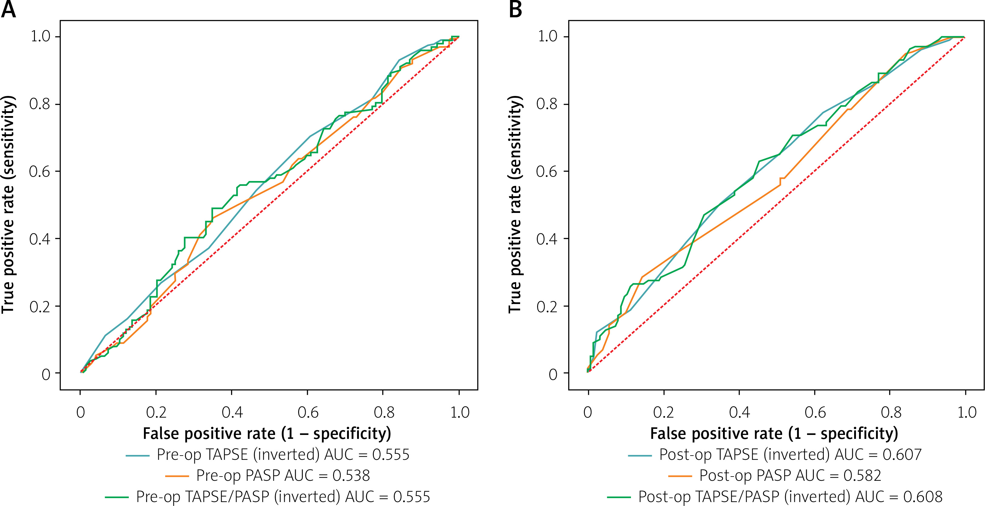
Figure 2
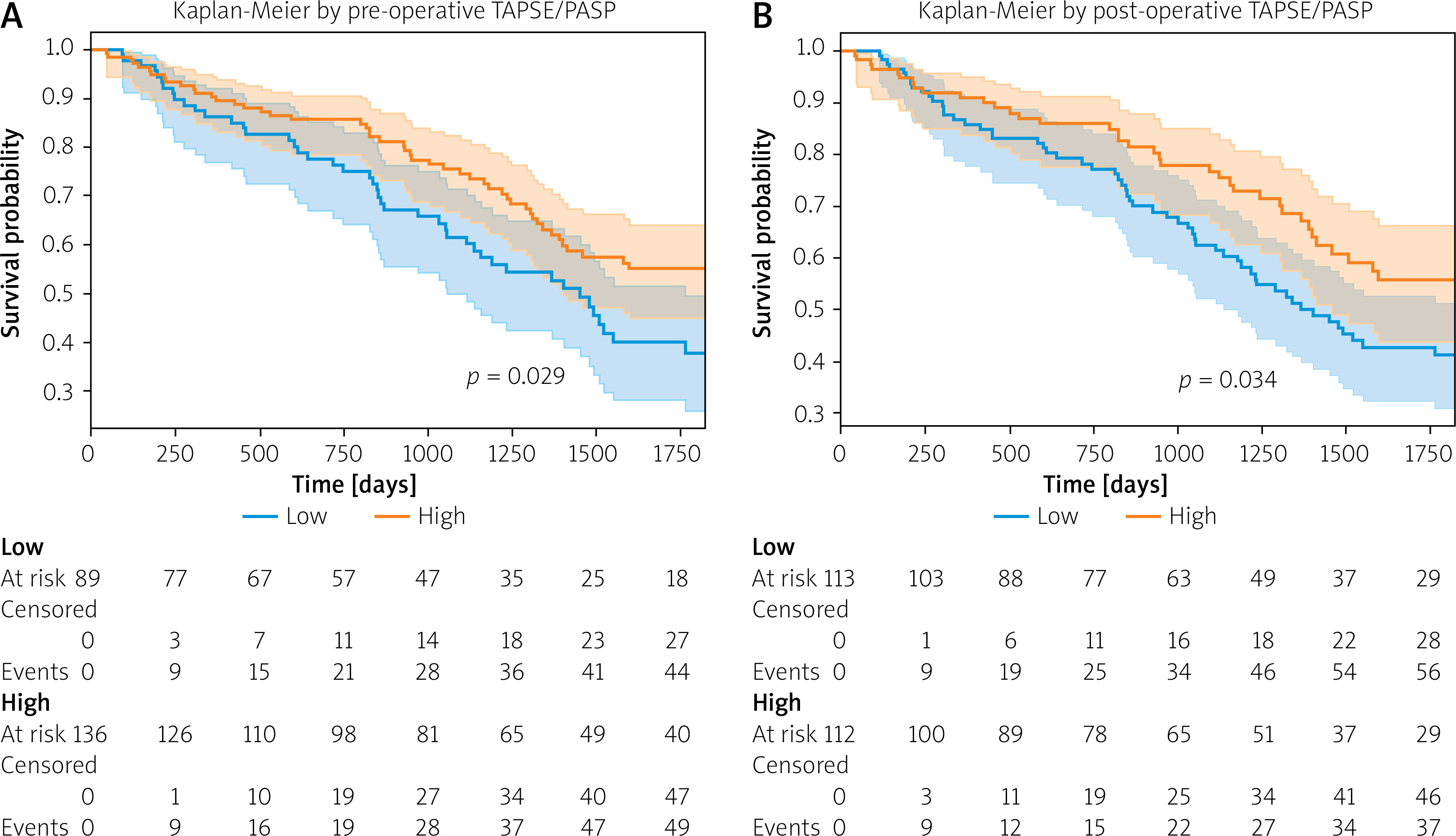
Kaplan–Meier survival analysis according to TAPSE/PASP ratio. A – Pre-procedural TAPSE/PASP ratio. B – Post-procedural TAPSE/PASP ratio
In model 2, which included the post-procedural TAPSE/PASP ratio instead of the baseline ratio, age (HR = 1.051, 95% CI: 1.024–1.078, p < 0.001), baseline aortic mean gradient (HR = 0.973, 95% CI: 0.956–0.990, p = 0.002), COPD (HR = 1.781, 95% CI: 1.207–2.628, p = 0.004), CAD (HR = 1.595, 95% CI: 1.002–2.537, p = 0.05), and the post-TAVI TAPSE/PASP ratio were independently associated with long-term mortality. The hazard ratio for post-TAVI TAPSE/PASP, expressed per 0.1 mm/mm Hg increase, was HR = 0.74, 95% CI: 0.55–0.99, p = 0.04, indicating that each 0.1 mm/mm Hg increment in discharge TAPSE/PASP was associated with a 26% reduction in the risk of subsequent long-term mortality (Table IV).
ROC curve analysis was performed as an exploratory analysis; however, given that standard ROC analysis does not account for censoring and follow-up time, these results should be interpreted with caution. The optimal cut-off for baseline TAPSE/PASP was 0.380 mm/mm Hg (sensitivity = 0.49, specificity = 0.65), with AUC values of 0.555, 0.538, and 0.555 for baseline TAPSE, PASP, and TAPSE/PASP, respectively. For post-TAVI TAPSE/PASP, the optimal cut-off was 0.514 mm/mm Hg (sensitivity = 0.63, specificity = 0.55), with AUC values of 0.607, 0.582, and 0.608 for post-TAVI TAPSE, PASP, and TAPSE/PASP, respectively. These AUC values indicate modest discriminative ability. TAPSE/PASP should therefore be regarded as a prognostic stratification marker rather than a standalone diagnostic test.
Kaplan–Meier survival analysis demonstrated significantly higher 5-year mortality among patients with low TAPSE/PASP ratios according to both baseline and post-procedural cut-off values (log-rank p = 0.029 for baseline TAPSE/PASP and p = 0.034 for post-TAVI TAPSE/PASP) (Figure 2).
Discussion
In this study, we evaluated the prognostic significance of RV–PA coupling assessed by the TAPSE/PASP ratio in patients with severe aortic stenosis and low left ventricular ejection fraction (LVEF < 50%) undergoing TAVI. The principal findings can be summarized as follows: First, the post-procedural TAPSE/PASP ratio was significantly higher in survivors compared with non-survivors. Second, the post-TAVI TAPSE/PASP ratio – evaluated as a landmark post-discharge risk marker – independently predicted subsequent long-term mortality after adjustment for relevant clinical and echocardiographic covariates. Third, the pre-procedural TAPSE/PASP ratio showed only a borderline, non-significant association in multivariable analysis and did not independently predict long-term mortality (p = 0.05). These findings indicate that persistent impairment of RV–PA coupling at discharge reflects residual cardiopulmonary burden and carries important prognostic implications in patients with LVEF < 50% undergoing TAVI.
Right ventricular function is a critical determinant of outcomes in patients with severe aortic stenosis undergoing transcatheter valve interventions. Chronic pressure overload caused by aortic stenosis leads to elevated left ventricular filling pressures, which subsequently result in pulmonary venous hypertension and progressive pulmonary vascular remodeling. This process increases right ventricular afterload and ultimately promotes right ventricular dysfunction. Previous studies have consistently demonstrated that impaired RV function is associated with increased mortality following TAVI. For example, Scheggi et al. reported that right ventricular dysfunction was independently associated with adverse outcomes in patients undergoing transcatheter valve replacement for severe aortic stenosis [10]. Similarly, observational cohorts have highlighted the prognostic relevance of right-sided hemodynamic parameters such as PASP and TAPSE in this population [11].
Pulmonary hypertension is a well-established predictor of adverse outcomes after TAVI. Alushi et al. demonstrated that PH confers a substantial prognostic burden even after valve replacement [12]. Subsequent studies emphasized that pulmonary pressure alone does not adequately reflect RV adaptation to afterload. However, isolated assessment of either pulmonary pressure or right ventricular contractility may not adequately capture the complex physiological interaction between the right ventricle and the pulmonary circulation. The concept of RV–PA coupling provides a more comprehensive representation of right heart adaptation to afterload by integrating RV contractile function with pulmonary vascular load. The TAPSE/PASP ratio has therefore been proposed as a practical echocardiographic surrogate of RV–PA coupling and has demonstrated prognostic value across a wide spectrum of cardiovascular conditions. Recent reviews and mechanistic analyses emphasize that RV–PA coupling represents a central determinant of hemodynamic stability in patients undergoing structural heart interventions.
Steffen et al. demonstrated that impaired RV–PA coupling is associated with worse outcomes across different AS phenotypes undergoing TAVI [13]. Similarly, Hakgor et al. reported that preoperative RV–PA coupling strongly predicts short- and long-term prognosis after TAVI [14]. Growing evidence suggests that RV–PA coupling plays an important role in the prognosis of patients undergoing TAVI. In a comprehensive review of transcatheter structural heart interventions, Theofilis et al. highlighted the importance of RV–PA interaction as a key determinant of clinical outcomes [15]. Similarly, Boxhammer et al. demonstrated that parameters reflecting right-heart performance, including TAPSE, pulmonary artery pressure, and RV–PA coupling, are strongly associated with survival in real-world TAVI cohorts [16].
Mendes et al. evaluated the impact of RV–PA coupling in a cohort of TAVI patients and reported that TAPSE/PASP was associated with outcomes in this population, with findings broadly consistent with the present study [17]. Meucci et al. prospectively examined the evolution of RV–PA coupling after TAVI and found that improvement in TAPSE/PASP following valve replacement was associated with better long-term outcomes, underscoring the dynamic nature of RV–PA coupling in this context [18].
Our findings are also consistent with recent studies specifically examining the TAPSE/PASP ratio in this setting. Tanyeri Uzel et al. reported that post-discharge RV–PA coupling assessed by TAPSE/PASP predicts long-term outcomes following TAVI [19]. The present study confirms and extends these observations in a cohort of patients with EF < 50%, a subgroup characterized by advanced myocardial remodeling, impaired ventricular interaction, and a higher burden of pulmonary vascular disease. In such patients, right ventricular dysfunction may represent a late manifestation of chronic cardiopulmonary remodeling and therefore provide incremental prognostic information beyond traditional left-sided parameters.
Beyond TAPSE-based assessment, right ventricular free wall longitudinal strain (RV-FWS) has emerged as a more sensitive early marker of RV dysfunction, capable of detecting subclinical impairment before changes in TAPSE become apparent. The RV-FWS/PASP ratio has been proposed as an alternative surrogate of RV–PA coupling. Lillo et al. and Parasca et al. demonstrated that RV-FWS/PASP provides early prognostic information in patients with severe aortic stenosis undergoing TAVI and may detect RV dysfunction before conventional parameters such as TAPSE deteriorate [20, 21]. Integration of strain-based RV–PA coupling indices alongside TAPSE/PASP may further enhance risk stratification in this population and represents an important avenue for future research.
A key observation of our study is that post-procedural TAPSE/PASP, assessed as a landmark marker at discharge, was independently associated with subsequent mortality, whereas the pre-procedural ratio showed only borderline significance (post TAVI TAPSE/PASP: HR = 0.74, 95% CI: 0.55–0.99, p = 0.04 vs. pre TAVI TAPSE/PASP: HR = 0.861, 95% CI: 0.740–1.002, p = 0.05). This suggests that the residual coupling status at discharge, rather than the pre-procedural baseline, more accurately reflects the integrated cardiopulmonary response to TAVI. Pre-procedural TAPSE/PASP may reflect the burden of chronic pressure overload but may not independently predict survival after valve replacement once other clinical and hemodynamic variables are accounted for. Although TAVI relieves left ventricular outflow obstruction and reduces afterload, improvement in pulmonary hemodynamics and right ventricular performance is variable. Some patients demonstrate substantial recovery of pulmonary pressures and RV function after valve replacement, whereas others exhibit persistent pulmonary hypertension and ongoing RV–PA uncoupling.
Prior studies evaluating pulmonary hypertension dynamics after TAVI have shown that persistent or incompletely reversible PH is associated with worse outcomes, supporting this interpretation [22]. Persistent RV–PA uncoupling following TAVI likely reflects several pathophysiological mechanisms. Chronic pulmonary vascular remodeling may lead to fixed increases in pulmonary vascular resistance that are not fully reversible after correction of the valvular lesion. In addition, prolonged right ventricular pressure overload may result in structural and functional myocardial changes that limit the capacity for RV recovery. Finally, ventricular interdependence and persistent left ventricular dysfunction may continue to influence right ventricular performance even after relief of the valvular obstruction. Consequently, post-discharge TAPSE/PASP may more accurately represent the residual cardiopulmonary burden after TAVI and therefore provide stronger prognostic information.
Patients with LVEF < 50% and low-flow states represent a particularly high-risk subgroup of aortic stenosis. In these patients, the interaction between impaired left ventricular contractility, pulmonary hypertension, and right ventricular dysfunction may contribute substantially to adverse outcomes despite successful valve replacement. As suggested by recent studies, low-flow physiology in aortic stenosis is not solely determined by left ventricular systolic dysfunction but also reflects complex hemodynamic interactions involving the right ventricle and pulmonary circulation [23]. Assessment of RV–PA coupling may therefore provide additional insight into the global cardiopulmonary status of these patients and help refine risk stratification beyond conventional echocardiographic parameters.
Patients with low-flow, low-gradient AS represent a particularly high-risk subgroup. Registry data from TOPAS-TAVI and subsequent analyses have shown higher mortality rates in these patients, particularly in paradoxical low-flow phenotypes [24]. Our findings suggest that RV–PA coupling may serve as a unifying prognostic marker across AS subtypes by capturing the downstream consequences of chronic left-sided pressure overload.
It is also noteworthy that pre-procedural PASP was identified as a predictor of acute kidney injury following TAVI in a recent study published in this journal [25], consistent with the systemic consequences of elevated right-sided pressures. Furthermore, myocardial strain and work analyses in patients with concomitant severe aortic stenosis and mitral regurgitation have highlighted the complexity of hemodynamic interactions in this population [26], which is particularly relevant to our cohort given the higher prevalence of severe mitral regurgitation among non-survivors.
The TAPSE/PASP ratio is a simple, widely available echocardiographic parameter obtainable during routine transthoracic echocardiography without additional cost or imaging modalities. Incorporating assessment of RV–PA coupling into the post-procedural evaluation of patients undergoing TAVI may provide additional prognostic information and help identify individuals with persistent hemodynamic impairment after a technically successful procedure.
Specifically, patients with a post-TAVI TAPSE/PASP ratio below the identified threshold of 0.514 mm/mm Hg at discharge may represent a higher-risk subgroup warranting closer follow-up. In practical terms, clinicians encountering such patients might consider: (1) more frequent post-discharge echocardiographic surveillance to monitor RV function and pulmonary pressures; (2) optimization of guideline-directed medical therapy for heart failure, including maximally tolerated doses of renin–angiotensin–aldosterone system inhibitors and β-blockers, with consideration of SGLT-2 inhibitors given their favorable effects on RV afterload and congestion; (3) early referral for specialist evaluation of pulmonary hypertension if elevated PASP persists at follow-up; and (4) multidisciplinary heart team discussion regarding mitral valve pathology, given the higher prevalence of severe MR in non-survivors. It must be explicitly acknowledged, however, that no randomized controlled trial data currently exist to support any specific intervention based solely on post-TAVI TAPSE/PASP thresholds. The proposed management strategies are, therefore, hypothesis-generating and should be validated in prospective studies.
Several limitations should be considered when interpreting the findings of this study. First, the retrospective design may introduce selection bias and unmeasured confounding factors. Second, the study was conducted at a single tertiary center, which may limit the generalizability of the results. Third, echocardiographic measurements were obtained from routine clinical practice and were not adjudicated by a centralized core laboratory. Fourth, ROC-based cut-off values were derived using standard methods that do not account for censoring; these should therefore be treated as exploratory findings. Fifth, although EuroSCORE II was not included in the final multivariable models due to collinearity with age, COPD, and renal function, residual confounding by unmeasured variables cannot be excluded. Sixth, RV-FWS was not systematically assessed in this cohort, which may limit comparison with strain-based RV–PA coupling indices.
Finally, although the sample size was moderate, larger prospective multicenter studies are required to confirm the prognostic thresholds of TAPSE/PASP and determine its incremental predictive value over established risk models used in TAVI populations.
Conclusions
In patients with severe aortic stenosis and low left ventricular ejection fraction (LVEF < 50%) undergoing TAVI, RV–PA coupling assessed by the TAPSE/PASP ratio provides important prognostic information. While the pre-procedural TAPSE/PASP ratio did not independently predict long-term mortality in multivariable analysis, a lower post-TAVI TAPSE/PASP ratio – evaluated as a landmark post-discharge risk marker among patients surviving to hospital discharge – independently predicted subsequent long-term mortality. These findings suggest that persistent RV–PA uncoupling at discharge reflects residual cardiopulmonary dysfunction and identifies patients who remain at increased risk despite technically successful TAVI. Incorporating TAPSE/PASP assessment into routine post-procedural echocardiographic evaluation may enhance risk stratification and guide closer clinical follow-up in this high-risk population.